Abstract
A 17-year old female presented with a chief complaint of melena and epigastric pain. She had a family history of colon cancer, her mother having been diagnosed with hereditary nonpolyposis colorectal carcinoma (HNPCC). After close examination including double-balloon enteroscopy, the patient was diagnosed with small bowel carcinoma, in spite of her young age. Here we report this rare case of small bowel carcinoma in a young patient with a family history of HNPCC.
Go to : 
References
1. Aarnio M, Mecklin JP, Aaltonen LA, Nyström-Lahti M, Järvinen HJ. Lifetime risk of different cancers in hereditary non-polyposis colorectal cancer (HNPCC) syndrome. Int J Cancer. 1995; 64:430–433.

2. Hampel H, Stephens JA, Pukkala E, et al. Cancer risk in hereditary nonpolyposis colorectal cancer syndrome: later age of onset. Gastroenterology. 2005; 129:415–421.

3. Lindor NM, Petersen GM, Hadley DW, et al. Recommendations for the care of individuals with an inherited predisposition to Lynch syndrome: a systematic review. JAMA. 2006; 296:1507–1517.
4. Anaya DA, Chang GJ, Rodriguez-Bigas MA. Extracolonic manifestations of hereditary colorectal cancer syndromes. Clin Colon Rectal Surg. 2008; 21:263–272.

5. Lynch HT, Smyrk TC, Lynch PM, et al. Adenocarcinoma of the small bowel in lynch syndrome II. Cancer. 1989; 64:2178–2183.

6. Schulmann K, Engel C, Propping P, Schmiegel W. Small bowel cancer risk in Lynch syndrome. Gut. 2008; 57:1629–1630.

7. Winawer S, Fletcher R, Rex D, et al. Gastrointestinal Consortium Panel. Colorectal cancer screening and surveillance: clinical guidelines and rationale-Update based on new evidence. Gastroenterology. 2003; 124:544–560.

8. Rodriguez-Bigas MA, Vasen HF, Lynch HT, et al. Characteristics of small bowel carcinoma in hereditary nonpolyposis colorectal carcinoma. International Collaborative Group on HNPCC. Cancer. 1998; 83:240–244.
Go to : 
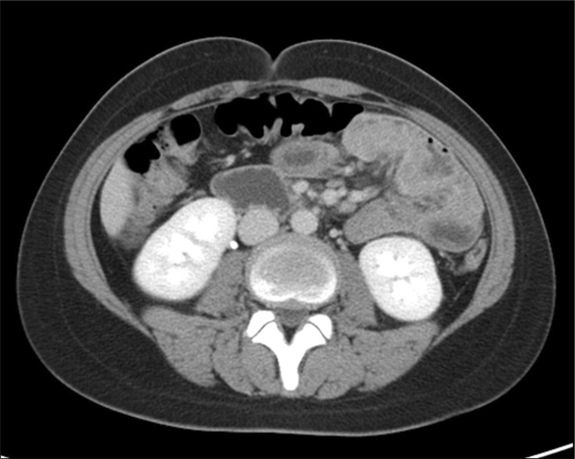 | Fig. 1.Abdominal CT finding. It revealed segmental eccentric wall thickening of the jejunum which indicated a pathology of lymphoma or adenocarcinoma. |
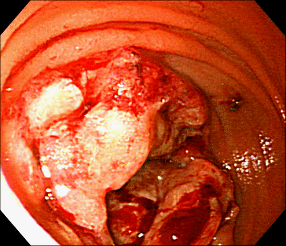 | Fig. 2.Endoscopic finding. A large cancerous lesion with friable bleeding mucosa was found in the proximal jejunum that almost obstructed the bowel. |
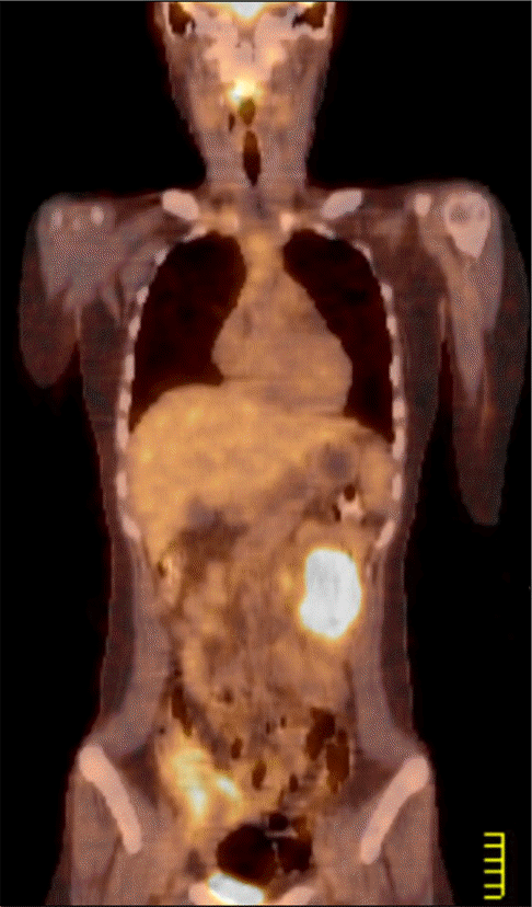 | Fig. 3.PET scan finding. It revealed that the jejunal mass showed intense hypermetabolism. There were no other abnormal metabolic lesions. |


 PDF
PDF ePub
ePub Citation
 Print
Print


 XML Download