Abstract
Fish bones are often ingested accidently. Most of them passes out through the gastrointestinal tract safely, but serious complications, such as perforation, abscess, obstruction, and bleeding in the gastrointestinal tract, can occur. An ingested fish bone can be easily removed by endoscopy, and surgery is rarely required. However, there may be complications related to the endoscopic procedure including mucosal laceration, bleeding, fever, and perforation. Here, we report a case of retroperitoneal hemorrhage developed after endoscopic removal of a fish bone stuck in the duodenal wall, and then resolved spontaneously by conservative care.
Go to : 
References
1. Eisen GM, Baron TH, Dominitz JA, et al. American Society for Gastrointestinal Endoscopy. Guideline for the management of ingested foreign bodies. Gastrointest Endosc. 2002; 55:802–806.

3. Hur H, Song KY, Jung SE, Jeon HM, Park CH. Laparoscopic removal of bone fragment causing localized peritonitis by intestinal perforation: a report of 2 cases. Surg Laparosc Endosc Percutan Tech. 2009; 19:e241–243.
4. Chen CK, Su YJ, Lai YC, Cheng HK, Chang WH. Fish bone-related intraabdominal abscess in an elderly patient. Int J Infect Dis. 2010; 14:e171–172.

5. Zhang S, Cui Y, Gong X, Gu F, Chen M, Zhong B. Endoscopic management of foreign bodies in the upper gastrointestinal tract in South China: a retrospective study of 561 cases. Dig Dis Sci. 2010; 55:1305–1312.

6. Li ZS, Sun ZX, Zou DW, Xu GM, Wu RP, Liao Z. Endoscopic management of foreign bodies in the upper-GI tract: experience with 1088 cases in China. Gastrointest Endosc. 2006; 64:485–492.

7. Kelly SL, Peters P, Ogg MJ, Li A, Smithers BM. Successful management of an aortoesophageal fistula caused by a fish bone–case report and review of literature. J Cardiothorac Surg. 2009; 4:21.

8. Choi J, Lee S, Moon J, Choi H. Fish bone induced aortic rupture treated with endovascular stent graft. Eur J Cardiothorac Surg. 2009; 35:360.

9. Berggreen PJ, Harrison E, Sanowski RA, Ingebo K, Noland B, Zierer S. Techniques and complications of esophageal foreign body extraction in children and adults. Gastrointest Endosc. 1993; 39:626–630.

10. Chan YC, Morales JP, Reidy JF, Taylor PR. Management of spontaneous and iatrogenic retroperitoneal haemorrhage: conservative management, endovascular intervention or open surgery? Int J Clin Pract. 2008; 62:1604–1613.

11. Kaw D, Malhotra D. Platelet dysfunction and end-stage renal disease. Semin Dial. 2006; 19:317–322.
12. Galea LA, Andrejevic P, Vassallo M. A strange case of acute abdomen. South Med J. 2009; 102:186–187.

13. Yoshimura H, Sasaki H. Retroperitoneal hemorrhage after diagnostic colonoscopy: an unusual complication. Am J Gastroenterol. 1999; 94:1992–1993.

Go to : 
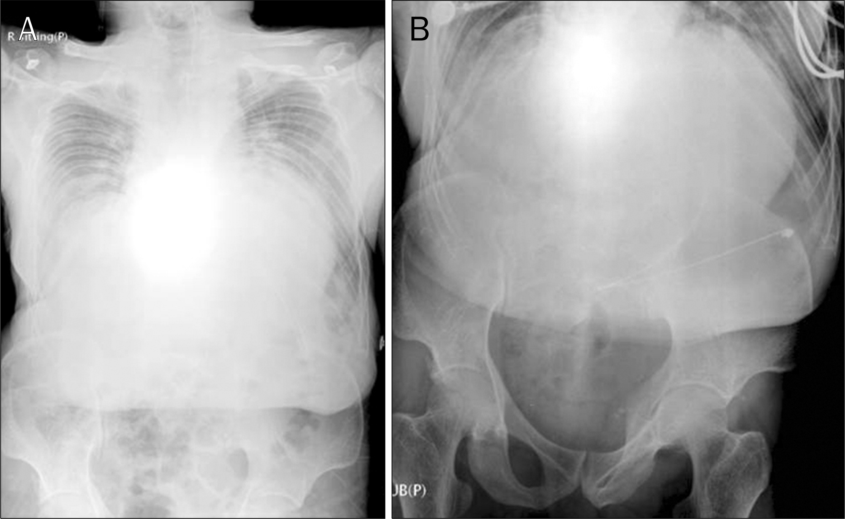
 | Fig. 1.Plain abdominal X-ray. It revealed severe kyphosis (A) and no other abnormalities in the abdomen on admission (B). |
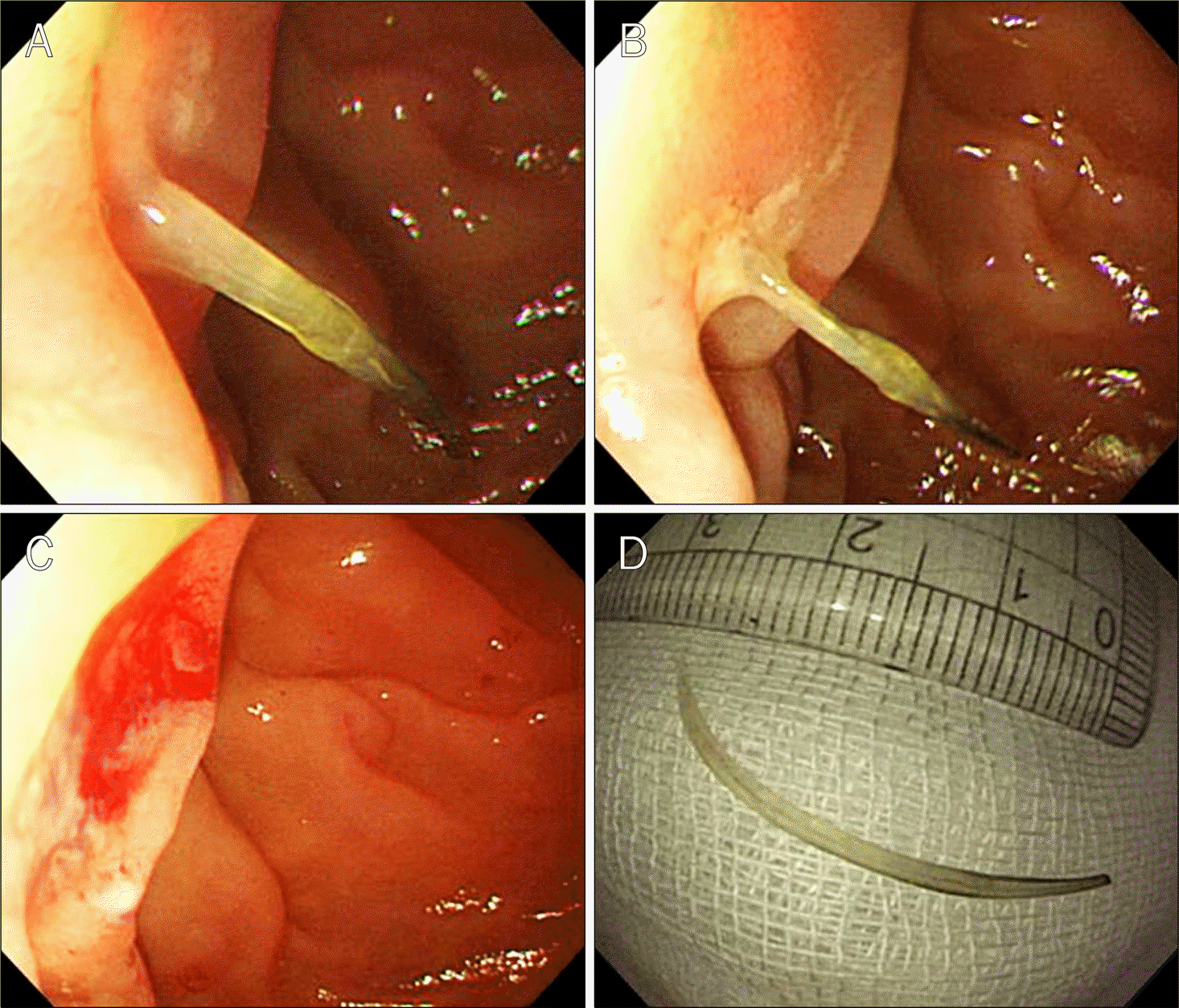
 | Fig. 2.Endoscopic findings. A fish bone was stuck in the duodenal wall (A, B). During endoscopy, the fish bone was removed by biopsy forceps (C). The fish bone was measured about 3 cm-in length (D). |
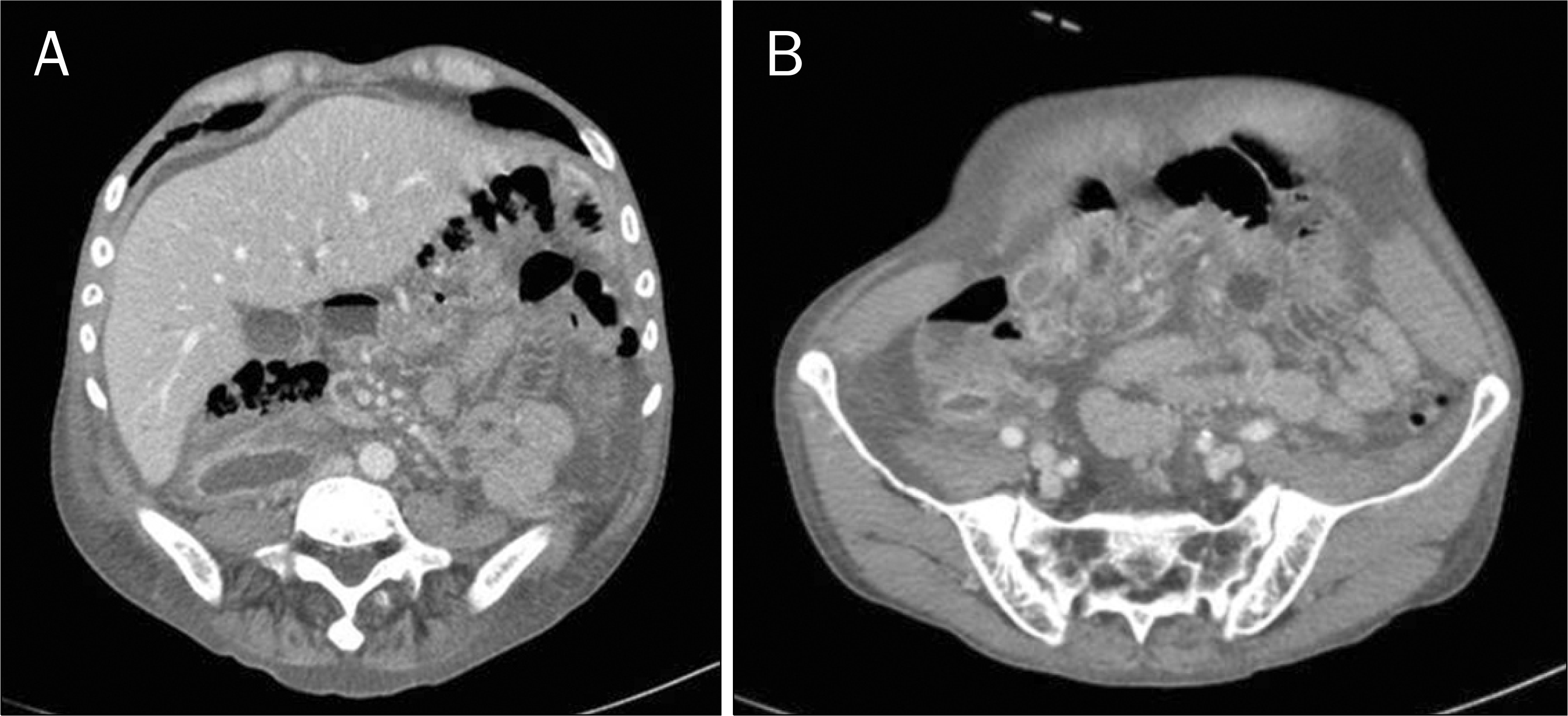
 | Fig. 3.Abdomen CT findings. One day after removing the fish bone, it showed retroperitoneal hemorrhage (white star). (A) Hemorrhage was noted around the second portion of the duodenum. (B) The hemorrhage extended to the retroperitoneal space of the pelvis. (C, D) A huge retroperitoneal hemorrhage was seen in the coronal view. |


 PDF
PDF ePub
ePub Citation
 Print
Print


 XML Download