Abstract
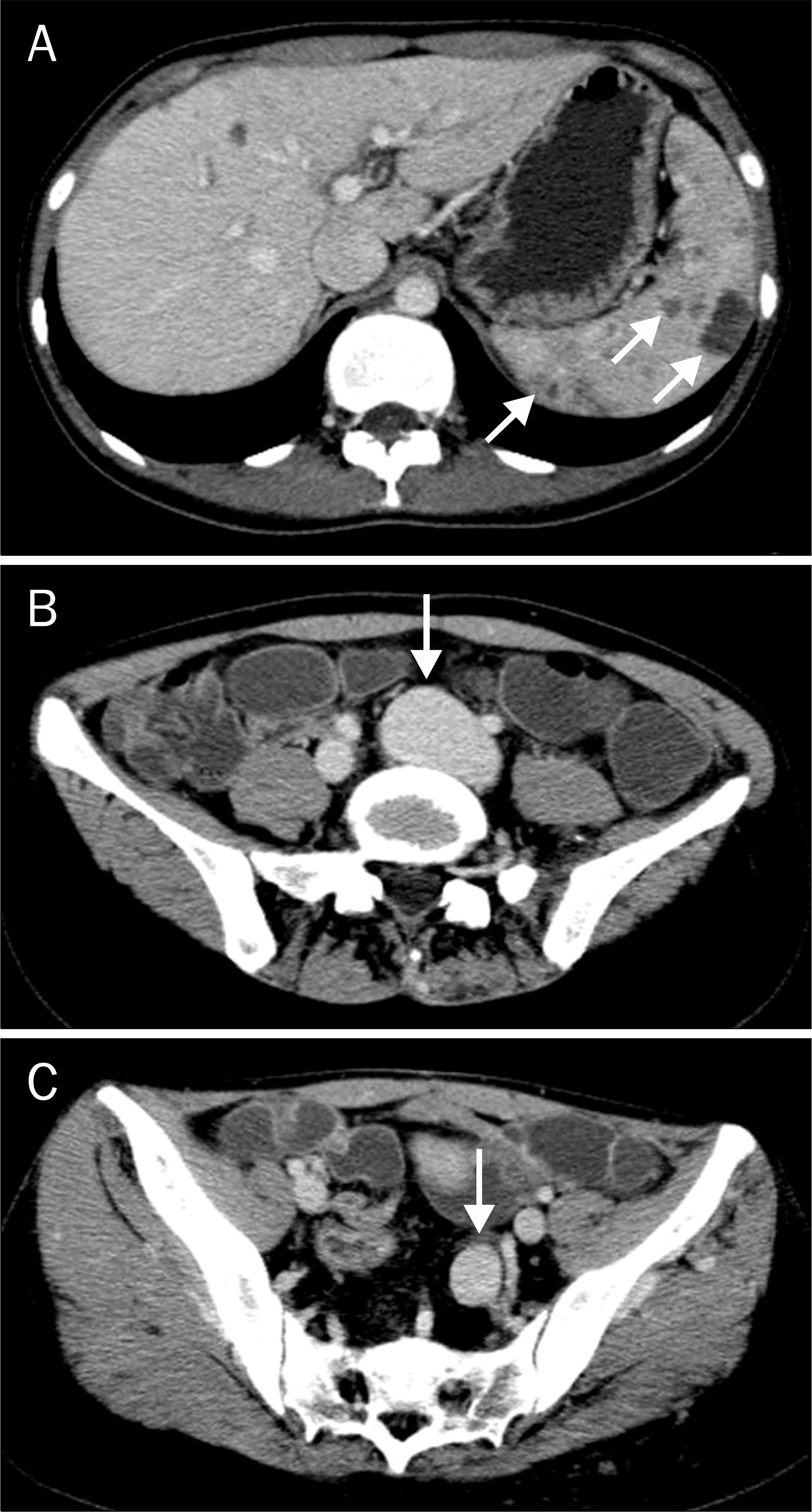
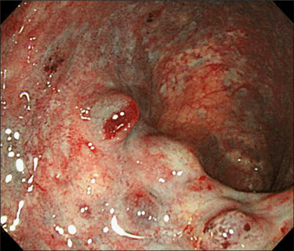
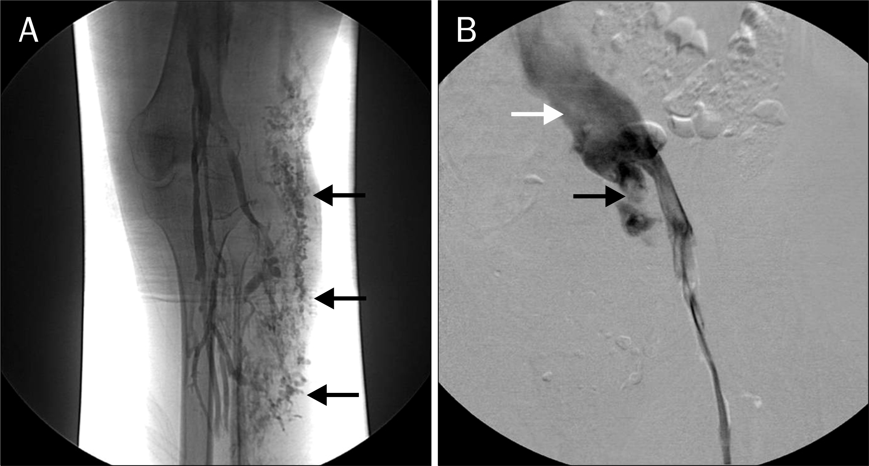
Klippel – Trenaunay syndrome (KTS) is characterized by a cutaneous vascular nevus of the involved extremity, bone and soft tissue hypertrophy of the extremity and venous malformations. We present a case of KTS with splenic hemangiomas and rectal varices. A 29-year-old woman was referred for intermittent hematochezia for several years. She had history with a number of operations for cutaneous and soft tissue hamangiomas since the age of one year old and for increased circumference of her left thigh during the last few months. Abdominal CT revealed multiple hemangiomas in the spleen, fusiform aneurysmal dilatation of the deep veins and soft tissue hemangiomas. There was no evidence of hepatosplenomegaly or liver cirrhosis. Colonoscopy revealed hemangiomatous involvement in the rectum. There were rectal varices without evidence of active bleeding. Upon venography of the left leg, we also found infiltrative dilated superficial veins in the subcutaneous tissue and aneurysmal dilatation of the deep veins. The patient was finally diagnosed with KTS, and treated with oral iron supplementation only, which has been tolerable to date. Intervention or surgery is not required. When gastrointestinal varices or hemangiomatous mucosal changes are detected in a young patient without definite underlying cause, KTS should be considered.
Go to : 
References
1. Klippel M, Trenaunay P. Du nævus variqueux ostéo-hyper-trophique. Arch Gen Med. 1900; 185:641–672.
2. Wilson CL, Song LM, Chua H, et al. Bleeding from cavernous an-giomatosis of the rectum in Klippel-Trenaunay syndrome: report of three cases and literature review. Am J Gastroenterol. 2001; 96:2783–2788.

3. Servelle M. Klippel and Trenaunay's syndrome. 768 operated cases. Ann Surg. 1985; 201:365–373.
4. Kim JH, Kim CW, Son DK, et al. A case of Klippel-Trenaunay-Weber syndrome presenting with esophageal and gastric varices bleeding. Korean J Gastroenterol. 2004; 43:137–141.
5. Servelle M, Bastin R, Loygue J, et al. Hematuria and rectal bleeding in the child with Klippel and Trenaunay syndrome. Ann Surg. 1976; 183:418–428.

6. Schmitt B, Posselt HG, Waag KL, Müller H, Bender SW. Severe hemorrhage from intestinal hemangiomatosis in Klippel-Trenaunay syndrome: pitfalls in diagnosis and management. J Pediatr Gastroenterol Nutr. 1986; 5:155–158.
7. Brown R, Ohri SK, Ghosh P, Jackson J, Spencer J, Allison D. Case report: jejunal vascular malformation in Klippel-Trenaunay syndrome. Clin Radiol. 1991; 44:134–136.

8. Capraro PA, Fisher J, Hammond DC, Grossman JA. KlippelTrenaunay syndrome. Plast Reconstr Surg. 2002; 109:2052–2060.

9. Natterer J, Joseph JM, Denys A, Dorta G, Hohlfeld J, de Buys Roessingh AS. Life-threatening rectal bleeding with KlippelTrenaunay syndrome controlled by angiographic embolization and rectal clips. J Pediatr Gastroenterol Nutr. 2006; 42:581–584.

10. Goenka MK, Kochhar R, Nagi B, Mehta SK. Rectosigmoid varices and other mucosal changes in patients with portal hypertension. Am J Gastroenterol. 1991; 86:1185–1189.
11. Kocaman O, Alponat A, Aygün C, et al. Lower gastrointestinal bleeding, hematuria and splenic hemangiomas in Klippel-Trenaunay syndrome: a case report and literature review. Turk J Gastroenterol. 2009; 20:62–66.
12. Yeoman LJ, Shaw D. Computerized tomography appearances of pelvic haemangioma involving the large bowel in childhood. Pediatr Radiol. 1989; 19:414–416.

13. Azouz EM. Hematuria, rectal bleeding and pelvic phleboliths in children with the Klippel-Trenaunay syndrome. Pediatr Radiol. 1983; 13:82–88.

14. Jindal R, Sullivan R, Rodda B, Arun D, Hamady M, Cheshire NJ. Splenic malformation in a patient with Klippel-Trenaunay syndrome: a case report. J Vasc Surg. 2006; 43:848–850.

15. Willcox TM, Speer RW, Schlinkert RT, Sarr MG. Hemangioma of the spleen: presentation, diagnosis and management. J Gastrointest Surg. 2000; 4:611–613.

16. Aggarwal K, Jain VK, Gupta S, Aggarwal HK, Sen J, Goyal V. Klippel-Trenaunay syndrome with a life threatening throm-boembolic event. J Dermatol. 2003; 30:236–240.
17. Baskerville PA, Ackroyd JS, Lea Thomas M, Browse NL. The Klippel-Trenaunay syndrome: clinical, radiological, and haemo-dynamic features and management. Br J Surg. 1985; 72:232–236.

18. Jang SH, Lee H, Han SH. Common peroneal nerve compression by a popliteal venous aneurysm. Am J Phys Med Rehabil. 2009; 88:947–950.

19. Baskerville PA, Ackroyd JS, Browse NL. The etiology of Klippel-Trenaunay syndrome. Ann Surg. 1985; 202:624–627.
Go to : 
 | Fig. 1.An abdominal CT scan revealed multiple hemangiomas in the spleen (A), and fusiform aneurysmal dilatation of the left common (B) and internal iliac veins (C). |


 PDF
PDF ePub
ePub Citation
 Print
Print


 XML Download