

 PDF
PDF ePub
ePub Citation
 Print
Print


Abstract
Lymphocytic ascites with low serum-ascites albumin gradient (SAAG) are observed mainly in tuberculous peritonitis, peritoneal carcinomatosis, and pancreatic disease. However, pelvic inflammatory disease (PID) induced generalized peritonitis causing diffuse ascites has been rarely described. We report a 26-year old female patient, who was diagnosed as generalized peritonitis with diffuse ascites due to Chlamydia trachomatis infection. Gynecologic examination did not show the clue of PID and in the analysis of ascites, low SAAG, predominant lymphocyte count and high level of adenosine deaminase were noted. Although the best impression was tuberculous peritonitis on the base of these findings, the laparoscopic finding was consistent with PID and the PCR for C. trachomatis infection in cervical swab was positive. This case suggests that C. trachomatis peritonitis should be considered as a rare cause of low SAAG and lymphocytic ascites in sexually active women and should be intensively evaluated including laparoscopic examination.
Go to : 
References
1. Hwangbo Y, Jung JH, Shim J, et al. Etiologic and laboratory analy-ses of ascites in patients who underwent diagnostic paracen-tesis. Korean J Hepatol. 2007; 13:185–195.
2. Weström L. Incidence, prevalence, and trends of acute pelvic inflammatory disease and its consequences in industrialized countries. Am J Obstet Gynecol. 1980; 138:880–892.

3. Heo H, Ha JY, Kim KW, et al. A report of pelvic inflammatory disease in the region of the Po-Hang. Korean J Obstet Gynecol. 2003; 46:1116–1120.
4. Votte-Lambert A, Joly JP, Becuwe C, Eb F, Capron JP, Dupas JL. Chlamydia Trachomatis peritonitis: another cause of protein-rich lymphocytic ascites. J Clin Gastroenterol. 1990; 12:341–343.
5. Kasper DL, Braunwald E, Fauci AS, Hauser SL, Longo DL, Jameson JL. Harrison's principles of internal medicines. 17th ed.New York: McGraw-Hill;2008. p. 821–831.
6. Paavonen J, Lehtinen M. Chlamydial pelvic inflammatory disease. Hum Reprod Update. 1996; 2:519–529.
7. Shin CJ. Chlamydia Trachomatis infection. Korean J Obstet Gynecol. 1992; 35:1561–1571.
8. Song JH, Song SR, Song JH, Jung YW, Min JW, Lee SS. A clinical evaluation of Chlamydia Trachomatis infection in women with pelvic inflammatory disease. Korean J Obstet Gynecol. 2005; 48:581–588.
9. Runyon BA. Ascites and spontaneous bacterial peritonitis. Feldman M, Friedman LS, Brandt LJ, editors. Sleisenger and Ford-tran's Gastrointestinal and Liver disease. 8th ed.Philadelphia: Saunders;2006. p. 1935–1965.
10. Demiroğ lu YZ, Turunç T, Aliş kan H, Colakoğ lu S, Arslan H. Primary peritonitis due to brucellosis mimicking tuberculous peritonitis. Turk J Gastroenterol. 2009; 20:135–137.
11. Müller-Schoop JW, Wang SP, Munzinger J, Schläpfer HU, Knoblauch M, Tammann RW. Chlamydia Trachomatis as possible cause of peritonitis and perihepatitis in young women. Br Med J. 1978; 1:1022–1024.
12. Wang HH, Kim MY, Kim JE, Kim YJ, Suh CH. CT differentiation of periappendiceal inflammation with appendicitis and pelvic inflammatory disease in woman with right lower quadrant pain. J Korean Radiol Soc. 2006; 55:83–89.
13. Shore A, Dosch HM, Gelfand EW. Role of adenosine deaminase in the early stages of precursor T cell maturation. Clin Exp Immunol. 1981; 44:152–155.
14. Lee JS, Kim KA, Lee WJ, et al. Diagnostic value of ascitic fluid adenosine deaminase activity for diagnosis of tuberculous peritonitis. Korean J Gastroenterol. 2003; 41:126–132.
15. Hankiewicz J, Michalski J. Adenosine deaminase in pregnancy and in some gynecological diseases. Enzymologia. 1971; 41:261–277.
16. Hillebrand DJ, Runyon BA, Yasmineh WG, Rynders GP. Ascitic fluid adenosine deaminase insensitivity in detecting tuberculous peritonitis in the United States. Hepatology. 1996; 24:1408–1412.
17. Bergmann JF, Bidart JM, George M, Beaugrand M, Levy VG, Bohuon C. Elevation of CA 125 in patients with benign and malig- nant ascites. Cancer. 1987; 59:213–217.
18. Wu JF, Li HJ, Lee PI, Ni YH, Yu SC, Chang MH. Tuberculous peritonitis mimicking peritonitis carcinomatosis: a case report. Eur J Pediatr. 2003; 162:853–855.
19. Jang DG, Choi JH, Park IY, Hwang SJ, Kim CJ, Kim CY. Laparoscopic finding of acute pelvic inflammatory disease. Korean J Obstet Gynecol. 2005; 48:750–754.
20. Bedioui H, Ksantini R, Nouira K, et al. Role of laparoscopic surgery in the etiologic diagnosis of exsudative ascites: a prospective study of 90 cases. Gastroenterol Clin Biol. 2007; 31:1146–1149.
Go to :
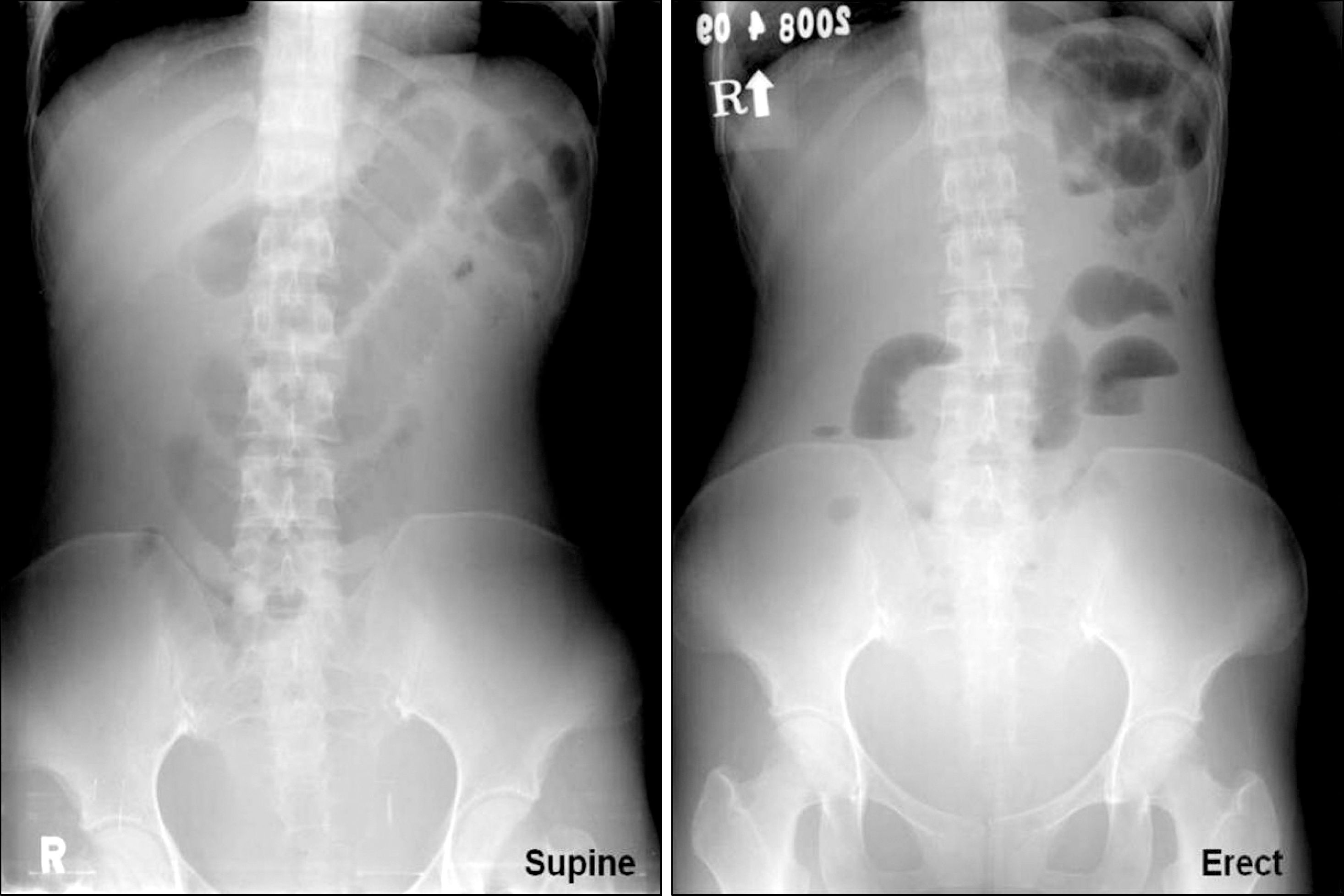 | Fig. 1.Simple abdominal x-ray. The picture showed small bowel ileus and hazziness suggesting ascites in the whole abdomen. |
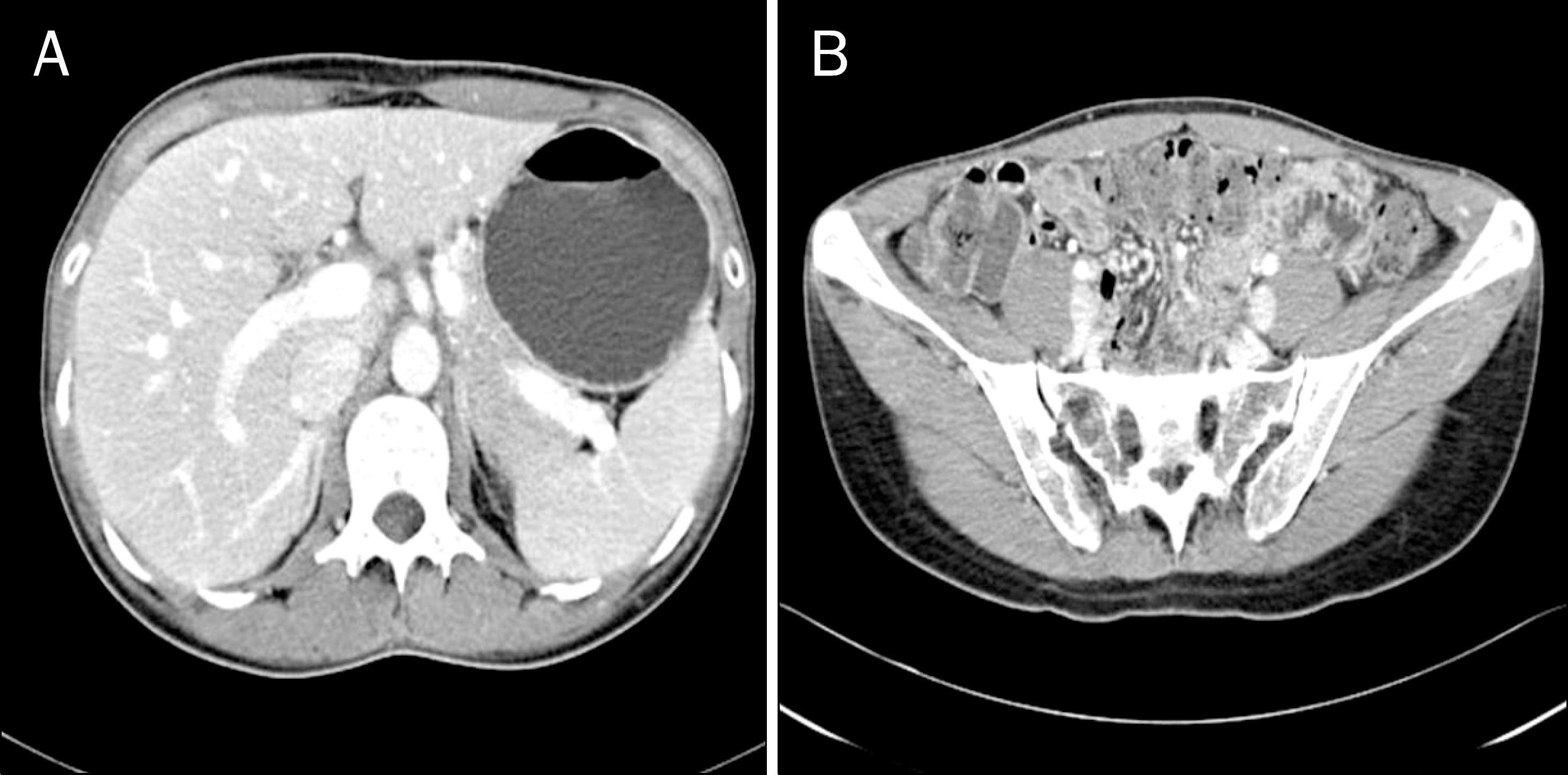
 | Fig. 2.Enhanced abdominopelvic CT scan. (A) The picture showed abrupt luminal narrowing (arrow) of the mid-ileum with proximal small bowel dilatation. (B) Ascites (arrow) was noted. (C) Omental nodular infiltration and peritoneal thickening (arrow heads) suggesting peritonitis was seen. (D) Swelling of both ovaries (arrows) was seen in the pelvis. |
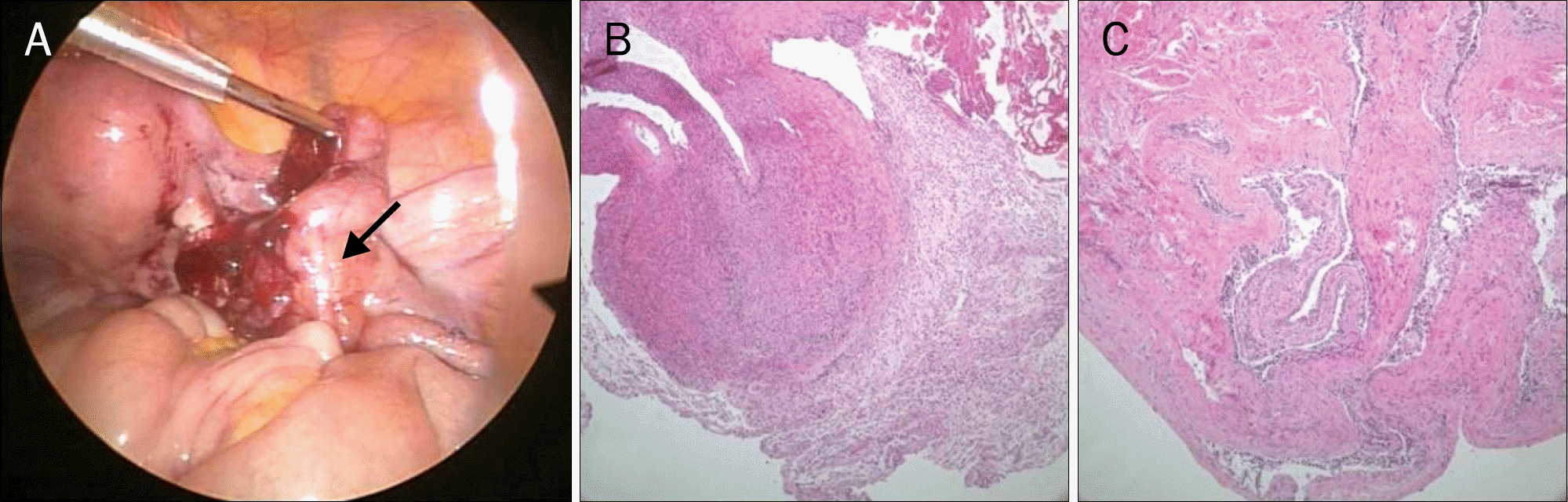 | Fig. 3.Laparoscopic finding and microscopic findings. (A) Laparoscopic examination showed slight swelling of both ovaries (arrow) without omental nodular infiltration and peritoneal thickening on the pelvis. (B) Ovarian (H&E, ×100) and (C) fimbrial tissue (H&E, ×40) with diffuse infiltrative plasma cells consisting with salpingo-oophoritis were noted on the laparoscopic biopsies. |
 XML Download