Abstract
Clostridium difficile (C. difficile) infection appears to be closely related to reactivation, diagnostic delay, and disease progression in patients with inflammatory bowel disease. However, whether C. difficile infection triggers the reactivation of inflammatory bowel disease or vice versa is not certain. We report a case of reactivated and progressed left ulcerative colitis following C. difficile infection in a 56-year-old woman. A series of endoscopic findings in this case report strongly supports a causative role of C. difficile infection on the reactivation and progression of ulcerative colitis.
References
1. Kethu SR. Extraintestinal manifestations of inflammatory bowel diseases. J Clin Gastroenterol. 2006; 40:467–475.

2. Satsangi J, Jewell DP, Bell JI. The genetics of inflammatory bowel disease. Gut. 1997; 40:572–574.

3. Bolton RP, Sherriff RJ, Read AE. Clostridium difficle associated diarrhea: a role in inflammatory bowel disease? Lancet. 1980; 1:383–384.
4. Meyers S, Mayer L, Bottone E, Desmond E, Janowitz HD. Occurrence of Clostridium difficile toxin during the course of inflammatory bowel disease. Gastroenterology. 1981; 80:697–700.
5. Meyer AM, Ramzan NN, Loftus EV Jr, Heigh RI, Leighton JA. The diagnostic yield of stool pathogen studies during relapses of inflammatory bowel disease. J Clin Gastroenterol. 2004; 38:772–775.

6. Mylonaki M, Langmead L, Pantes A, Johnson F, Rampton DS. Enteric infection in relapse of inflammatory bowel disease: im-portance of microbiological examination of stool. Eur J Gastroenterol Hepatol. 2004; 16:775–778.
7. Issa M, Vijayapal A, Graham MB, et al. Impact of Clostridium difficile on inflammatory bowel disease. Clin Gastroenterol Hepatol. 2007; 5:345–351.
8. Ananthakrishnan AN, McGinley EL, Binion DG. Excess hospital-isation burden associated with Clostridium difficile in patients with inflammatory bowel disease. Gut. 2008; 57:205–210.
9. Rodemann JF, Dubberk ER, Reske KA, Seo da H, Stone CD. Incidence of Clostridium difficile infection in inflammatory bowel disease. Clin Gastroenterol Hepatol. 2007; 5:339–344.
10. Issa M, Ananthakrishnan AN, Binion DG. Clostridium difficile and inflammatory bowel disease. Inflamm Bowel Dis. 2008; 14:1432–1442.
11. Miller DL, Sedlack JD, Holt RW. Perforation complicating ri-fampin-associated pseudomembranous enteritis. Arch Surg. 1989; 124:1082.

12. Wang A, Takeshima F, Ikeda M, et al. Ulcerative colitis complicating pseudomembranous colitis of the right colon. J Gastroenterol. 2002; 37:309–312.

13. Hookman P, Barkin JS. Clostridium difficile associated infection, diarrhea and colitis. World J Gastroenterol. 2009; 15:1554–1580.
14. Bartlett JG. Clinical practice. Antibiotic-associated diarrhea. N Engl J Med. 2002; 346:334–339.
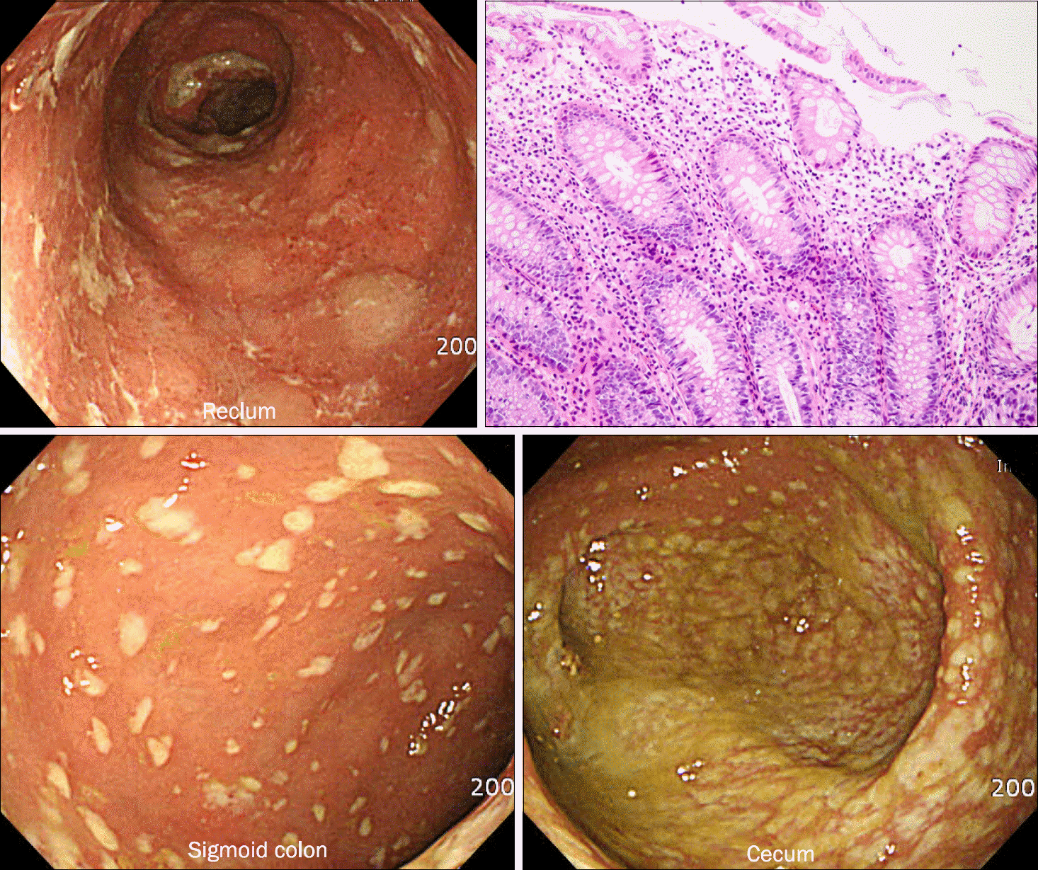
Fig. 1.
Initial colonoscopic and pathologic findings. Typical findings suggestive of ulcerative proctitis were noted on the rectal mucosa, while numerous yellowish plaques were scattered proximally starting from the sigmoid colon to cecal base. Well-demarcated active ulceration was noted at upper rectum. In the lower rectum, nonspecific colitis with chronic and acute inflammatory cells in lamina propria was noted. There was no architectural distortion (H&E,×200).

Fig. 2.
Colonoscopic findings 10 days after oral vancomycin administration. The rectal and proximal mucosal lesions were nearly normalized except for a healing ulcerative lesion at the upper rectum.

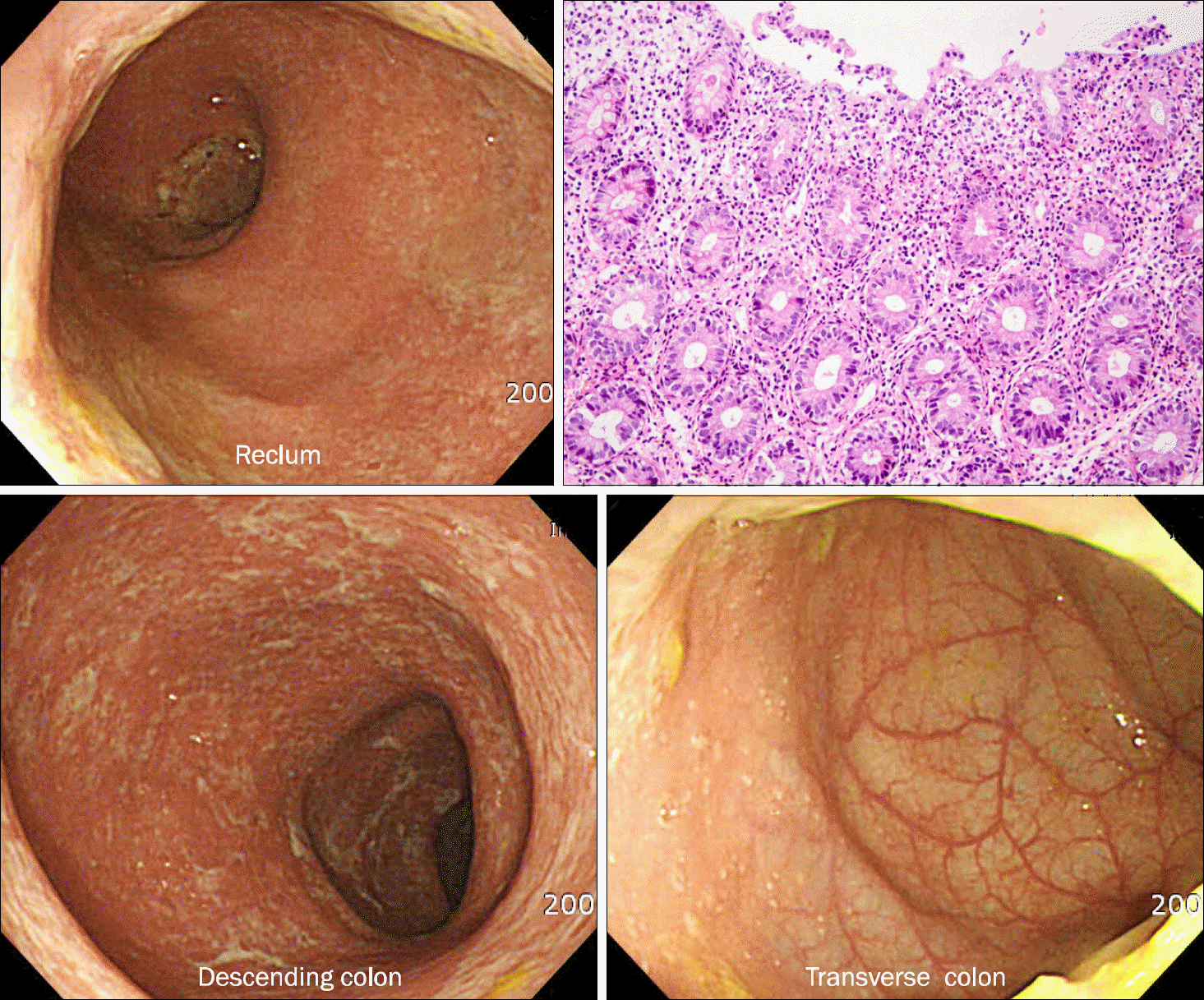
Fig. 3.
Colonoscopic findings 20 days after the cessation of oral vancomycin. Diffuse mucosal erosions, loss of vascularity, and mucosal granularity were noted in a continuous fashion from the lower rectum to distal transverse colon. Abrupt transition to normal mucosa was also noted at the distal transverse colon, suggesting of left-sided ulcerative colitis. Ulcer was still present at the upper rectum. No pseudomembrane was observed. In sigmoid colon, the lamina propria contained a dense infiltrate of pro-minent neutrophils, plasma cells and lymphocytes. The epithelium appear-ed mucin depleted however, histolo-gic feature of active colitis including cryptitis or crypt abscess was not present (H&E, ×200).


 PDF
PDF ePub
ePub Citation
 Print
Print


 XML Download