

 PDF
PDF ePub
ePub Citation
 Print
Print


References
1. Stranks GJ, Mathai JT, Rowe-Jones DC. Primary malignant melanoma of the oesophagus: case report and review of surgical pathology. Gut. 1991; 32:828–830.

2. Li B, Lei W, Shao K, et al. Characteristics and prognosis of primary malignant melanoma of the esophagus. Melanoma Res. 2007; 17:239–242.
3. Chalkiadakis G, Wihlm JM, Morand G, Weill-Bousson M, Witz JP. Primary malignant melanoma of the esophagus. Ann Thorac Surg. 1985; 39:472–475.
4. Simpson NS, Spence RA, Biggart JD, Cameron CH. Primary malignant melanoma of the oesophagus. J Clin Pathol. 1990; 43:82–83.
5. Uetsuka H, Naomoto Y, Fujiwara T, et al. Primary malignant melanoma of the esophagus: long-term survival following pre- and postoperative adjuvant hormone/chemotherapy. Dig Dis Sci. 2004; 49:1646–1651.
Go to : 
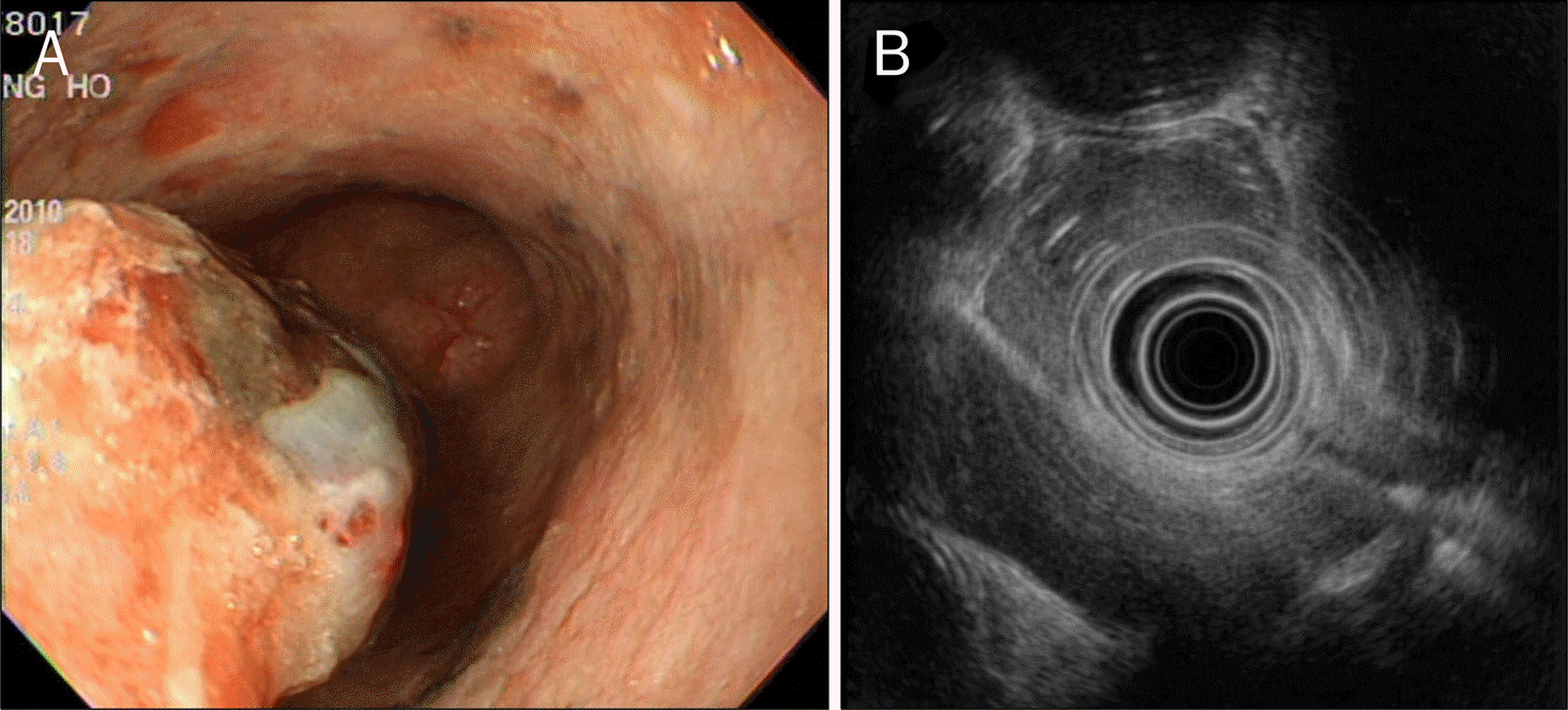 | Fig. 1.Endoscopic and EUS finding. (A)3 cm sized polypoid mass with central black pigmentation was noted in mid-esophagus. (B) Segmental wall thickening with submucosal invasion was observed. |

 XML Download