Abstract
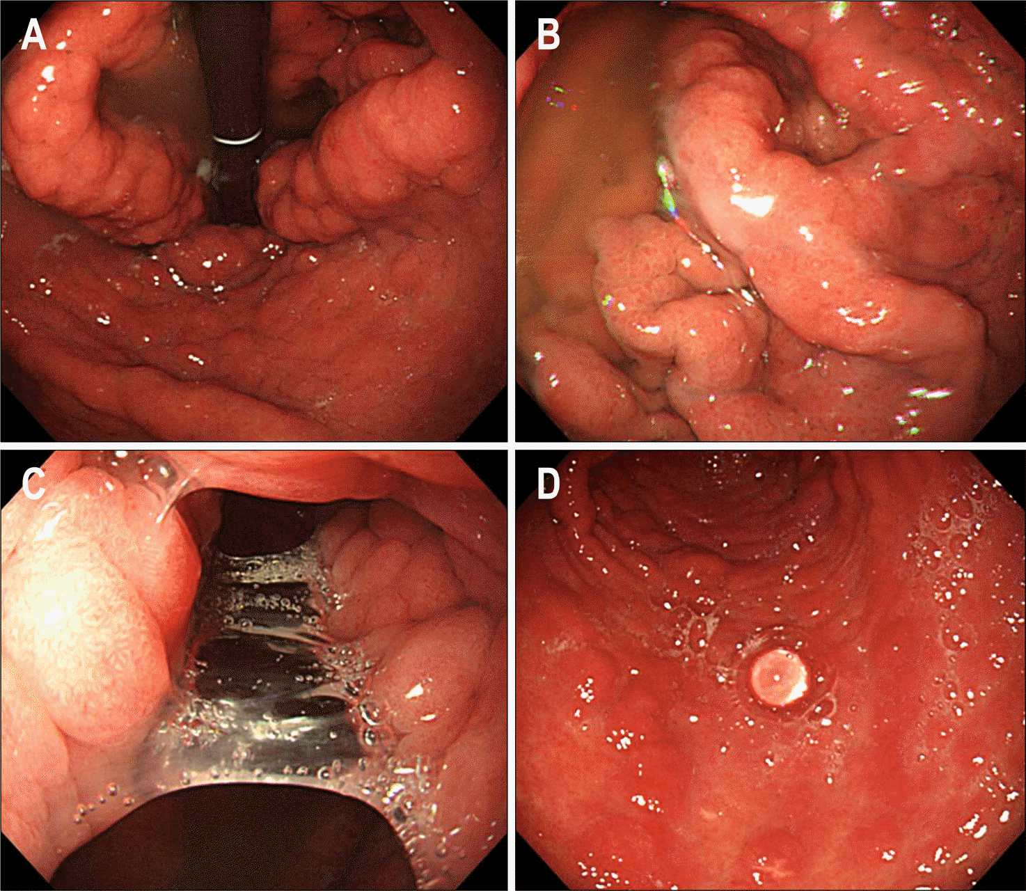
Menetrier's disease is a rare entity characterized by large, tortuous gastric mucosal folds. The mucosal folds in Menetrier's disease are often most prominent in the body and fundus. Histologically, massive foveolar hyperplasia (hyperplasia of surface and glandular mucous cells) is noted, which replaces most of the chief and parietal cells. Profuse mucus is usually observed during the endoscopy but there have been few cases that show interesting endoscopic findings such as mucus bridge or water pearl. Herein, we report a case of Menetrier's disease showing mucus bridge by excessive mucus observed during the endoscopy.
Go to : 
References
1. Menetrier P. Des polyadenomes gastriques et de leurs rapports avec le cancer de I'estomac. Arch Physiol Norm Pathol. 1888; 32:236–262.
2. Valle John D. Peptic Ulcer Disease and Related Disorders. Fauci AS, Braunwald E, Kasper DL, Hauser SL, Longo DL, Jameson JL, Loscalzo J, editors. Harrison's principles of internal medicine. Vol 2. 17th ed.New York: McGraw Hill;2008. p. 1871–1872.
3. Romano M, Polk WH, Awad JA, et al. Transforming growth factor alpha protection against drug-induced injury to the rat gastric mucosa in vivo. J Clin Invest. 1992; 90:2409–2421.

4. Dempsey PJ, Goldenring JR, Soroka CJ, et al. Possible role of transforming growth factor alpha in the pathogenesis of Ménétrier's disease: supportive evidence form humans and transgenic mice. Gastroenterology. 1992; 103:1950–1963.
5. Takagi H, Jhappan C, Sharp R, Merlino G. Hypertrophic gastropathy resembling Ménétrier's disease in transgenic mice overexpressing transforming growth factor alpha in the stomach. J Clin Invest. 1992; 90:1161–1167.

6. Megged O, Schlesinger Y. Cytomegalovirus-associated protein-losing gastropathy in childhood. Eur J Pediatr. 2008; 167:1217–1220.

7. Lim YJ, Rhee PL, Kim YH, et al. Clinical features of Menetrier's disease in Korea. Korean J Gastrointest Endosc. 2000; 21:909–916.
8. Ginès A, Pellise M, Fernández-Esparrach G, et al. Endoscopic ultrasonography in patients with large gastric folds at endoscopy and biopsies negative for malignancy: predictors of malignant disease and clinical impact. Am J Gastroenterol. 2006; 101:64–69.
9. Songür Y, Okai T, Watanabe H, Motoo Y, Sawabu N. Endosonographic evaluation giant gastric folds. Gastrointest Endosc. 1995; 41:468–474.
10. Scott HW Jr, Shull HJ, Law DH 4th, Burko H, Page DL. Surgical management of Menetrier's disease with protein-losing gastropathy. Ann Surg. 1975; 181:765–777.

11. Jun DW, Kim DH, Kim SH, et al. Ménétrier's disease associated with herpes infection: response to treatment with acyclovir. Gastrointest Endosc. 2007; 65:1092–1095.

12. Jung JH, Hong SJ, Lee MS. Resolution of Menetrier's disease after Helicobacter pylori eradication. Korean J Gastroenterol. 2006; 48:1–3.
13. Lim HJ, Park SJ, Seo JA, Moon W, Kim KJ, Park MI. A case of rapidly improved Menetrier's disease after short-term treatment with proton pump inhibitor. Korean J Gastrointest Endosc. 2008; 36:83–86.
Go to : 
 | Fig. 1.Endoscopic findings. (A) Tortuous enlarged gastric folds were noted. (B) The surface of gastric mucosa was glossy due to overlying mucus. (C) Mucus bridge between gastric folds was observed. (D) Water was not dispersed due to thick mucus, which looked like a pearl. |
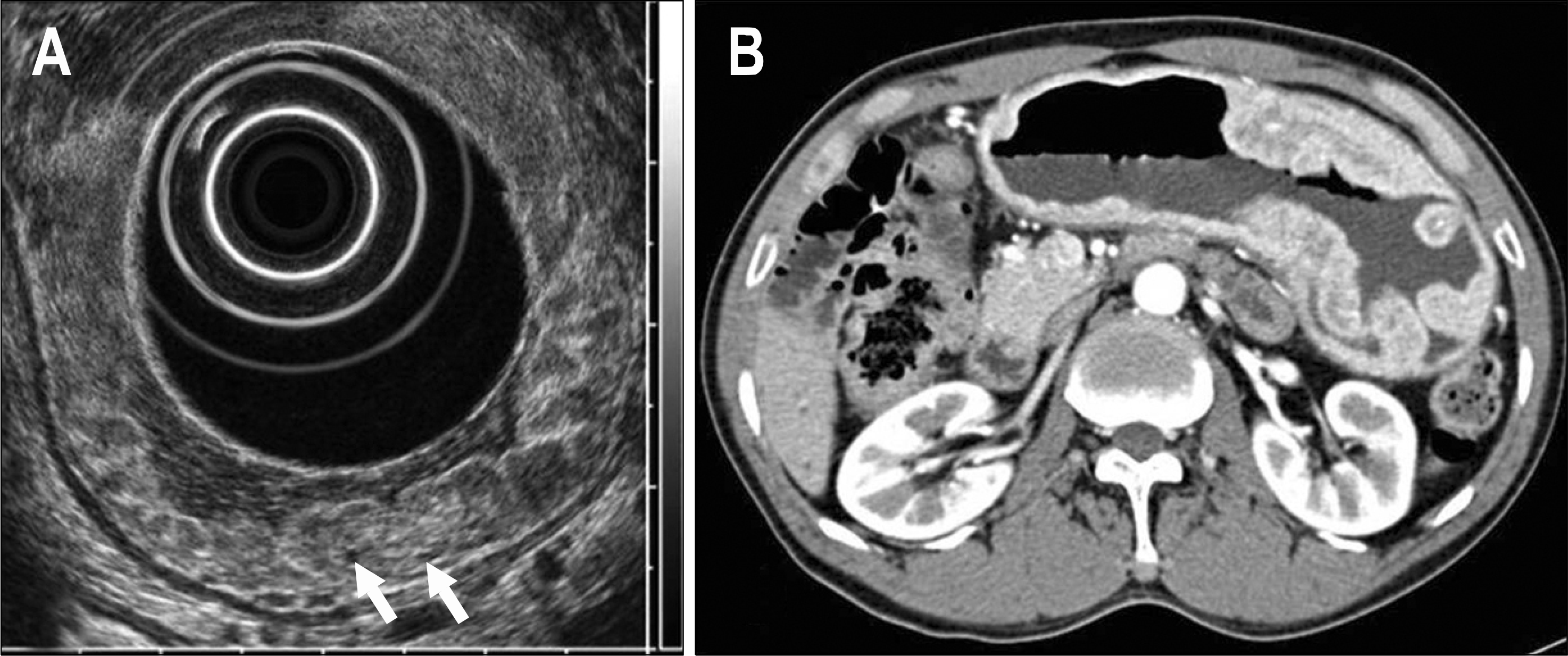
 | Fig. 2.(A) Endosonographic finding. The gastric folds were enlarged due to thickening of only the second layer. Some cystic areas were seen in the thickened second layer (arrows). (B) Abdominal CT finding. It showed markedly enlarged gastric folds. |
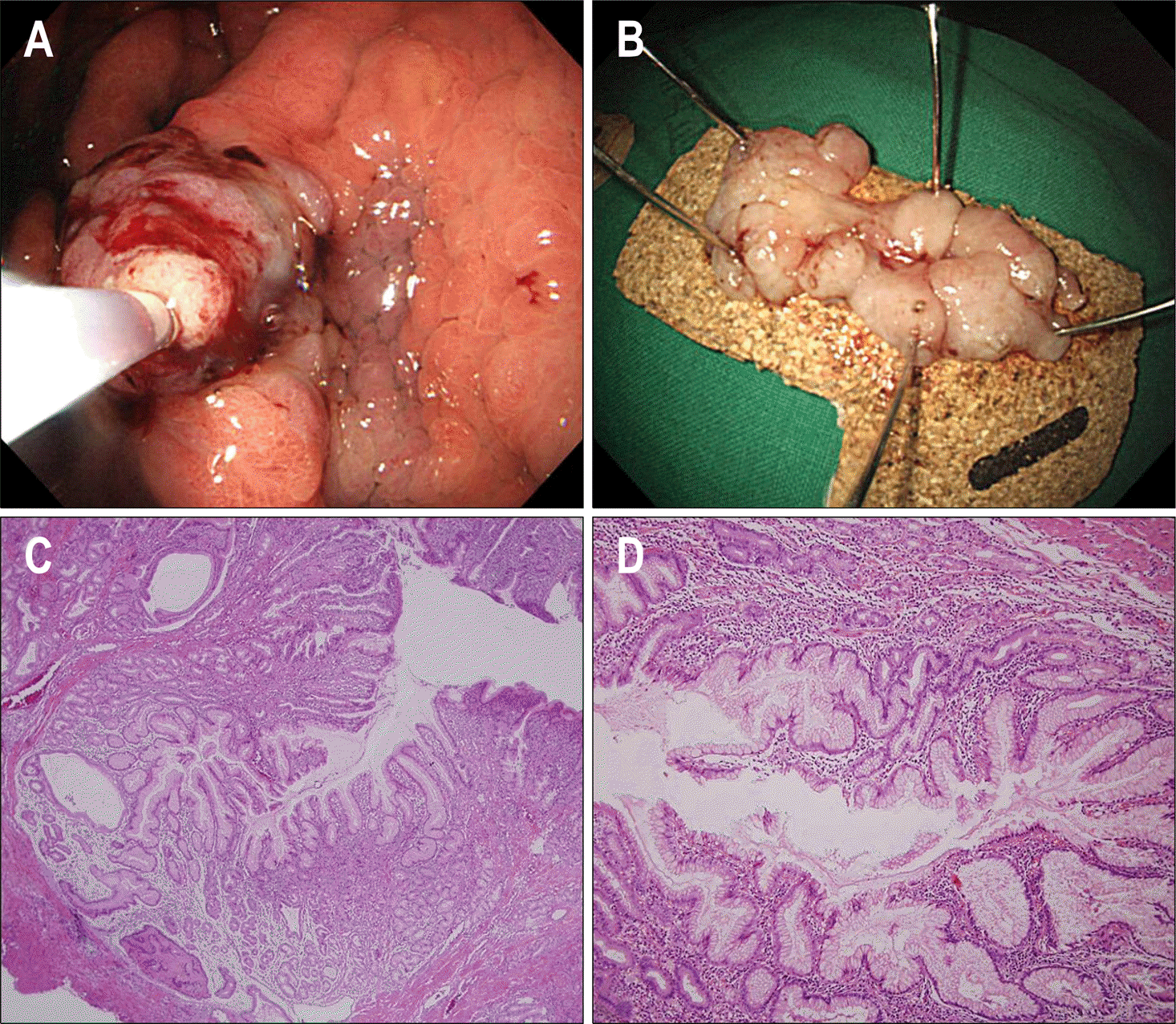
 | Fig. 3.Gross and microscopic findings. (A) Strip biopsy was performed for definite diagnosis. (B) The resected specimen showed nodular thickening of gastric mucosa. (C) At low power, foveolar hyperplasia was a marked feature. The foveola was elongated and had a corkscrew appearance (H&E, ×40). (D) The lamina propria was moderately infiltrated by inflammatory cells and a mild degree of intraepithelial lymphocytosis was present (H&E, ×100). |
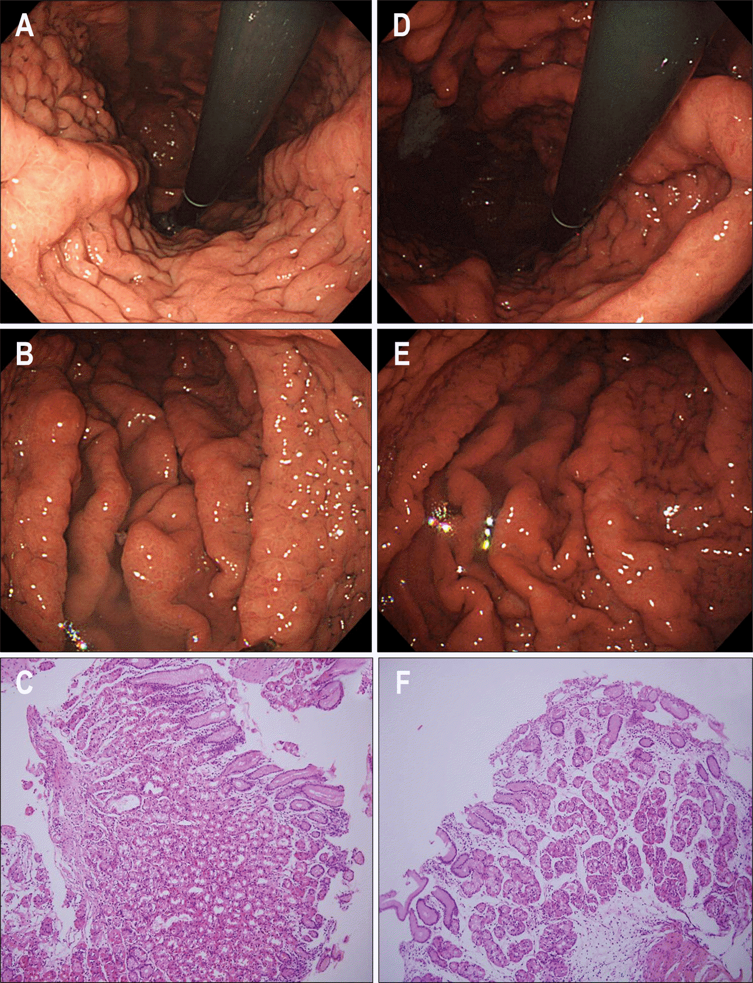
 | Fig. 4.Follow-up endoscopic and microscopic findings. (A, B, C) After 6 months of treatment with H. pylori eradication and proton pump inhibitor, the thickening of mucosal folds and the degree of foveolar hyperplasia was decreased (H&E, ×100). (D, E, F) After 18 months of treatment, the thickening of mucosal folds and the degree of foveolar hyperplasia was more decreased (H&E, ×100). |


 PDF
PDF ePub
ePub Citation
 Print
Print


 XML Download