Abstract
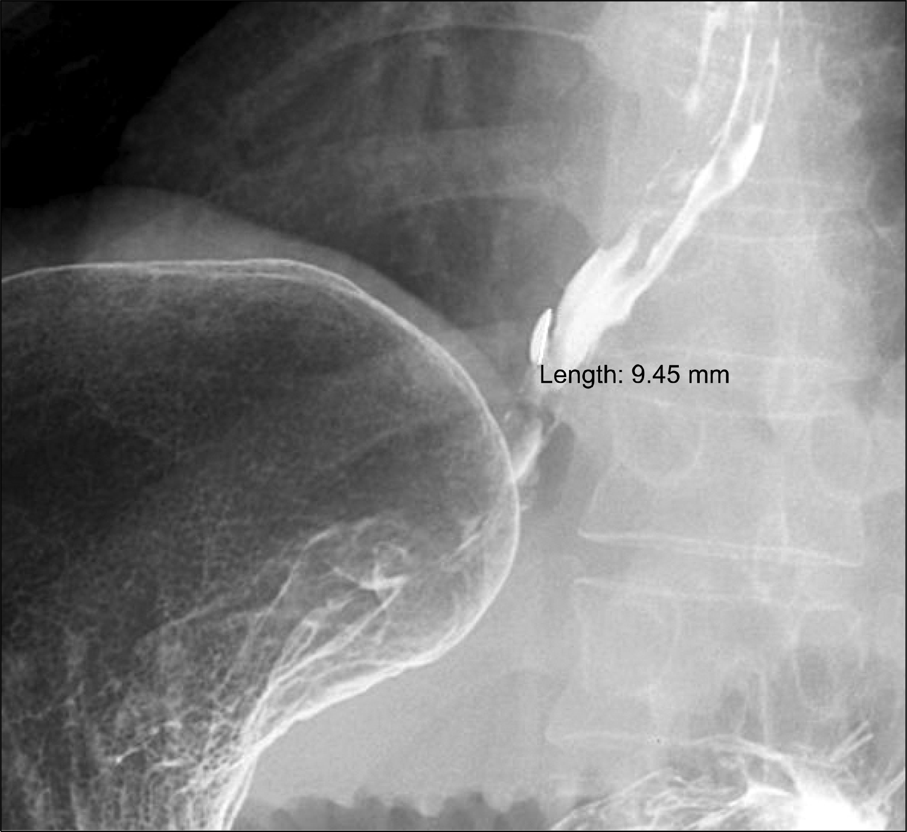
Intravariceal injection of N-butyl-2-cyanoacrylate is widely used for the hemostasis of bleeding gastric varices, but not routinely for esophageal variceal hemorrhage because of various complications such as pyrexia, bacteremia, deep ulceration, and pulmonary embolization. We report a rare case of esophageal sinus formation after cyanoacrylate obliteration therapy for uncontrolled bleeding from post-endoscopic variceal ligation (EVL) ulcer. A 50-year-old man with alcoholic liver cirrhosis presented with hematemesis. Emergent esophagogastroscopy revealed bleeding from large esophageal varices with ruptured erosion, and bleeding was initially controlled by EVL, but rebleeding from the post-EVL ulcer occurred at 17th day later. Although we tried again EVL and the injections of 5% ethanolamine oleate at paraesophageal varices, bleeding was not controlled. Therefore, we administered 1 mL cyanoacrylate diluted with lipiodol and bleeding was controlled. Three months after the endoscopic therapy, follow-up endoscopy showed medium to large-sized esophageal varices and sinus at lower esophagus. Barium esophagog-raphy revealed an outpouching in esophageal wall and endoscopic ultrasonography demonstrated an ostium with sinus. It is noteworthy that esophageal sinus can be developed as a rare late complication of endoscopic cyanoacrylate obliteration therapy.
Go to : 
References
1. Park DK, Um SH, Lee JW, et al. Clinical significance of variceal hemorrhage in recent years in patients with liver cirrhosis and esophageal varices. J Gastroenterol Hepatol. 2004; 19:1042–1051.

2. Garcia-Tsao G, Sanyal AJ, Grace ND, Carey W. Practice Guidelines Committee of the American Association for the Study of Liver Diseases; Practice Parameters Committee of the American College of Gastroenterology. Prevention and management of gastroesophageal varices and variceal hemorrhage in cirrhosis. Hepatology. 2007; 46:922–938.

3. Huang YH, Yeh HZ, Chen GH, et al. Endoscopic treatment of bleeding gastric varices by N-butyl-2-cyanoacrylate (Histoacryl) injection: long-term efficacy and safety. Gastrointest Endosc. 2000; 52:160–167.

4. Kim YS, Cho SW, Hahm KB, et al. Esophagus, stomach & intestine; hemostatic effect of histoacryl for band: induced esophageal ulcer bleeding. Korean J Gastrointest Endosc. 1997; 17:119–124.
5. Lee GH, Shin YJ, Ko YY, et al. N-utyl–yanoacrylate (Histoacryl) in the treatment of esophageal variceal bleeding: comparison with band ligation. Korean J Hepatol. 1999; 5:306–313.
7. Tan YM, Goh KL, Kamarulzaman A, et al. Multiple systemic embo-lisms with septicemia after gastric variceal obliteration with cyanoacrylate. Gastrointest Endosc. 2002; 55:276–278.

8. Park WG, Yeh RW, Triadafilopoulos G. Injection therapies for variceal bleeding disorders of the GI tract. Gastrointest Endosc. 2008; 67:313–323.

9. Petersen B, Barkun A, Carpenter S, et al. Tissue adhesives and fibrin glues. Gastrointest Endosc. 2004; 60:327–333.

10. D'Imperio N, Piemontese A, Baroncini D, et al. Evaluation of un-diluted N-butyl-2-cyanoacrylate in the endoscopic treatment of upper gastrointestinal tract varices. Endoscopy. 1996; 28:239–243.
11. Oho K, Iwao T, Sumino M, Toyonaga A, Tanikawa K. Ethanolamine oleate versus butyl cyanoacrylate for bleeding gastric varices: a nonrandomized study. Endoscopy. 1995; 27:349–354.

12. Lux G, Retterspitz M, Stabenow-Lohbauer U, Langer M, Altendorf-Hofmann A, Bozkurt T. Treatment of bleeding esophageal varices with cyanoacrylate and polidocanol, or polidoca-nol alone: results of a prospective study in an unselected group of patients with cirrhosis of the liver. Endoscopy. 1997; 29:241–246.

13. Thakeb F, Salama Z, Salama H, Abdel Raouf T, Abdel Kader S, Abdel Hamid H. The value of combined use of N-butyl-2-cyanoa-crylate and ethanolamine oleate in the management of bleeding esophagogastric varices. Endoscopy. 1995; 27:358–364.

14. Feretis C, Dimopoulos C, Benakis P, Kalliakmanis B, Apostolidis N. N-butyl-2-cyanoacrylate (Histoacryl) plus sclerotherapy versus sclerotherapy alone in the treatment of bleeding esophageal varices: a randomized prospective study. Endoscopy. 1995; 27:355–357.

15. Jutabha R, Jensen DM, Egan J, Machicado GA, Hirabayashi K. Randomized, prospective study of cyanoacrylate injection, sclerotherapy, or rubber band ligation for endoscopic hemostasis of bleeding canine gastric varices. Gastrointest Endosc. 1995; 41:201–205.

Go to : 
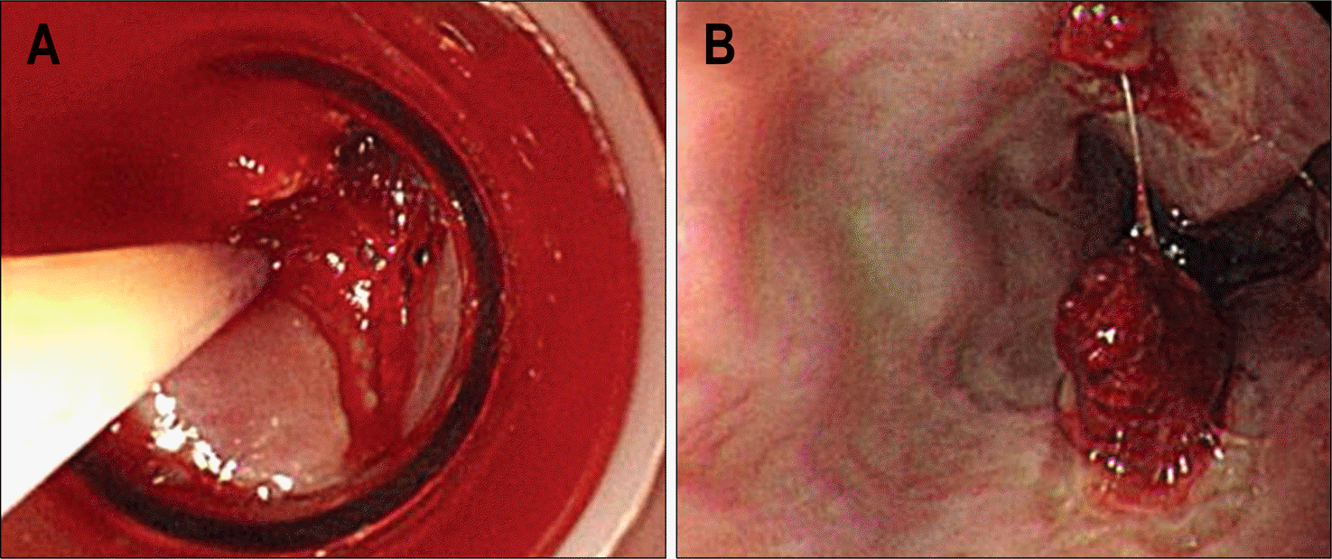
 | Fig. 1.Endoscopic findings. (A) It showed post-EVL ulcer bleeding and hemostatic trial by endoscopic sclerotherapy with total 5 mL (2 and 3 ml) of 5% ethanolamine oleate at two sites of paraesophageal varices. (B) It showed successful hemostasis of post-EVL ulcer bleeding after injection of total 1 ml of mixed solution of cyanoacrylate (0.5 mL) diluted with lipiodol (0.5 mL). EVL: endoscopic variceal ligation. |
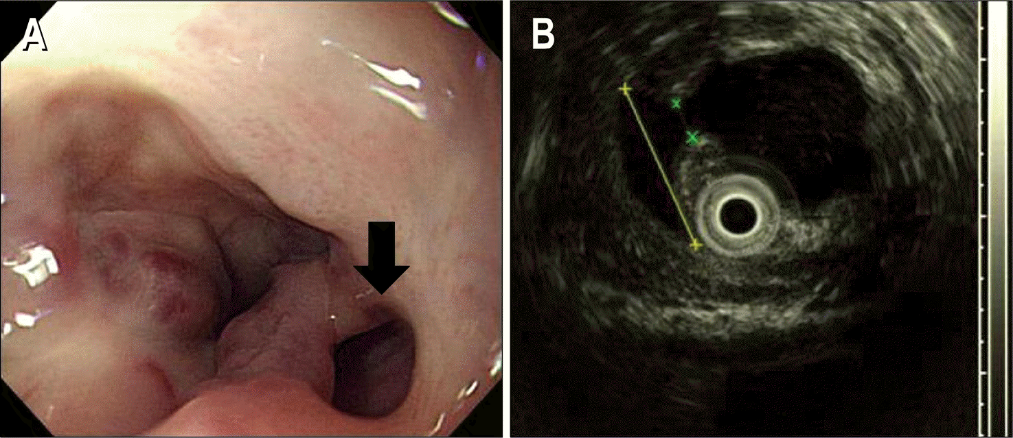
 | Fig. 2.Endoscopic finding of esophageal varices about 3 months after cyanoacrylate injection (A) and endoscopic ultrasongraphic finding of esophageal sinus (B). (A) It showed remained esophageal varices and a esophageal sinus (black arrow) at the lower esophagus. (B) The 10.9 mm sized sinus is indicated by yellow-colored x-marks and the orifice of sinus was 2.4 mm indicated by green-colored x-marks. |


 PDF
PDF ePub
ePub Citation
 Print
Print


 XML Download