Journal List > Korean J Gastroenterol > v.56(2) > 1006762

Park, Kim, and Lee: A Case of Chronic Intestinal Pseudoobstruction Accompanied by Parkinson's Disease

REFERENCES

1. Antonucci A, Fronzoni L, Cogliandro L, et al. Chronic intestinal pseudoobstruction. World J Gastroenterol. 2008; 14:2953–2961.
crossref
2. Myung SJ. Clinical features and management of chronic intestinal pseudoobstruction. Korean J Neurogastroenterol Motil. 2008; 14:1–6.
3. De Giorgio R, Sarnelli G, Corinaldesi R, Stanghellini V. Advances in our understanding of the pathology of chronic intestinal pseudoobstruction. Gut. 2004; 53:1549–1552.
crossref
4. Pfeiffer RF. Gastrointestinal dysfunction in Parkinson's disease. Lancet Neurol. 2003; 2:107–116.
crossref
5. Kupsky WJ, Grimes MM, Sweeting J, Bertsch R, Cote LJ. Parkinson's disease and megacolon: concentric hyaline in-clusions (Lewy bodies) in enteric ganglion cells. Neurology. 1987; 37:1253–1255.
crossref
6. Singaram C, Ashraf W, Gaumnitz EA, et al. Dopaminergic defect of enteric nervous system in Parkinson's disease patients with chronic constipation. Lancet. 1995; 346:861–864.
crossref
7. Choo KY, Choi MG, Choi H, et al. A case of colonic pseudoobstruction in a patient with Parkinson's disease. Korean J Gastrointest Motil. 2001; 7:251–256.
8. Natale G, Pasquali L, Ruggieri S, Paparelli A, Fornai F. Parkinson's disease and the gut: a well known clinical association in need of an effective cure and explanation. Neurogastroenterol Motil. 2008; 20:741–749.
crossref
9. Howard LM, Markus H. Pseudoobstruction secondary to an-ticholinergic drugs in Parkinson's disease. Postgrad Med J. 1992; 68:70–71.
crossref

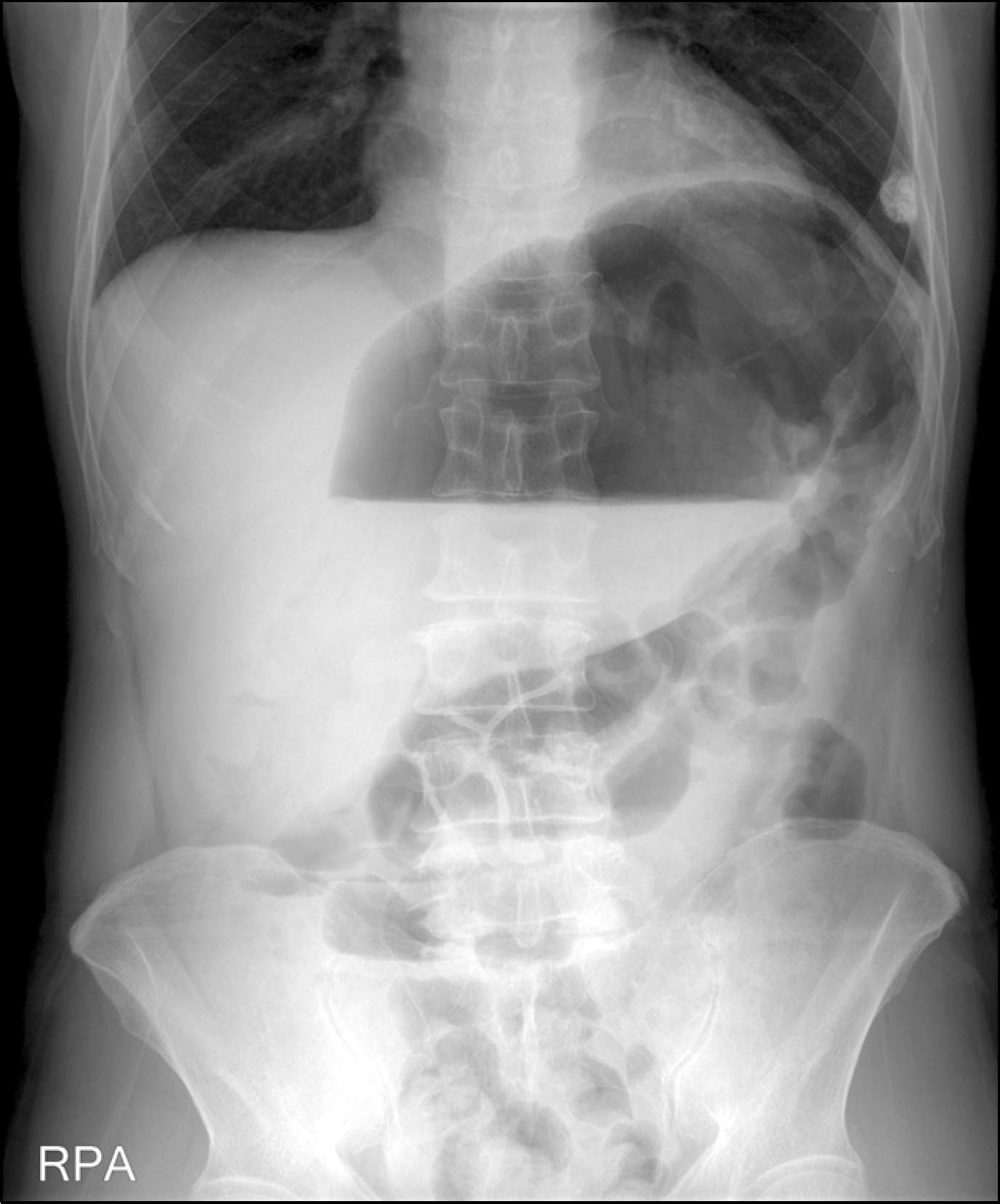
Fig. 1.
Simple abdomen view. It shows marked gaseous dis-tension of transverse colon.
kjg-56-65f1.tif
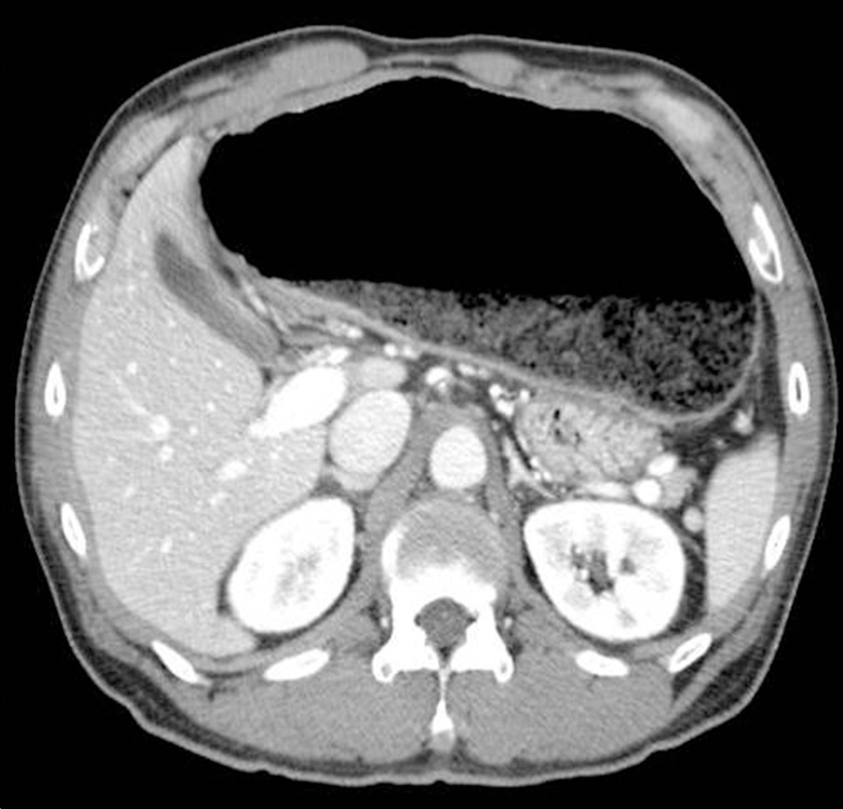
Fig. 2.
Abdominal CT scan finding. It shows marked dilated ascending and transverse colon without distal obstructive lesion.
kjg-56-65f2.tif
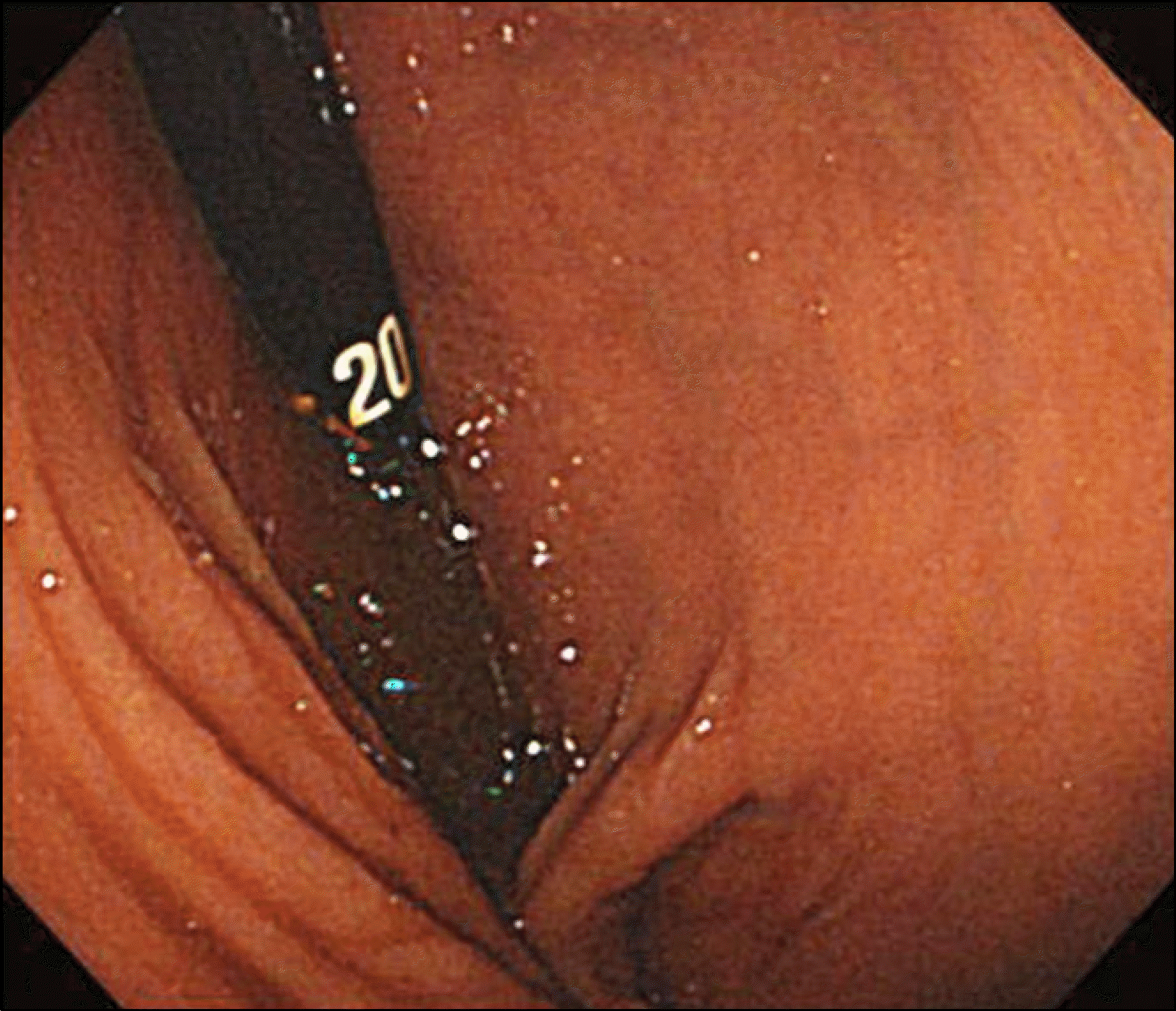
Fig. 3.
Colonoscopy finding. It shows luminal dilatation in the transverse colon.
kjg-56-65f3.tif
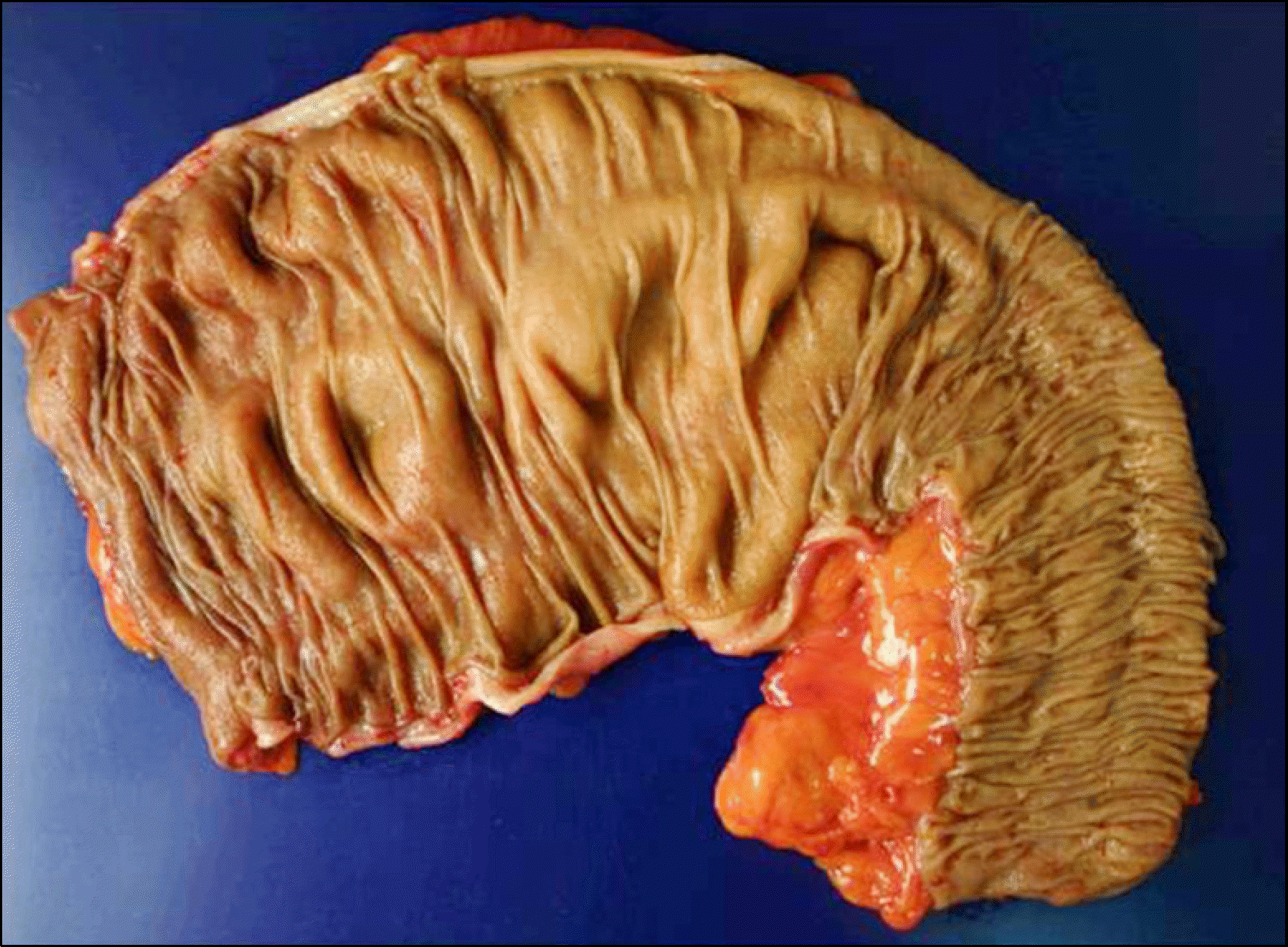
Fig. 4.
Gross finding. It shows near total colectomy specimen, consisting of the ascending, transverse, and descending colon.
kjg-56-65f4.tif
Fig. 5.
Microscopic findings. It shows normal looking submucosal Meissner's (white arrow) (A) and intermuscular myenteric Auerbach's autonomic nerve plexuses (white arrow) (B) (H&E, ×200).
kjg-56-65f5.tif
TOOLS
Similar articles