

 PDF
PDF ePub
ePub Citation
 Print
Print


Abstract
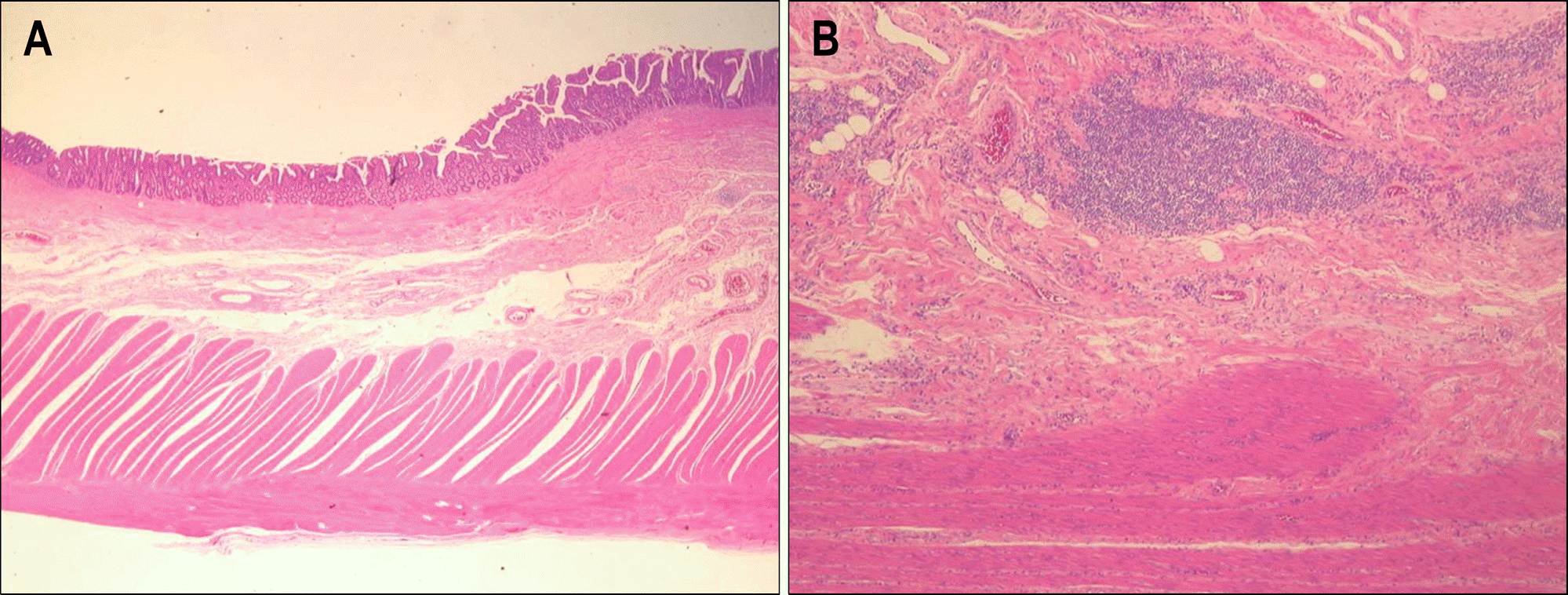
Chronic non-granulomatous jejunoileitis is a rare disease characterized by malabsorption, abdominal pain, and diarrhea that causes shallow ulcers in the small bowel. The etiology of chronic non-granulomatous jejunolieitis re-mains unknown. A 69-year-old man complained of abdominal pain and lower extremity edema. A 99m-Tc albumin scan showed increased radioactivity at the left upper quadrant, suggesting protein-losing enteropathy. A small bowel follow-through did not disclose any lesions. Wireless capsule endoscopy revealed several small bowel ulcers and strictures. A jejunoileal segmentectomy with end-to-end anastomosis was performed, and the histologic examination revealed non-granulomatous ulcers with focal villous atrophy. Ruling out all other possible diagnoses, we diagnosed our patient with chronic non-granulomatous ulcerative jejunoileitis. Postoperatively, the patient's abdominal pain and lower extremity edema improved, and the serum albumin normalized. This is the first case of chronic non-granulomatous ulcerative jejunoileitis localized by wireless capsule endoscopy and treated successfully with segment resection.
REFERENCES
1. Moon H, Kim YS, Chung YJ, et al. Three cases of chronic nongranulomatous ulcerative jejunoileitis. Korean J Gastroenterol. 1999; 34:399–405.
2. LePane CA, Barkin JS, Parra J, Simon T. Ulcerative jejunoileitis: a complication of celiac sprue simulating Crohn's disease diagnosed with capsule endoscopy (PillCam). Dig Dis Sci. 2007; 52:698–701.

3. Sturniolo GC, Di Leo V, Vettorato MG, D'Inca R. Clinical relevance of small-bowel findings detected by wireless capsule endoscopy. Scand J Gastroenterol. 2005; 40:725–733.
4. Ruan EA, Komorowski RA, Hogan WJ, Soergel KH. Nongranulomatous chronic idiopathic enterocolitis: clinico-pathologic profile and response to corticosteroids. Gastroenterology. 1996; 111:629–637.
5. Perera DR, Weinstein WM, Rubin CE. Symposium on pathol-ogy of the gastrointestinal tract-Part II. Small intestinal biopsy. Hum Pathol. 1975; 6:157–217.
6. Jeffries GH, Steinberg H, Sleisenger MH. Chronic ulcerative (nongranulomatous) jejunitis. Am J Med. 1968; 44:47–59.
7. Mills PR, Brown IL, Watkinson G. Idiopathic chronic ulcerative enteritis. Report of five cases and review of the literature. Q J Med. 1980; 49:133–149.
8. Ersoy O, Harmanci O, Aydinli M, Sivri B, Bayraktar Y. Capability of capsule endoscopy in detecting small bowel ulcers. Dig Dis Sci. 2009; 54:136–141.
9. Barkin JS, Friedman S. Wireless capsule endoscopy requiring surgical intervention. The world's experience. Am J Gastroenterol. 2002; 97:A83.
10. Ho KK, Joyce AM. Complications of capsule endoscopy. Gastrointest Endosc Clin N Am. 2007; 17:169–178.
11. Moy L, Levine J. Wireless capsule endoscopy in the pediatric age group: experience and complications. J Pediatr Gastroenterol Nutr. 2007; 44:516–520.
12. Delvaux M, Ben Soussan E, Laurent V, Lerebours E, Gay G. Clinical evaluation of the use of the M2A patency capsule system before a capsule endoscopy procedure, in patients with known or suspected intestinal stenosis. Endoscopy. 2005; 37:801–807.
13. Toy E, Rojany M, Sheikh R, Mann S, Prindiville T. Capsule endoscopy's impact on clinical management and outcomes: a single-center experience with 145 patients. Am J Gastroenterol. 2008; 103:3022–3028.
14. Idiopathic chronic ulcerative enteritis. Lancet. 1980; 2:1118–1119.
Fig. 1.
99m-Tc albumin scan finding. At 2 hours, increased ra-dioactivities at the left upper quadrant (arrowheads) were noticed.
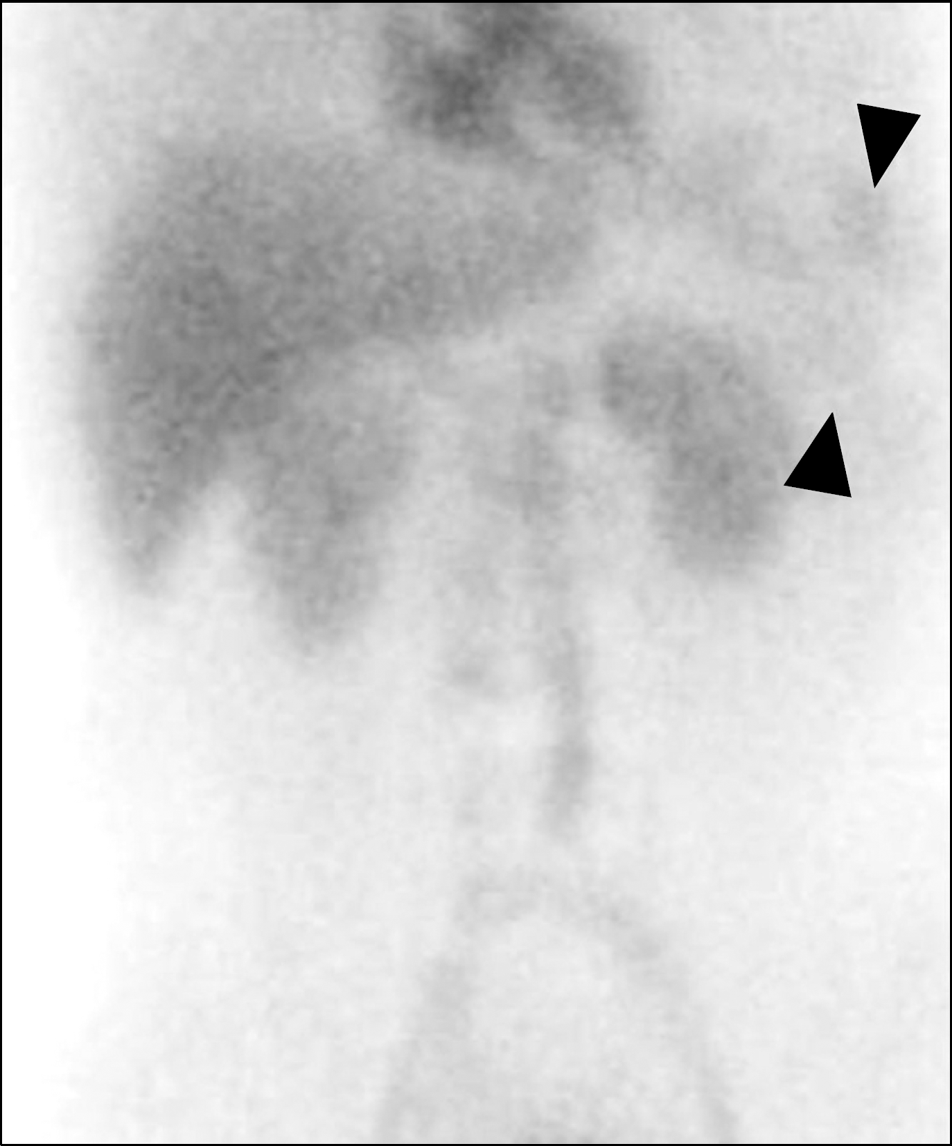
Fig. 2.
Wireless capsule endoscopy findings. (A) It revealed multiple geographic and shallow ulcerations with villous atrophy on the distal jejunum. (B) It showed stenotic area, which caused capsule retention in the proximal ileum.
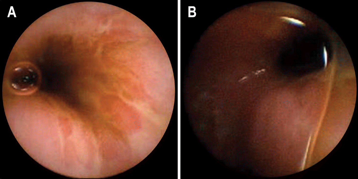
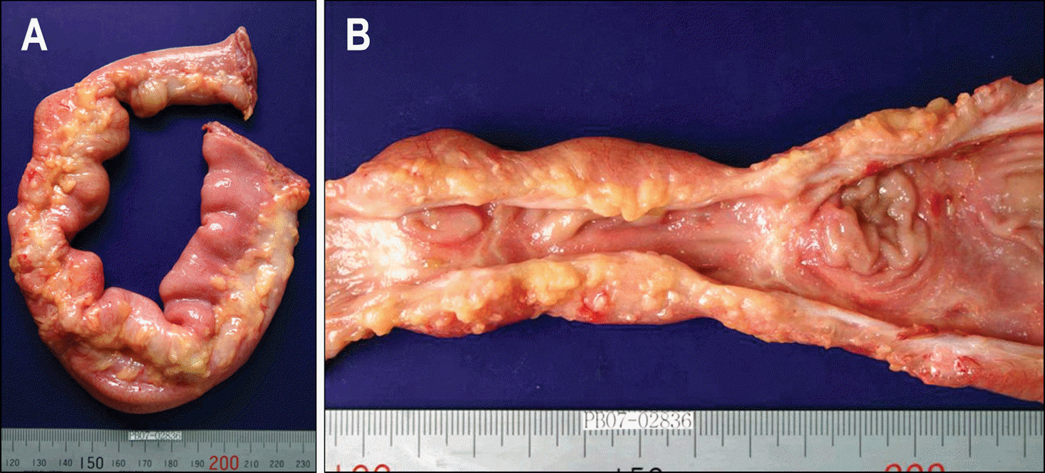
Fig. 3.
The gross findings of resected specimen. (A) It showed a dilated proximal bowel segment 4.2 cm in diameter. The mesenteric fat tissue was mildly adher-ent to the serosal surface focally. (B) On opening, several shallow transverse or oval ulcerations were observed with two white fi-brous stenotic foci.
 XML Download