Abstract
Background/Aims
Disease activity in ulcerative colitis (UC) is generally assessed using symptoms, laboratory da-ta, endoscopic findings, and histology of the biopsy specimens. In this study, we compared disease activity of UC as determined by clinical features and endoscopic findings, and aimed to assess the clinical usefulness of Doppler sonography.
Methods
The duplex Doppler sonography of superior mesenteric artery (SMA) and inferior mesenteric artery (IMA) of 10 patients with clinically inactive UC and 20 patients with active UC were evaluated by one radiologist who was blinded to clinical information. Peak systolic velocity (PSV), end diastolic velocity (EDV), resistance index (RI), and pulsatility index (PI) of the SMA and IMA were evaluated. All patients underwent biochemical and endoscopic evaluations thereafter. Correlation between disease activity by the Truelove-Witts classification and the Mayo scoring system was measured, and we compared hemodynamic parameters between active and inactive UC.
Results
Correlation rate of disease activity between these two scoring systems was 93.3%. Flow velocities (PSV, p<0.001 and EDV, p=0.03) and PI (p=0.03) were significantly higher in patients with active UC than inactive UC. PSVs of the SMA and IMA were also significantly correlated with disease severity. The active UC could be accurately diagnosed using Doppler sonography (AUC=0.83; 95% confidence interval 0.68-0.99).
REFERENCES
1. Langholz E, Munkholm P, Davidsen M, Binder V. Course of ulcerative colitis: analysis of changes in disease activity over years. Gastroenterology. 1994; 107:3–11.

2. Ando T, Nishio Y, Watanabe O, et al. Value of colonoscopy for prediction of prognosis in patients with ulcerative colitis. World J Gastroenterol. 2008; 14:2133–2138.

3. Edwards FC, Truelove SC. The course and prognosis of ulcerative colitis. Gut. 1963; 4:299–315.
4. Selby W. The natural history of ulcerative colitis. Baillieres Clin Gastroenterol. 1997; 11:53–64.
5. Hiwatashi N, Yao T, Watanabe H, et al. Longterm follow-up study of ulcerative colitis in Japan. J Gastroenterol. 1995; 30(suppl 8):13–16.
6. Truelove SC, Witts LJ. Cortisone in ulcerative colitis; final report on a therapeutic trial. Br Med J. 1955; 2:1041–1048.
7. Naber AH, de Jong DJ. Assessment of disease activity in inflammatory bowel disease; relevance for clinical trials. Neth J Med. 2003; 61:105–110.
8. Vermeire S, Van Assche G, Rutgeerts P. Laboratory markers in IBD: useful, magic, or unnecessary toys? Gut. 2006; 55:426–431.

9. Solem CA, Loftus EV Jr, Tremaine WJ, Harmsen WS, Zinsmeister AR, Sandborn WJ. Correlation of C-reactive protein with clinical, endoscopic, histologic, and radiographic activity in inflammatory bowel disease. Inflamm Bowel Dis. 2005; 11:707–712.

10. Hulté n L, Lindhagen J, Lundgren O, Fasth S, Ahré n C. Regional intestinal blood flow in ulcerative colitis and Crohn's disease. Gastroenterology. 1977; 72:388–396.

11. Ludwig D, Wiener S, Brü ning A, et al. Mesenteric blood flow is related to disease activity and risk of relapse in ulcerative colitis: a prospective follow up study. Gut. 1999; 45:546–552.

12. Di Sabatino A, Armellini E, Corazza GR. Doppler sonography in the diagnosis of inflammatory bowel disease. Dig Dis. 2004; 22:63–66.

13. Siğ irci A, Baysal T, Kutlu R, Aladağ M, Saraç K, Harput-luoğ lu H. Doppler sonography of the inferior and superior mesenteric arteries in ulcerative colitis. J Clin Ultrasound. 2001; 29:130–139.
14. Fillinger MF, Schwartz RA. Volumetric blood flow measurement with color Doppler ultrasonography: the importance of visual clues. J Ultrasound Med. 1993; 12:123–130.

15. Schroeder KW, Tremaine WJ, Ilstrup DM. Coated oral 5-ami-nosalicylic acid therapy for mildly to moderately active ulcerative colitis. A randomized study. N Engl J Med. 1987; 317:1625–1629.
16. Azzolini F, Pagnini C, Camellini L, et al. Proposal of a new clinical index predictive of endoscopic severity in ulcerative colitis. Dig Dis Sci. 2005; 50:246–251.

17. Rachmilewitz D. Coated mesalazine (5-aminosalicylic acid) versus sulphasalazine in the treatment of active ulcerative colitis: a randomised trial. BMJ. 1989; 298:82–86.

18. Baron JH, Conell AM, Lennard-Jones JE. Variation between observers in describing mucosal appearances in proctocolitis. Br Med J. 1964; 1:89–92.

19. Lee SD, Cohen RD. Endoscopy in inflammatory bowel disease. Gastroenterol Clin North Am. 2002; 31:119–132.

20. R⊘seth AG, Aadland E, Jahnsen J, Raknerud N. Assessment of disease activity in ulcerative colitis by faecal calprotectin, a novel granulocyte marker protein. Digestion. 1997; 58:176–180.

21. de Lange T, Larsen S, Aabakken L. Interobserver agreement in the assessment of endoscopic findings in ulcerative colitis. BMC Gastroenterol. 2004; 4:9.

22. Higgins PD, Schwartz M, Mapili J, Zimmermann EM. Is endoscopy necessary for the measurement of disease activity in ulcerative colitis? Am J Gastroenterol. 2005; 100:355–361.

23. Menees S, Higgins P, Korsnes S, Elta G. Does colonoscopy cause increased ulcerative colitis symptoms? Inflamm Bowel Dis. 2007; 13:12–18.

24. Gabay C, Kushner I. Acute-phase proteins and other systemic responses to inflammation. N Engl J Med. 1999; 340:448–454.

25. Zlonis M. The mystique of the erythrocyte sedimentation rate. A reappraisal of one of the oldest laboratory tests still in use. Clin Lab Med. 1993; 13:787–800.

26. Hilliard NJ, Waites KB. CME: C-reactive protein and ESR; What can one tell you that the other can't? Contemp Pediatr. 2002; 19:64–74.
27. Ha JS, Lee JS, Kim HJ, et al. Comparative usefulness of erythrocyte sedimentation rate and C-reactive protein in assessing the severity of ulcerative colitis. Korean J Gastroenterol. 2006; 48:313–320.
Fig. 1.
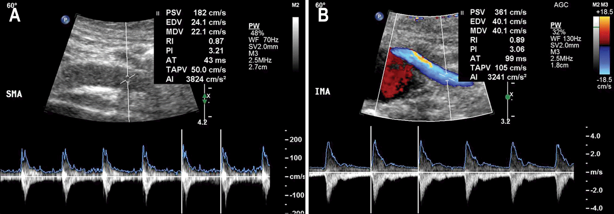
Duplex doppler sonographic examination. (A) The sampling cursor was placed within the lumen of the vessel, 2-3 cm distal to its origin, and the angle between the ultrasound beam and the SMA is maintained at 60 o. (B) Duplex Doppler sonography of the IMA was performed with 60 o of the insonation angle. The hemodynamic parameters were calculated automatically by the spectral analyzer of the ultrasound system.

Fig. 2.
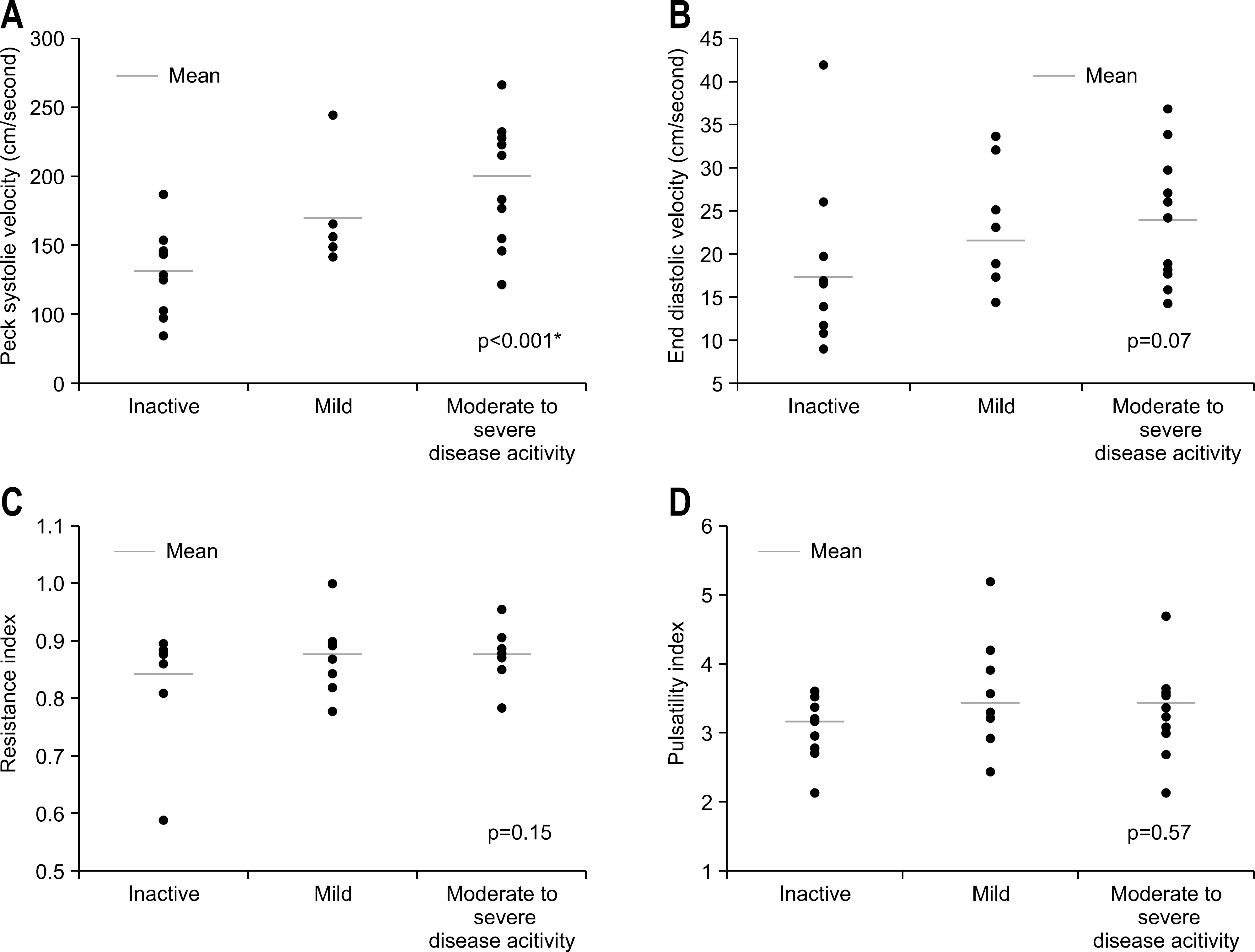
Hemodynamic parameters of SMA by doppler sonography. (Peak systolic velocity, PSV (A), End diastolic velocity, EDV (B), Resistance index, RI (C), Pulsatility index, PI (D)). When the PSV, EDV, PI and RI were measured according to disease severity classi-fied by the Mayo scoring system, only PSV was significantly correlated with disease severity (p<0.001). SMA, superior mesenteric artery; PSV, peak systolic velocity; EDV, end diastolic velocity; RI, resistance index; PI, pulsatility index.

Table 1.
Comparison of the Parameters Measured by Doppler Sonography between the Active and Inactive Ulcerative Colitis Patients
Table 2.
Comparison of the Parameters Measured by Doppler Sonography according to the Disease Severity Categorized by Truelove Witts Classification


 PDF
PDF ePub
ePub Citation
 Print
Print


 XML Download