

 PDF
PDF ePub
ePub Citation
 Print
Print


Abstract
Background/Aims
We aimed to determine the concordance rate and clinical predictors of eosinophilic esophagitis (EoE) in patients with endoscopically suspected eosinophilic esophagitis (ESEoE) findings.
Methods
From June 2006 to December 2009 in Gangnam Severance Hospital, we prospectively enrolled the patients of “endoscopically suspected eosinophilic esophagitis (ESEoE)”, and then we retrospectively reviewed and analyzed clinical features and endoscopic findings.
Results
We found 17 patients of ESEoE, and 5 of them were finally confirmed as an EoE by histology (diagnostic concordance rate 29.4%). We added two more patients previously diagnosed as EoE and compared patients of EoE+ (n=7) with EoE− (n=12). Mean age was 56.0 (range 36-70) and 51.0 (range 36-68) years old, respectively. In EoE+ group, there were 5 males and 2 females and 6 males and 6 females in EoE- group. The symptoms of EoE+ patients were dysphagia (n=5), food impaction (n=3), foreign body sensation in esophagus (n=2), chest pain (n=1), and heartburn (n=1). EoE− patients complained food impaction (n=5), heartburn (n=4), chest pain (n=2), foreign body sensation in esophagus (n=2), and dysphagia (n=1). The endoscopic findings of EoE+ patients were furrows (n=6), rings (n=5), exudates or nodules (n=3), and friability (n=1). EoE− patients showed rings (n=10) and furrows(n=7). Univariative analysis showed that ‘a symptom of dysphagia’, ‘presence of exudates or nodules’, ‘more than 2 suggestive endoscopic findings’ were significantly different between two groups.
Go to : 
REFERENCES
1. Landres RT, Kuster GG, Strum WB. Eosinophilic esophagitis in a patient with vigorous achalasia. Gastroenterology. 1978; 74:1298–1301.

2. Attwood SE, Smyrk TC, Demeester TR, Jones JB. Esophageal eosinophilia with dysphagia. A distinct clinicopathologic syndrome. Dig Dis Sci. 1993; 38:109–116.
3. Kim JW, Park JS, Kim YH, et al. Secondary achalasia by eosinophilic esophagitis. Korean J Gastrointest Endosc. 2002; 25:198–202.
4. Lee B, Park H, Yoon H, Kim HK, Kim HS. Three cases of eosinophilic esophagitis with dysphagia as a chief complaint. Korean J Gastrointest Endosc. 2008; 36:145–149.
5. Furuta GT, Liacouras CA, Collins MH, et al. Eosinophilic esophagitis in children and adults: a systematic review and consensus recommendations for diagnosis and treatment. Gastroenterology. 2007; 133:1342–1363.
6. Potter JW, Saeian K, Staff D, et al. Eosinophilic esophagitis in adults: an emerging problem with unique esophageal features. Gastrointest Endosc. 2004; 59:355–361.
7. Straumann A, Spichtin HP, Grize L, Bucher KA, Beglinger C, Simon HU. Natural history of primary eosinophilic esophagitis: a follow-up of 30 adult patients for up to 11.5 years. Gastroenterology. 2003; 125:1660–1669.
8. Croese J, Fairley SK, Masson JW, et al. Clinical and endoscopic features of eosinophilic esophagitis in adults. Gastrointest Endosc. 2003; 58:516–522.
9. Prasad GA, Talley NJ, Romeo Y, et al. Prevalence and predictive factors of eosinophilic esophagitis in patients presenting with dysphagia: a prospective study. Am J Gastroenterol. 2007; 102:2627–2632.
10. Veerappan GR, Perry JL, Duncan TJ, et al. Prevalence of eosinophilic esophagitis in an adult population undergoing upper endoscopy: a prospective study. Clin Gastroenterol Hepatol. 2009; 7:420–426.
11. Yu YH, Jo YJ, Jung MY, et al. Prevalence of eosinophlic esophagitis with dysphagia and reflux related symptoms in Korean patients. Korean J Neurogastroenterol Motil. 2009; 15:15–22.
12. Park HJ. Eosinophilic esophagitis. Korean J Gastroenterol. 2007; 50:286–291.
13. Sgouros SN, Bergle C, Mantides A. Eosinophilic esophagitis in adults: what is the clinical significance? Endoscopy. 2006; 38:515–520.
14. Dellon ES, Gibbs WB, Fritchie KJ, et al. Clinical, endoscopic, and histologic findings distinguish eosinophilic esophagitis from gastroesophageal reflux disease. Clin Gastroenterol Hepatol. 2009; 7:1305–1313.
15. Putnam PE, Rothenberg ME. Eosinophilic esophagitis: concepts, controversies, and evidence. Curr Gastroenterol Rep. 2009; 11:220–225.
16. Lim JR, Gupta SK, Croffie JM, et al. White specks in the esophageal mucosa: An endoscopic manifestation of non-reflux eosinophilic esophagitis in children. Gastrointest Endosc. 2004; 59:835–838.
17. Katzka DA. Demographic data and symptoms of eosinophilic esophagitis in adults. Gastrointest Endosc Clin N Am. 2008; 18:25–32.
Go to :
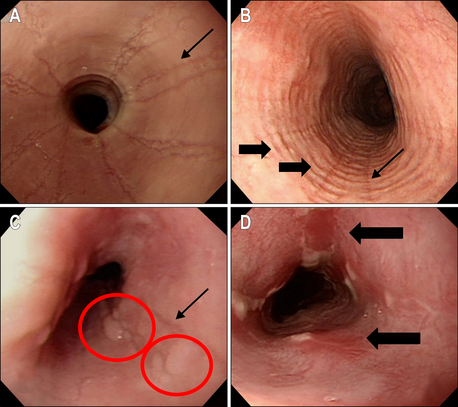 | Fig. 1.Endoscopic findings of eosinophilic esophagitis (EoE+) cases. Typical endoscopic findings of eosinophilic esophagitis patients including (A) linear furrows (thin arrow), (EoE+ No.3), (B) linear furrows (thin arrow) and circular rings (thick arrows), (EoE+ No.6), (C) linear furrows (thin arrow) and several nodules (red circles), (EoE+ No.1), (D) several linear shearings (thick arrows), (EoE+ No.5). Patient number is listed in Table 1. |
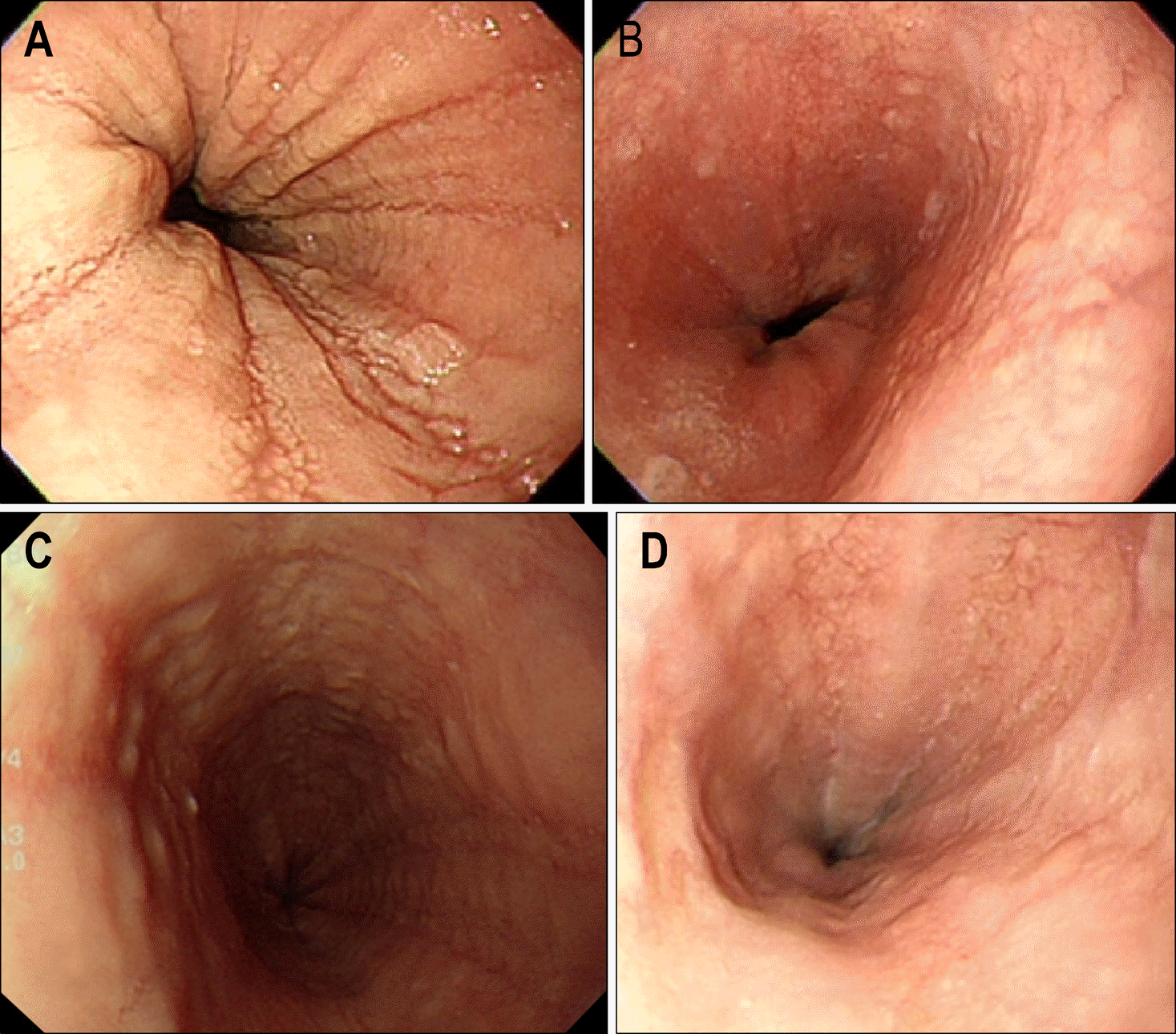 | Fig. 2.Initial & follow-up endoscopic findings of eosinophilic esophagitis (EoE+) patients after the treatment. (A) It showed longitudinal furrows (EoE+ No.7) and his symptoms were relieved after proton pump inhibitor (PPI) treatment. (B) It showed endoscopically improved findings 3 months later. (C) It showed longitudinal furrows and circular rings (EoE+ No.4). She had no response to 2 months PPI treatment, so we prescribed a flucati-sone inhaler (125 ug/puff, 1,000 ug/D) for 4 weeks. Symptoms were relieved after taking a PPI for 6 more weeks. (D) It showed slightly improved findings 2 years later. Patient number is listed in Table 1. |
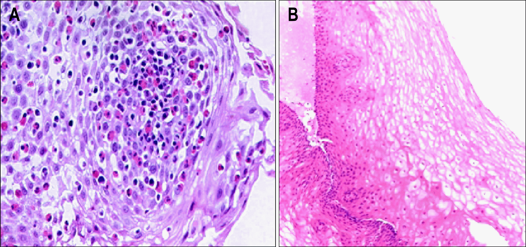 | Fig. 3.Initial and follow-up histologic findings of eosinophilic esophagitis (EoE+) patient. (A) Initial microscopic finding of EoE+ No.4 patient. It showed massive infiltrations of eosinophilis on the esophageal mucosa. (B) Microscopic finding of EoE+ No.4 patient 2 years after the treatment. It showed normal esophageal epithelium without eosinophils. Patient number is listed in Table 1. |
Table 1.
List of Patients with Eosinophilic Esophagitis
| No. | Sex | Age | Date | Indication of endoscopy | Esophageal endoscopic findings | Allergy history | Treatment & Follow-up |
|---|---|---|---|---|---|---|---|
| 1∗ | M | 67 | 2002.2. | Food impaction | Nodules | Food allergy | Follow-up loss |
| Globus sensation | B. asthma | ||||||
| Rt. Chest pain | |||||||
| 2† | M | 70 | 2005.9. | Esophgeal nodules | Nodules | Follow-up loss | |
| 3 | M | 48 | 2006.6. | Dysphagia | Ring | Follow-up loss | |
| Furrow | |||||||
| Whitish discoloration | |||||||
| 4 | F | 54 | 2007.4. | Dysphagia | Ring | SPT(−) | PPI & Steroid inhaler |
| Food impaction | Furrow | ||||||
| 5 | F | 68 | 2007.10. | Dysphagia | Linear shearing (friability) | PPI | |
| Sore throat | Exudate | Follow-up loss | |||||
| 6 | M | 36 | 2008.11. | Check-up | Ring | SPT(+) | PPI |
| Odynophagia & Dysphagia | Furrow | ||||||
| Nodules | |||||||
| 7 | M | 46 | 2009.3. | Check-up | Ring | Allergic history(+), | PPI |
| Furrow | SPT(+) |
Table 2.
Clinical Features & Endoscopic Findings of Endoscopically Suspected Eosinophilic Esophagitis Patients
| EoE+ (n=7) | EoE− (n=12) | p-value | ||||
|---|---|---|---|---|---|---|
| Sex | % | % | ||||
| Male | 5 | 71.4 | 6 | 50.0 | ||
| Female | 2 | 28.6 | 6 | 50.0 | ||
| Mean age (yr) | 56.0 | (36-70) | 51.0 | (36-68) | ||
| Presenting symptom | ||||||
| Dysphagia | 5 | 71.4 | 1 | 8.3 | <0.05∗ | |
| Food impaction | 3 | 42.9 | 5 | 41.7 | ||
| Heartburn | 1 | 14.3 | 4 | 33.3 | ||
| Chest pain | 1 | 14.3 | 2 | 16.7 | ||
| Foreign body sensation | 2 | 28.6 | 2 | 16.7 | ||
| Allergic history | 2 | 28.6 | 3 | 25.0 | ||
| Endoscopic findings | ||||||
| Furrow | 6 | 85.7 | 7 | 58.3 | ||
| Ring | 5 | 71.4 | 10 | 83.3 | ||
| Exudates/Nodules | 3 | 42.9 | 0 | 0 | <0.05∗ | |
| Friability | 1 | 14.3 | 0 | 0 | ||
| ≥2 Endoscopic findings | 6 | 85.7 | 6 | 50.0 | <0.05∗ | |
 XML Download