

 PDF
PDF ePub
ePub Citation
 Print
Print


Abstract
Hemorrhagic acalculous cholecystitis is an extremely rare but potentially fatal disease if detection is delayed. Its risk factors include critical illness, diabetes, malignant disease, uremia, and bleeding diathesis. This is the first case report in which hemorrhagic acalculous cholecystitis not accompanied by any risk factor. We herein present a case of hemorrhagic acalculous cholecystitis in a previously healthy patient who suffered from acute abdomen.
Go to : 
REFERENCES
1. Pandya R, O'Malley C. Hemorrhagic cholecystitis as a complication of anticoagulant therapy: role of CT in its diagnosis. Abdom Imaging. 2008; 33:652–653.

2. Lai YC, Tarng DC. Hemorrhagic acalculous cholecystitis: an unusual location of uremic bleeding. J Chin Med Assoc. 2009; 72:484–487.
3. Tikkakoski T. Acalculous cholecystitis in a previously healthy man. Duodecim. 2001; 117:279–281.
4. Parithivel VS, Gerst PH, Banerjee S, Parikh V, Albu E. Acute acalculous cholecystitis in young patients without pre-disposing factors. Am Surg. 1999; 65:366–368.
5. Chinn DH, Miller EI, Piper N. Hemorrhagic cholecystitis. Sonographic appearance and clinical presentation. J Ultrasound Med. 1987; 6:313–317.
6. Kwun KB, Lee SJ, Kim JH, Roh SG, Seo WS. Acute acalculous cholecystitis. Korean J Gastroenterol. 1992; 24:1087–1093.
7. Savoca PE, Longo WE, Zucker KA, McMillen MM, Modlin IM. The increasing prevalence of acalculous cholecystitis in outpatients. Results of a 7-year study. Ann Surg. 1990; 211:433–437.
8. Taoka H. Experimental study on the pathogenesis of acute acalculous cholecystitis, with special reference to the roles of microcirculatory disturbances, free radicals and membrane- bound phospholipase A2. Gastroenterol Jpn. 1991; 26:633–644.
9. Gremmels JM, Kruskal JB, Parangi S, Kane RA. Hemorrhagic cholecystitis simulating gallbladder carcinoma. J Ultrasound Med. 2004; 23:993–995.
10. Oshiba S, Hata S, Okamoto S. A plasminogen activator in mammalian bile. Jpn J Physiol. 1969; 19:212–219.
11. Moon W, Sohn JH, Jang MH, et al. A case of acute cholecystitis secondary to hemobilia after percutaneous liver biopsy. Korean J Gastroenterol. 2006; 47:72–76.
12. Johnson LB. The importance of early diagnosis of acute acal-culus cholecystitis. Surg Gynecol Obstet. 1987; 164:197–203.
13. Devine RM, Farnell MB, Mucha P Jr. Acute cholecystitis as a complication in surgical patients. Arch Surg. 1984; 119:1389–1393.
14. Kim KH, Sung CK, Park BK, Kim WK, Oh CW, Kim KS. Percutaneous gallbladder drainage for delayed laparoscopic cholecystectomy in patients with acute cholecystitis. Am J Surg. 2000; 179:111–113.
Go to :
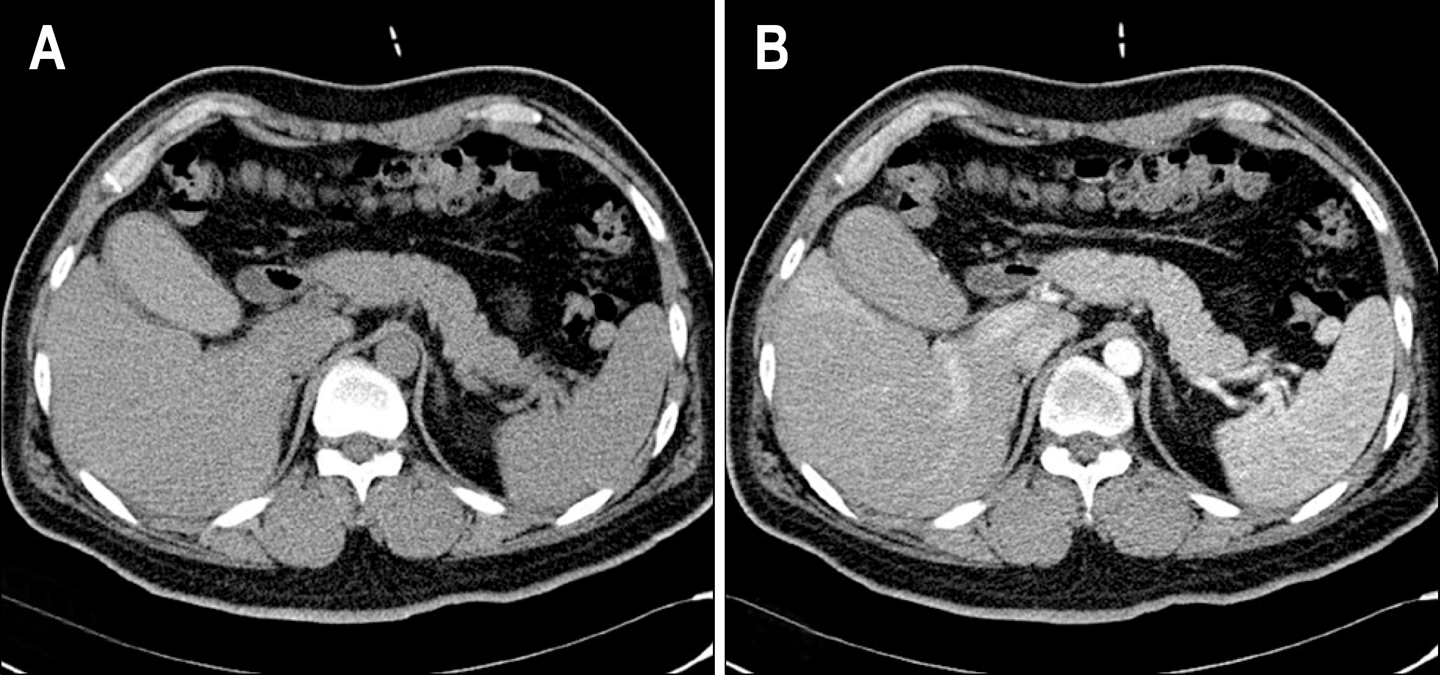 | Fig. 1.Computed tomography finding. It showed highly attenuated, homogenous materials with near blood density in the gallbladder at both pre-enhance and arterial phase. It showed a diffuse gallbladder wall thickening and hyperemia of peri gallbladder liver parenchyma on early arterial phase. This finding suggested the possibility of hemorrhagic cholecystitis (A: pre-enhance phase, B: aterial phase). |
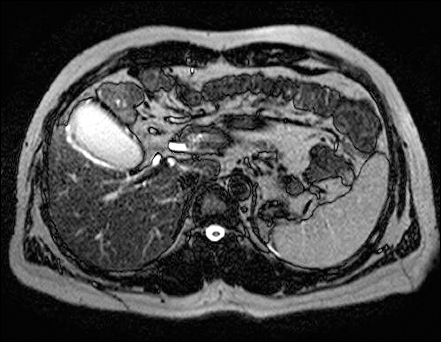 | Fig. 3.Magnetic resonance finding. It showed a distended gallbladder and diffuse gallbladder wall thickening and high signal intensity materials filled the gallbladder lumen on T2 weighted image. |

 XML Download