

 PDF
PDF ePub
ePub Citation
 Print
Print


Abstract
Endoscopic methods such as endoscopic mucosal resection or endoscopic submucosal dissection (ESD) have been increasingly used for the treatment of gastric adenoma and early gastric cancer. Especially, ESD is very useful since it allows en bloc resection of large lesions. Bleeding and perforation are well known as common complications after ESD. However, there is no report of acute myocardial infarction associated with ESD. We report a case of acute myocardial infarction which was detected immediately after ESD.
Go to : 
REFERENCES
1. Gotoda T, Yamamoto H, Soetikno RM. Endoscopic submucosal dissection of early gastric cancer. J Gastroenterol. 2006; 41:929–942.

2. Chung IK, Lee JH, Lee SH, et al. Therapeutic outcomes in 1000 cases of endoscopic submucosal dissection for early gastric neoplasms: Korean ESD Study Group multicenter study. Gastrointest Endosc. 2009; 69:1228–1235.
3. Kim JJ, Lee JH, Jung HY, et al. EMR for early gastric cancer in Korea: a multicenter retrospective study. Gastrointest Endosc. 2007; 66:693–700.
4. Yachimski P, Hur C. Upper endoscopy in patients with acute myocardial infarction and upper gastrointestinal bleeding: results of a decision analysis. Dig Dis Sci. 2009; 54:701–711.
5. Lee EK, Jeon SW, Oh JT, et al. The feasibility and safety of endoscopic submucosal dissection for gastric neoplasm in elderly Korean patients. Korean J Gastrointest Endosc. 2009; 38:323–331.
6. Min BH, Lee JH, Kim JJ, et al. Clinical outcomes of endoscopic submucosal dissection (ESD) for treating early gastric cancer: comparison with endoscopic mucosal resection after circumferential precutting (EMR-P). Dig Liver Dis. 2009; 41:201–209.
7. Kakushima N, Fujishiro M. Endoscopic submucosal dissection for gastrointestinal neoplasms. World J Gastroenterol. 2008; 14:2962–2967.
8. Cao Y, Liao C, Tan A, Gao Y, Mo Z, Gao F. Meta-analysis of endoscopic submucosal dissection versus endoscopic mucosal resection for tumors of the gastrointestinal tract. Endoscopy. 2009; 41:751–757.
9. Habr-Gama A, Waye JD. Complications and hazards of gastrointestinal endoscopy. World J Surg. 1989; 13:193–201.
10. Shahmir M, Schuman BM. Complications of fiberoptic endoscopy. Gastrointest Endosc. 1980; 26:86–91.
11. Yusuf S, Hawken S, Ounpuu S, et al. Effect of potentially modifiable risk factors associated with myocardial infarction in 52 countries (the INTERHEART study): case-control study. Lancet. 2004; 364:937–952.
12. Jung HY, Choi KD, Song HJ, Lee GH, Kim JH. Risk management in endoscopic submucosal dissection using needle knife in Korea. Dig Endosc. 2007; 19(suppl 1):S5–S8.
13. Becker RC, Scheiman J, Dauerman HL, et al. Management of platelet-directed pharmacotherapy in patients with athero-sclerotic coronary artery disease undergoing elective endoscopic gastrointestinal procedures. Am J Gastroenterol. 2009; 104:2903–2917.
14. Eisen GM, Baron TH, Dominitz JA, et al. Guideline on the management of anticoagulation and antiplatelet therapy for endoscopic procedures. Gastrointest Endosc. 2002; 55:775–779.
15. Lee CT, Huang SP, Cheng TY, et al. Factors associated with myocardial infarction after emergency endoscopy for upper gastrointestinal bleeding in high-risk patients: a prospective observational study. Am J Emerg Med. 2007; 25:49–52.
16. Haffner SM, Lehto S, Rö nnemaa T, Pyö rä lä K, Laakso M. Mortality from coronary heart disease in subjects with type 2 diabetes and in nondiabetic subjects with and without prior myocardial infarction. N Engl J Med. 1998; 339:229–234.
17. Ridker PM, Cushman M, Stampfer MJ, Tracy RP, Hennekens CH. Inflammation, aspirin, and the risk of cardiovascular disease in apparently healthy men. N Engl J Med. 1997; 336:973–979.
18. Assmann G, Cullen P, Schulte H. Simple scoring scheme for calculating the risk of acute coronary events based on the 10-year follow-up of the prospective cardiovascular Munster (PROCAM) study. Circulation. 2002; 105:310–315.
19. Anderson KM, Odell PM, Wilson PW, Kannel WB. Cardiovascular disease risk profiles. Am Heart J. 1991; 121:293–298.
20. Mehta SK, Frutkin AD, Lindsey JB, et al. Bleeding in patients undergoing percutaneous coronary intervention: the development of a clinical risk algorithm from the National Cardiovascular Data Registry. Circ Cardiovasc Interv. 2009; 2:222–229.
Go to :
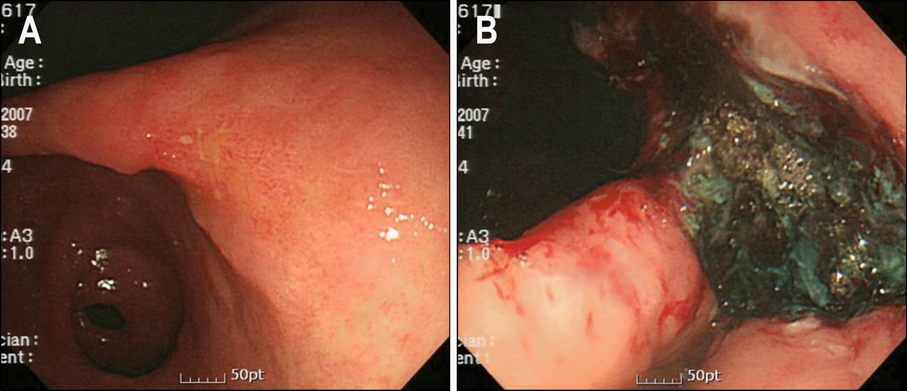 | Fig. 1.Endoscopic findings. (A) About 3×3 cm sized elevated lesion with central depression and vague margin was noted in the posterior side of gastric angle. (B) Iatrogenic ulcer after dissection was noted. |
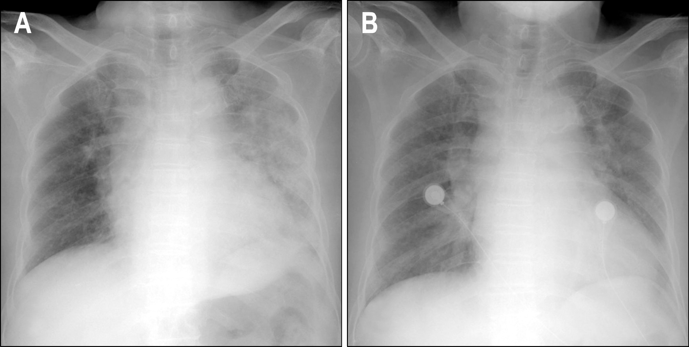 | Fig. 2.Chest AP views. (A) Patchy consolidation was noted in the diffuse left lung field after procedure. (B) On the 2 nd admission day, the improvement of patchy consolidation was noted. |
 | Fig. 3.ECG findings. (A) Abnormal Q waves and elevated ST segments were noted in aV L lead, and ST segments depressions in II, III, aV F, and V4-6 leads. (B) On the 4 th admission day, in spite of persistent abnormal Q waves in aV L lead, normalized ST segments were observed in all leads. |
 XML Download