Abstract
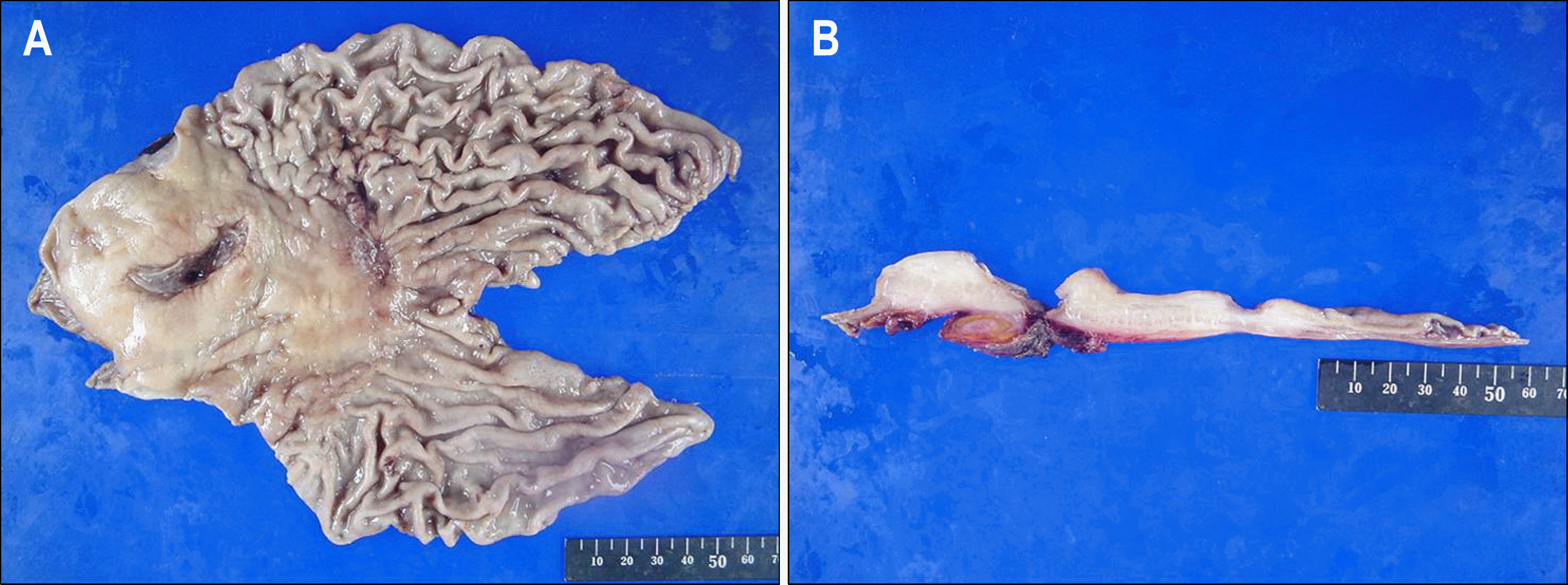
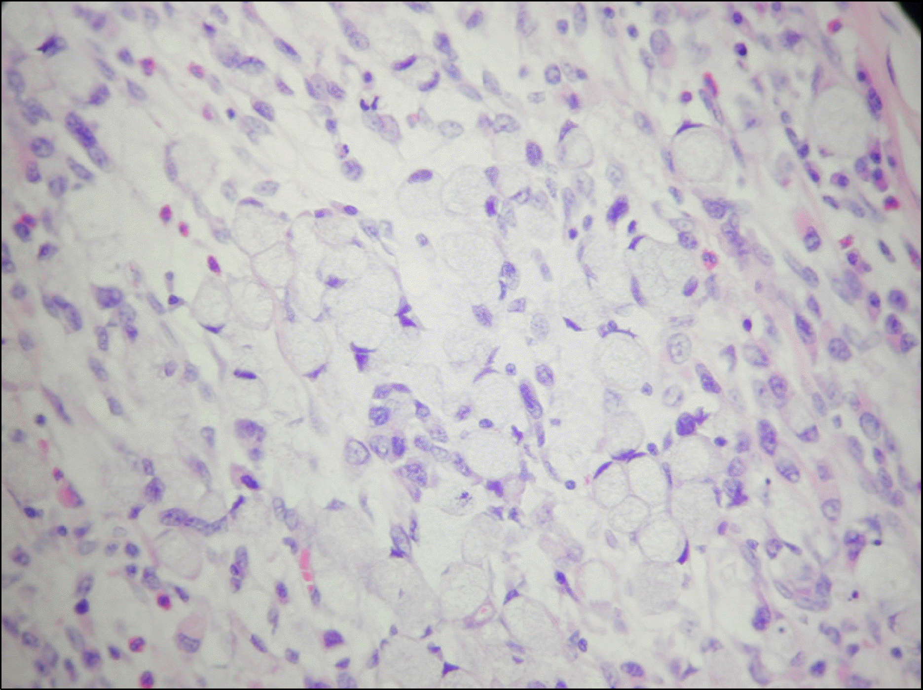
Intrahepatic abscess is an unusual complication of peptic ulcer disease. We present a case of gastric cancer in which the ulcer penetrated into the left lobe of liver with subsequent abscess and fistula formation. Esophagogastroduodenoscopy confirmed ulcers and a fistula opening in the antrum. Abdominal computed tomo-gram showed a subcapsular liver abscess adjacent to the gastric antrum. Subtotal gastrectomy with curettage of the fistulous tract was performed. The final diagnosis was the signet ring cell gastric carcinoma complicating subcapsular liver abscess. To our knowledge, this is the first reported case in Korea.
REFERENCES
1. Norris JR, Haubrich WS. The incidence and clinical features of penetration in peptic ulceration. JAMA. 1961; 178:386–389.

2. Allard JC, Kuligowska E. Percutaneous treatment of an intrahepatic abscess caused by a penetrating duodenal ulcer. J Clin Gastroenterol. 1987; 9:603–606.
4. Andrup H. Liver abscess caused by a penetrating prepyloric gastric ulcer. Ugeskr Laeger. 1988; 150:1173–1174.
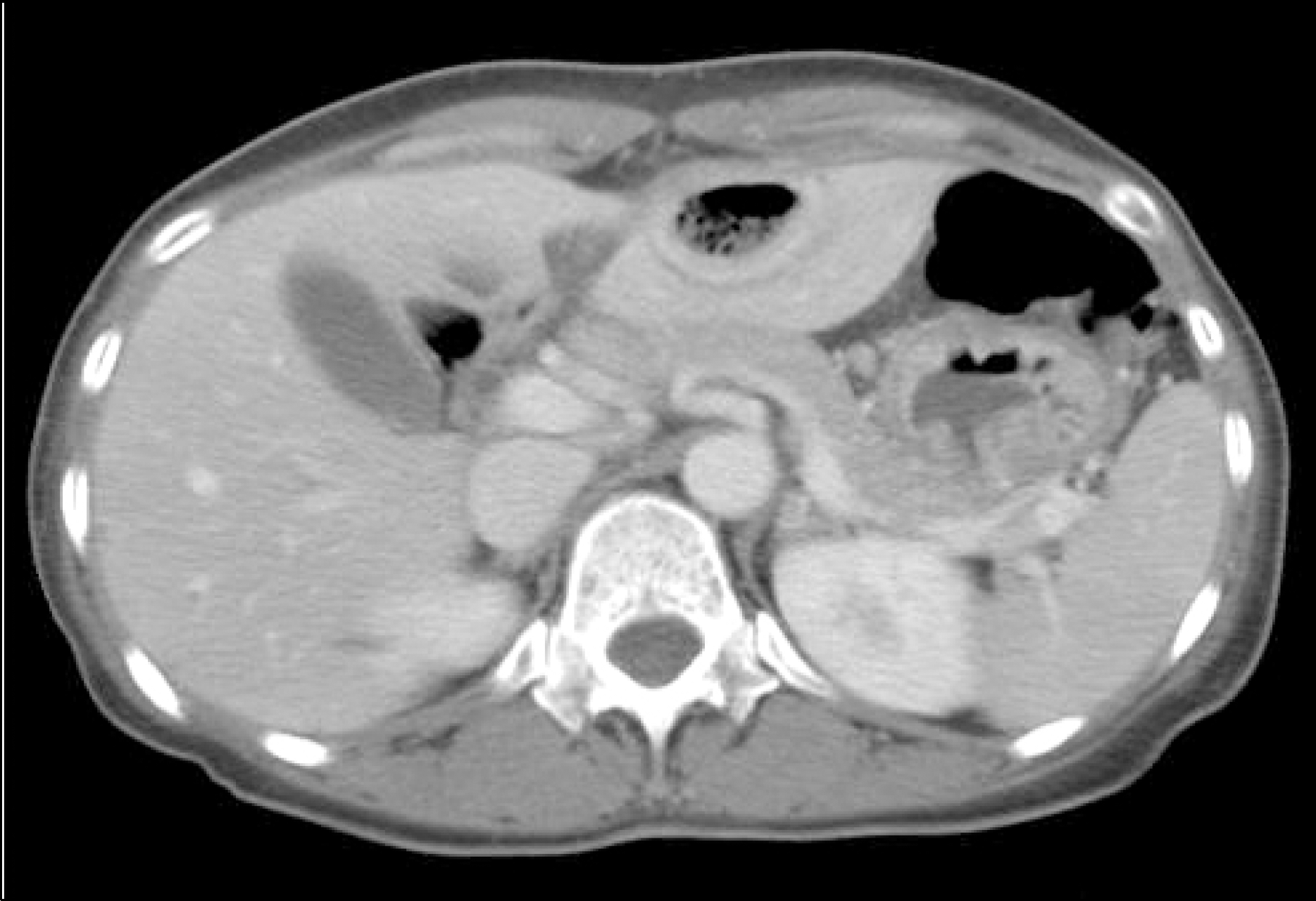
Fig. 1.
Initial abdominal CT scan showed a subcapsular liver abscess in the left lobe. Note that there was no evidence of free air.

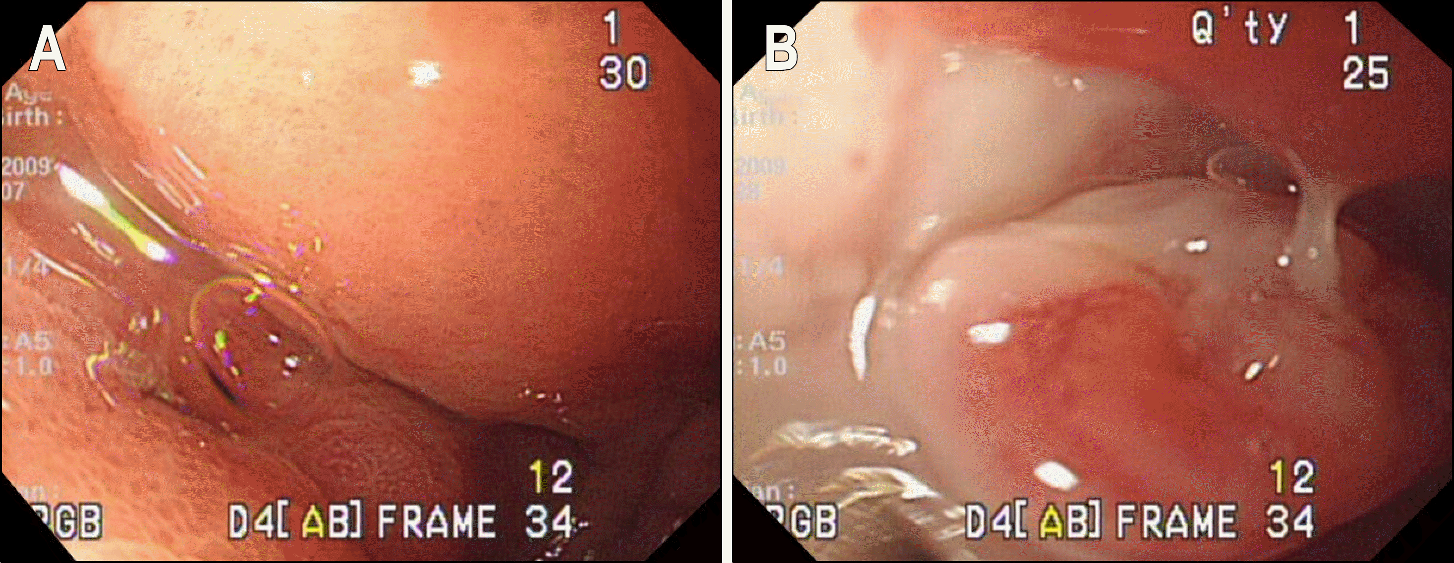
Fig. 2.
Endoscopic findings. (A) Air bubbles were leaking in the antral wall during the initial endoscopy. (B) During the follow-up examination, a fistula opening within the ulcer base was noticed. Note that there was pus around the orifice.


 PDF
PDF ePub
ePub Citation
 Print
Print


 XML Download