REFERENCES
1. Kang HJ, Park DY, Kim KH, Song GA, Lauwers GY. Pathologic diagnosis of gastric epithelial neoplasia. Korean J Gastroenterol. 2008; 52:273–280.
2. Abraham SC, Park SJ, Lee JH, Mugartegui L, Wu TT. Genetic alterations in gastric adenomas of intestinal and foveolar phenotypes. Mod Pathol. 2003; 16:786–795.

3. Abraham SC, Montgomery EA, Singh VK, Yardley JH, Wu TT. Gastric adenomas: intestinal-type and gastric-type adenomas differ in the risk of adenocarcinoma and presence of background mucosal pathology. Am J Surg Pathol. 2002; 26:1276–1285.
5. Vieth M, Kushima R, Borchard F, Stolte M. Pyloric gland adenoma: a clinicopathological analysis of 90 cases. Virchows Arch. 2003; 442:317–321.

6. Chen ZM, Scudiere JR, Abraham SC, Montgomery E. Pyloric gland adenoma: an entity distinct from gastric foveolar type adenoma. Am J Surg Pathol. 2009; 33:186–193.
7. Chung IS, Chung KW, Sun HS, et al. Clinicopathologic evaluation of endoscopic mucosal resection of early gastric carcinomas and gastric adenomas. Korean J Gastroenterol. 1996; 16:15–24.
8. Kushima R, Mü ller W, Stolte M, Borchard F. Differential p53 protein expression in stomach adenomas of gastric and intestinal phenotypes: possible sequences of p53 alteration in stomach carcinogenesis. Virchows Arch. 1996; 428:223–227.

9. Kushima R, Vieth M, Borchard F, Stolte M, Mukaisho K, Hattori T. Gastric-type well-differentiated adenocarcinoma and pyloric gland adenoma of the stomach. Gastric Cancer. 2006; 9:177–184.

10. Jung CK, Song KY, Park G, et al. Mucin phenotype and CdX2 expression as prognostic factors in gastric carcinomas. Korean J Pathol. 2007; 41:139–148.
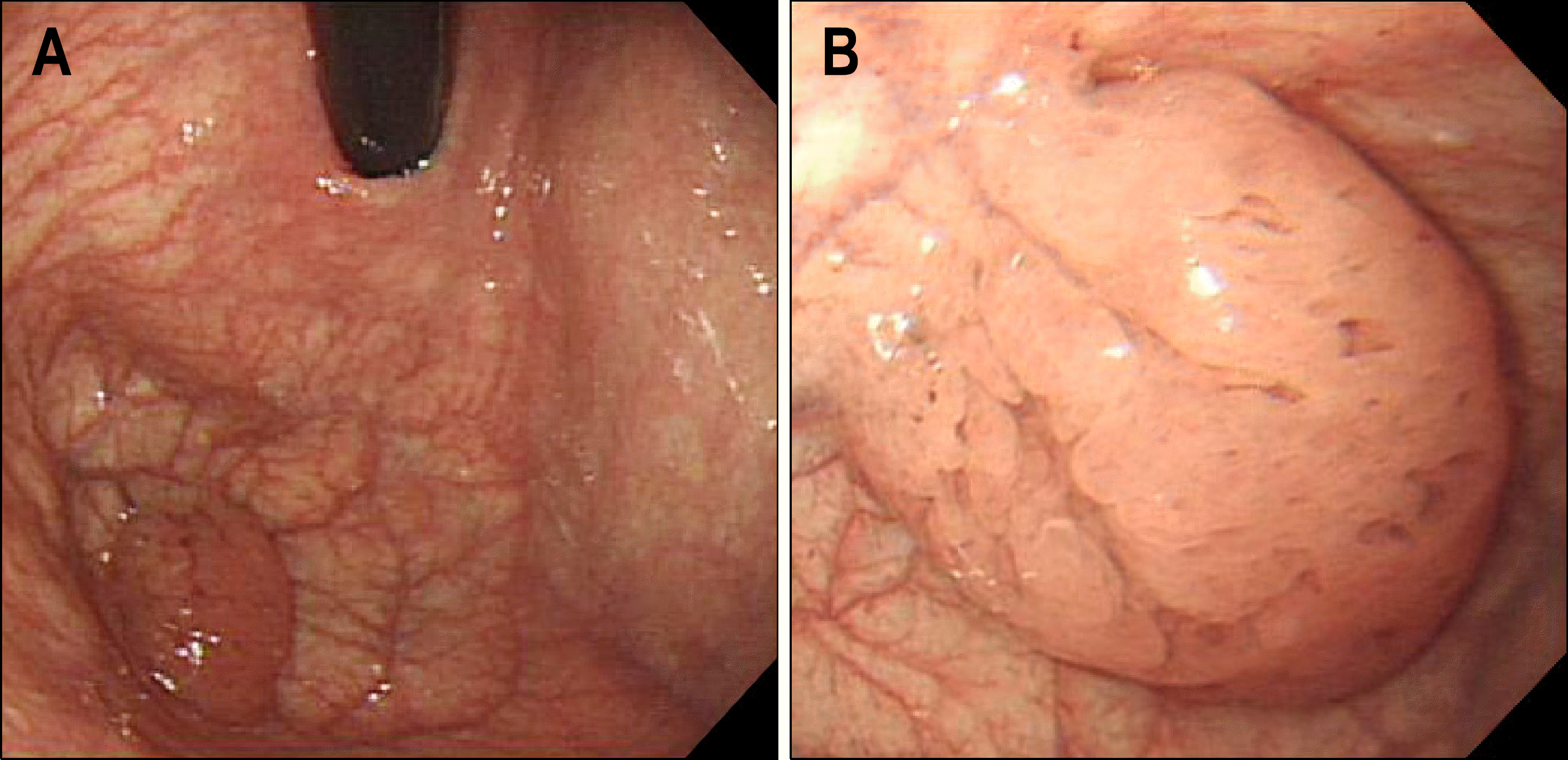
Fig. 1.
Endoscopic findings. (A) About 3 cm sized polypoid mass sessile has been detected at gastric fundus. (B) The mass showed cri-briform or sponge-like surface full of tiny holes. Its surface appeared to be greasy with transparent mucus.

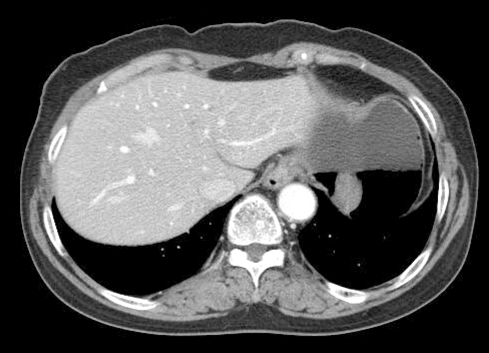
Fig. 2.
Abdominal CT showed a 3.5 cm sized polypoid mass with a short stalk in the gastric fundus. Lymph node involve-ment or distant metastasis was not observed.

Fig. 3.
Gross findings. (A) Polypoid lesion was detected at the resected stomach. (B) Polypoid mass was connected to the stomach by a short stalk with 1.5 cm sized base.

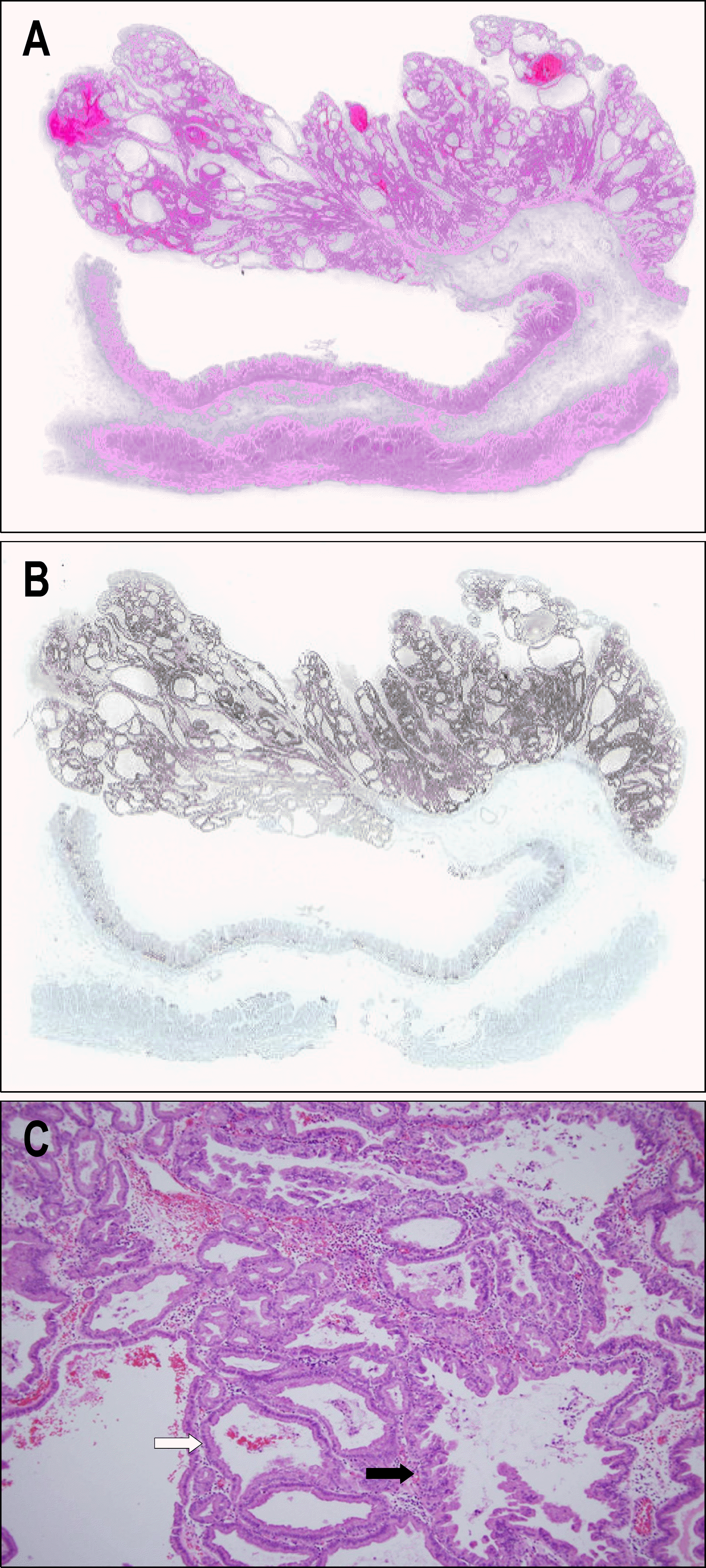
Fig. 4.
Microscopic findings. (A) Polypoid mass consisted of cystically dilated tubules which are not fused or irregularly branched (H&E stain, ×1). (B) Epithelial glands except gastric foveolar epithelum were strongly stained for MUC6 (×1). (C) Pyloric gland adenoma was composed of closely packed pyloric gland type tubules with a monolayer of cuboidal columnar epithelial cells containing pale to eosinophilic cytoplasm (white arrow). Pyloric gland adenocarcinoma had a nuclear atypism, ser-rated epithelial arrangement, and focal complex branching of tubules (black arrow) (H&E stain, ×100).


 PDF
PDF ePub
ePub Citation
 Print
Print


 XML Download