Abstract
Background/Aims
Eradication of Helicobacter pylori (H. pylori) is accepted as initial treatment of stage I E1 gastric mucosa associated lymphoid tissue (MALT) lymphoma. However, 10-20% of gastric low grade MALT lymphomas are unresponsive to H. pylori eradication treatment. The aim of this study was to find out the predictive factors of complete remission of gastric MALT lymphoma after H. pylori eradication.
Methods
From 1995 to 2006, consecutive 95 patients with modified Ann Arbor stage I E1 gastric MALT lymphoma were enrolled, and their medical records were reviewed. The patients were initially treated by H. pylori eradication. The complete remission was determined by endoscopic and histologic finding.
Results
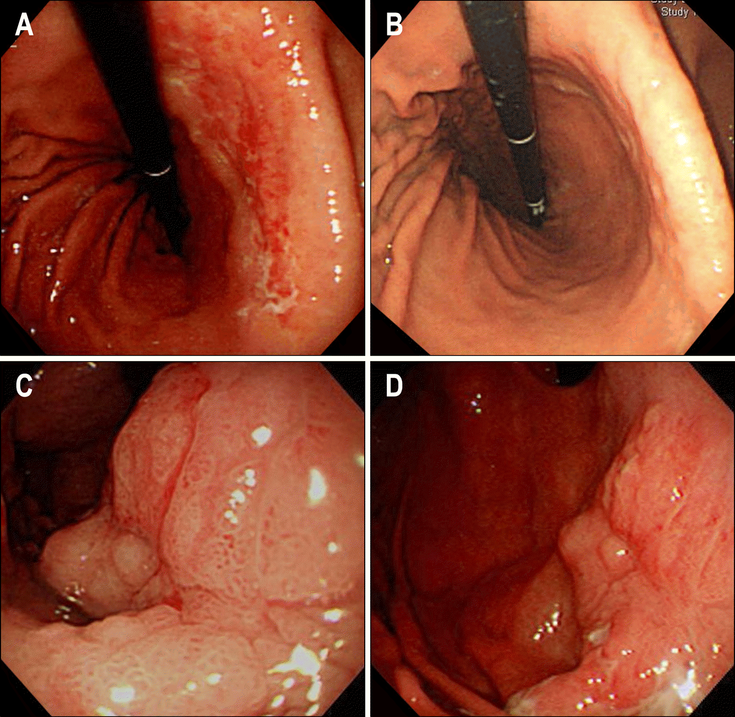
Eighty eight patients (92.6%) achieved complete remission after H. pylori eradication therapy. Mean follow up time for these patients was 40±25 months. Seven patients (7.4%) failed to achieve complete remission. There was no significant difference in the age, sex, endoscopic appearance, and large cell component between the remission group and failure group. Among 66 patients with distal tumor, 65 patients (98.5%) achieved complete remission. On the other hand, among 13 patient with proximal tumor, 9 patients (69.2%) achieved complete remission (p=0.001). The odds ratio of proximal tumor for H. pylori eradication failure was 28.9 (95% CI=2.9-288.0).
Go to : 
REFERENCES
1. Isaacson P, Wright DH. Malignant lymphoma of mucosa-associated lymphoid tissue. A distinctive type of B-cell lymphoma. Cancer. 1983; 52:1410–1416.

2. Muller AF, Maloney A, Jenkins D, et al. Primary gastric lymphoma in clinical practice 1973-1992. Gut. 1995; 36:679683.

3. Wotherspoon AC, Finn TM, Isaacson PG. Trisomy 3 in low-grade B-cell lymphomas of mucosa-associated lymphoid tissue. Blood. 1995; 85:2000–2004.

4. Eidt S, Stolte M, Fischer R. Helicobacter pylori gastritis and primary gastric non-Hodgkin's lymphomas. J Clin Pathol. 1994; 47:436–439.
5. Wotherspoon AC, Ortiz-Hidalgo C, Falzon MR, Isaacson PG. Helicobacter pylori-associated gastritis and primary B-cell gastric lymphoma. Lancet. 1991; 338:1175–1176.
6. Nakamura S, Yao T, Aoyagi K, Iida M, Fujishima M, Tsuneyoshi M. Helicobacter pylori and primary gastric lymphoma. A histopathologic and immunohistochemical analysis of 237 patients. Cancer. 1997; 79:3–11.
7. Karat D, O'Hanlon DM, Hayes N, Scott D, Raimes SA, Griffin SM. Prospective study of Helicobacter pylori infection in primary gastric lymphoma. Br J Surg. 1995; 82:1369–1370.
8. Parsonnet J, Hansen S, Rodriguez L, et al. Helicobacter pylori infection and gastric lymphoma. N Engl J Med. 1994; 330:1267–1271.
9. Bayerdorffer E, Neubauer A, Rudolph B, et al. Regression of primary gastric lymphoma of mucosa-associated lymphoid tissue type after cure of Helicobacter pylori infection. MALT Lymphoma Study Group. Lancet. 1995; 345:1591–1594.
10. Roggero E, Zucca E, Pinotti G, et al. Eradication of Helicobacter pylori infection in primary low-grade gastric lymphoma of mucosa-associated lymphoid tissue. Ann Intern Med. 1995; 122:767–769.
11. Savio A, Zamboni G, Capelli P, et al. Relapse of low-grade gastric MALT lymphoma after Helicobacter pylori eradication: true relapse or persistence? Longterm post-treatment follow-up of a multicenter trial in the north-east of Italy and evaluation of the diagnostic protocol's adequacy. Recent Results Cancer Res. 2000; 156:116–124.
12. Wotherspoon AC, Doglioni C, Diss TC, et al. Regression of primary low-grade B-cell gastric lymphoma of mucosa-associated lymphoid tissue type after eradication of Helicobacter pylori. Lancet. 1993; 342:575–577.
13. Ruskone-Fourmestraux A, Lavergne A, Aegerter PH, et al. Predictive factors for regression of gastric MALT lymphoma after anti-Helicobacter pylori treatment. Gut. 2001; 48:297–303.
14. Levy M, Copie-Bergman C, Traulle C, et al. Conservative treatment of primary gastric low-grade B-cell lymphoma of mucosa-associated lymphoid tissue: predictive factors of response and outcome. Am J Gastroenterol. 2002; 97:292–297.

15. Hong SS, Jung HY, Choi KD, et al. A prospective analysis of low-grade gastric MALT lymphoma after Helicobacter pylori eradication. Helicobacter. 2006; 11:569–573.
16. A clinical evaluation of the International Lymphoma Study Group classification of non-Hodgkin's lymphoma. The Non- Hodgkin's Lymphoma Classification Project. Blood. 1997; 89:3909–3918.
17. Genta RM, Hamner HW, Graham DY. Gastric lymphoid fol-licles in Helicobacter pylori infection: frequency, distribution, and response to triple therapy. Hum Pathol. 1993; 24:577–583.
18. Hussell T, Isaacson PG, Crabtree JE, Spencer J. The response of cells from low-grade B-cell gastric lymphomas of mucosa-associated lymphoid tissue to Helicobacter pylori. Lancet. 1993; 342:571–574.
19. Hussell T, Isaacson PG, Crabtree JE, Spencer J. Helicobacter pylori-specific tumour-infiltrating T cells provide contact dependent help for the growth of malignant B cells in low-grade gastric lymphoma of mucosa-associated lymphoid tissue. J Pathol. 1996; 178:122–127.
20. Sackmann M, Morgner A, Rudolph B, et al. Regression of gastric MALT lymphoma after eradication of Helicobacter pylori is predicted by endosonographic staging. MALT Lymphoma Study Group. Gastroenterology. 1997; 113:1087–1090.
21. Nakamura S, Matsumoto T, Suekane H, et al. Predictive value of endoscopic ultrasonography for regression of gastric low grade and high grade MALT lymphomas after eradication of Helicobacter pylori. Gut. 2001; 48:454–460.
22. Steinbach G, Ford R, Glober G, et al. Antibiotic treatment of gastric lymphoma of mucosa-associated lymphoid tissue. An uncontrolled trial. Ann Intern Med. 1999; 131:88–95.

23. de Jong D, Boot H, van Heerde P, Hart GA, Taal BG. Histological grading in gastric lymphoma: pretreatment criteria and clinical relevance. Gastroenterology. 1997; 112:1466–1474.

24. de Jong D, Boot H, Taal B. Histological grading with clinical relevance in gastric mucosa-associated lymphoid tissue (MALT) lymphoma. Recent Results Cancer Res. 2000; 156:27–32.

25. Liu H, Ruskon-Fourmestraux A, Lavergne-Slove A, et al. Resistance of t(11; 18) positive gastric mucosa-associated lymphoid tissue lymphoma to Helicobacter pylori eradication therapy. Lancet. 2001; 357:39–40.
26. Liu H, Ye H, Dogan A, et al. T(11; 18)(q21;q21) is associated with advanced mucosa-associated lymphoid tissue lymphoma that expresses nuclear BCL10. Blood. 2001; 98:1182–1187.
27. Kim JS, Chung SJ, Choi YS, et al. Helicobacter pylori eradication for low-grade gastric mucosa-associated lymphoid tissue lymphoma is more successful in inducing remission in distal compared to proximal disease. Br J Cancer. 2007; 96:1324–1328.
28. Negrini R, Lisato L, Zanella I, et al. Helicobacter pylori infection induces antibodies cross-reacting with human gastric mucosa. Gastroenterology. 1991; 101:437–445.
29. Greiner A, Marx A, Heesemann J, Leebmann J, Schmausser B, Muller-Hermelink HK. Idiotype identity in a MALT-type lymphoma and B cells in Helicobacter pylori associated chronic gastritis. Lab Invest. 1994; 70:572–578.
Go to : 
 | Fig. 1.Different treatment response to H. pylori eradication according to site. (A, B) A case of gastric MALT lymphoma located on the distal part of stomach showing complete response 3 months after H. pylori eradication (A: before treatment, B: after treatment). (C, D) A case of gastric MALT lymphoma located on the proximal part of stomach showing no improvement during follow-up of 9 months after H. pylori eradication (C: before treatment, D: after treatment). |
Table 1.
Baseline Characteristics and Risk Factor Analysis of Failure of MALT-lymphoma Remission


 PDF
PDF ePub
ePub Citation
 Print
Print


 XML Download