Abstract
Fitz-Hugh-Curtis syndrome has been described as focal perihepatitis accompanying pelvic inflammatory disease caused by Neisseria gonorrhea and Chlamydia trachomatis. The highest incidence occurs in young, sexually active females. However, the syndrome has been reported to occur infrequently in males, according to the foreign literature. The predominant symptoms are right upper quadrant pain and tenderness, and pleuritic right sided chest pain. The clinical presentation is similar in men and women. In women, the spread of infection to liver capsule is thought to occur directly from infected fallopian tube via the right paracolic gutter. In men, hematogenous and lymphatic spread is thought to be postulated. Recently, we experienced a case of Fitz-Hugh-Curtis syndrome occurred in a man. As far as we know, it is the first report in Korea, and we report a case with a review of the literature.
Go to : 
REFERENCES
2. Fitz-Hugh T. Acute gonococcic peritonitis of the right upper quadrant in women. JAMA. 1934; 102:2094–2096.

3. Mü ller-Schoop JW, Wang SP, Munzinger J, Schlä pfer HU, Knoblauch M, Ammann RW. Chlamydia trachomatis as pos-sible cause of peritonitis and perihepatitis in young women. Br Med J. 1978; 1:1022–1024.
4. Joo SH, Kim MJ, Lim JS, Kim JH, Kim KW. CT diagnosis of Fitz-Hugh and Curtis syndrome: value of the arterial phase scan. Korean J Radiol. 2007; 8:40–47.

5. Kimball MW, Knee S. Gonococcal perihepatitis in a male. The Fitz-Hugh–Curtis syndrome. N Engl J Med. 1970; 282:1082–1083.
6. Francis TI, Osoba AO. Gonococcal hepatitis (Fitz-Hugh-Curtis syndrome) in a male patient. Brit J Vener Dis. 1972; 48:187–188.
8. Davidson AC, Hawkins DA. Pleuritic pain: Fitz Hugh Curtis syndrome in a man. Br Med J (Clin Res Ed). 1982; 284:808.

9. Winkler WP, Kotler DP, Saleh J. Fitz-Hugh and Curtis syndrome in a homosexual man with impaired cell mediated immunity. Gastrointest Endosc. 1985; 31:28–30.

10. Owens S, Yeko TR, Bloy R, Maroulis GB. Laparoscopic treatment of painful perihepatic adhesions in Fitz-Hugh-Curtis syndrome. Obstet Gynecol. 1991; 78:542–543.
11. Counselman FL. An unusual presentation of Fitz-Hugh-Curtis syndrome. J Emerg Med. 1994; 12:167–170.

12. Lee SC, Nah BG, Kim HS, et al. Two cases of Fitz-Hugh-Curtis syndrome in acute phase. Korean J Gastroenterol. 2005; 45:137–142.
13. Chung HJ, Choi KY, Cho YJ, et al. Ten cases of Fitz-Hugh-Curtis syndrome. Korean J Gastroenterol. 2007; 50:328–333.
14. Yang HW, Jung SH, Han HY, et al. Clinical feature of Fitz-Hugh-Curtis syndrome: analysis of 25 cases. Korean J Hepatol. 2007; 14:178–184.

15. Ham YC, Lee KL, Shin DG, et al. Clinical experience of Fitz-Hugh-Curtis syndrome. J Korean Surg Soc. 2009; 76:36–42.
16. Wang SP, Eschenbach DA, Holmes KK, Wager G, Grayston JT. Chlamydia trachomatis infection in Fitz-Hugh-Curtis syndrome. Am J Obstet Gynecol. 1980; 138:1034–1038.
17. Ross JD, Jensen JS. Mycoplasma genitalium as a sexually transmitted infection: implications for screening, testing, and treatment. Sex Transm Infect. 2006; 82:269–271.
18. Taylor-Robinson D, Horner PJ. The role of Mycoplasma genitalium in non-gonococcal urethritis. Sex Transm Infect. 2001; 77:229–231.
19. Nishie A, Yoshimitsu K, Irie H, et al. Fitz-Hugh-Curtis syndrome. Radiologic manifestation. J Comput Assist Tomogr. 2003; 27:786–791.
Go to : 
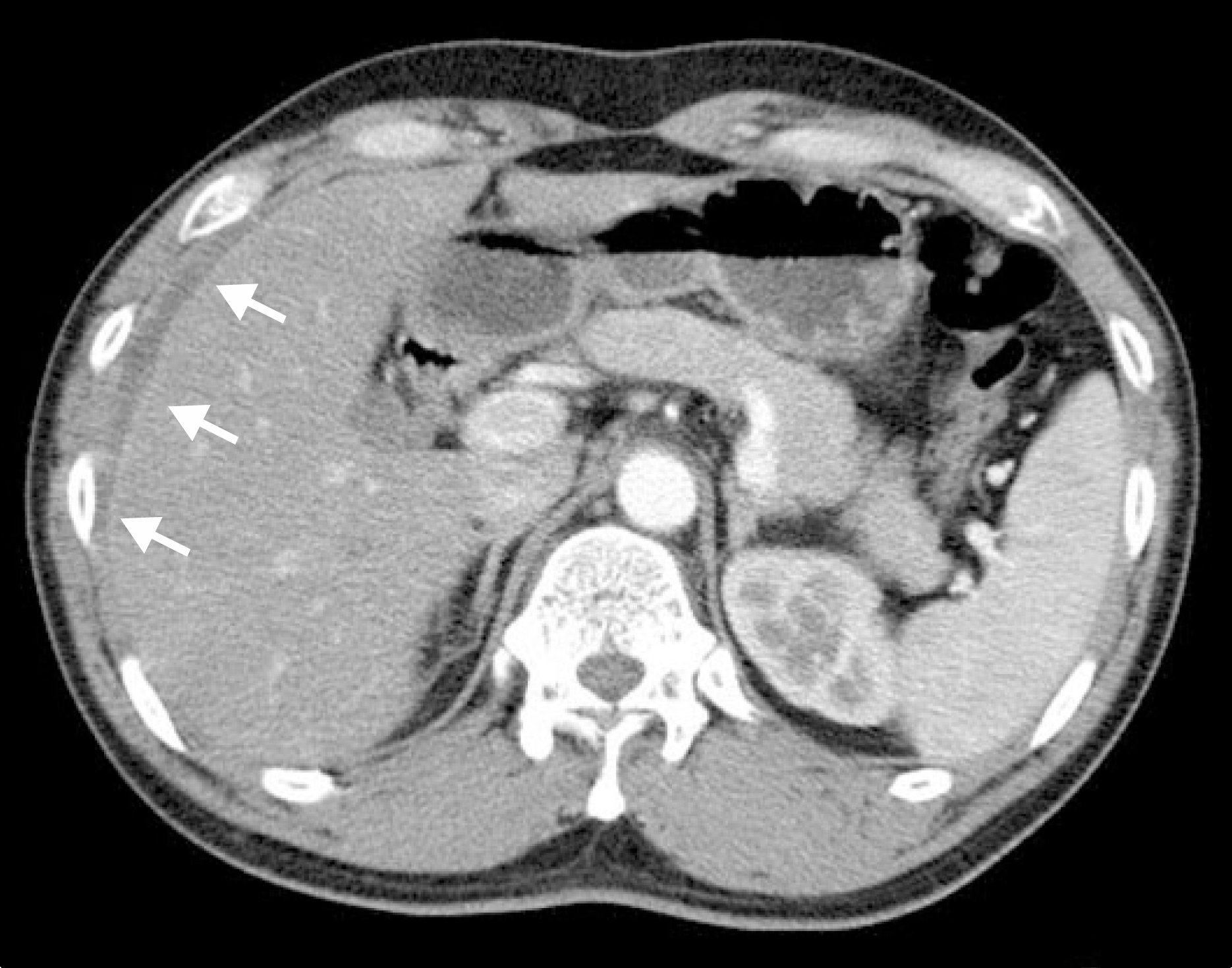
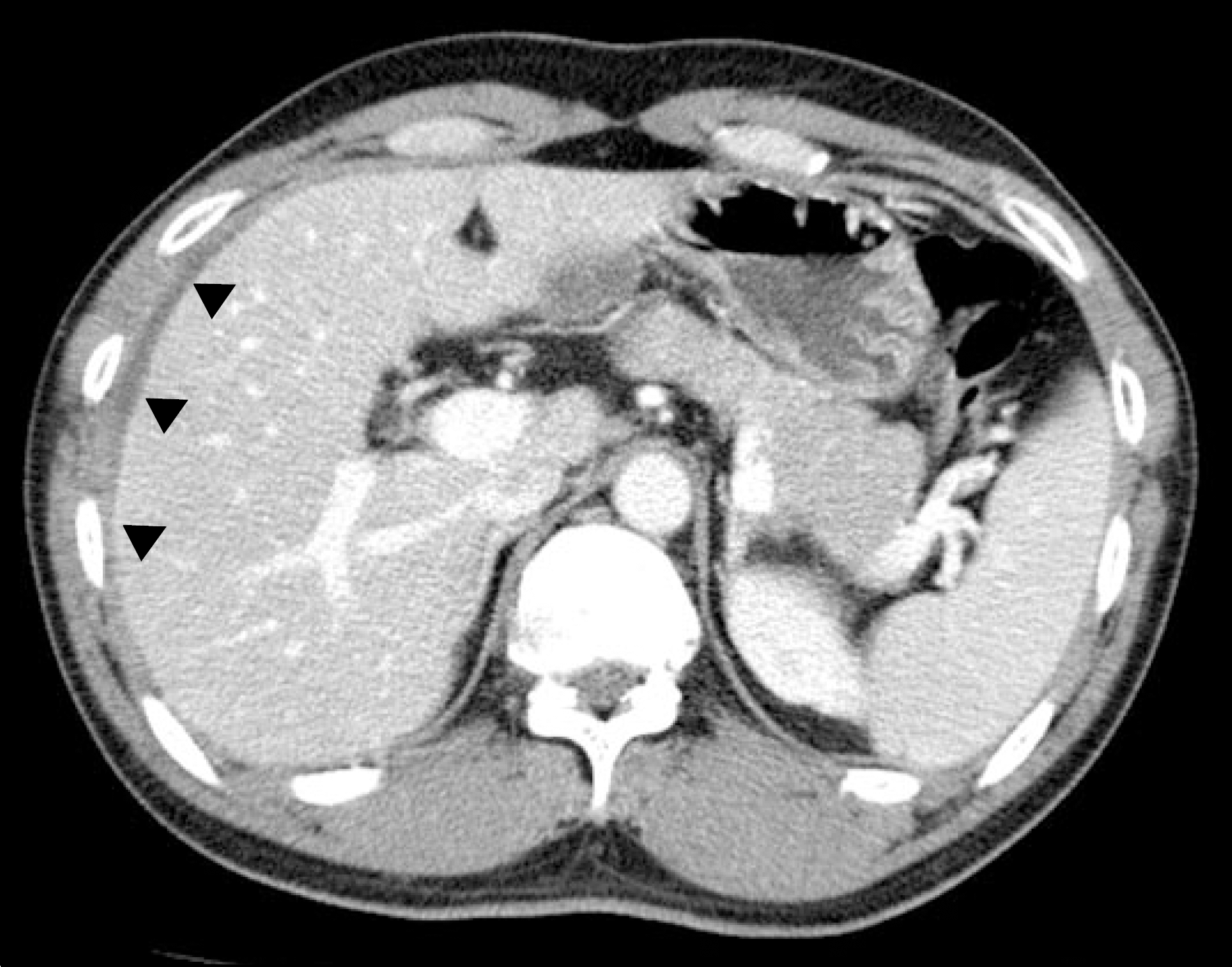
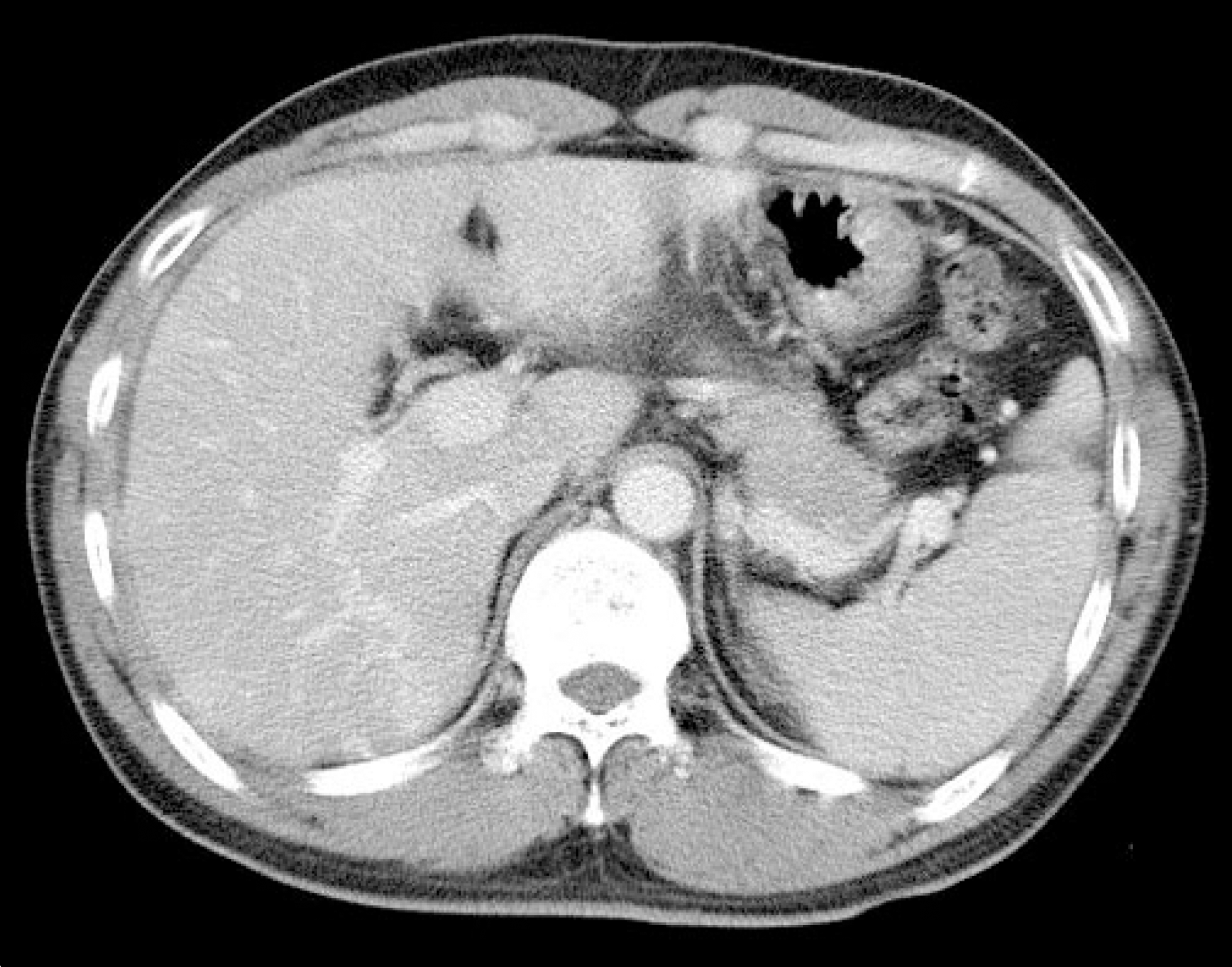
 | Fig. 1.Abdominal CT finding. On arterial phase image, linear capsular enhancement (white arrow) was noted at the arterial surface of the medial segment, and above it, between the liver and parietal peritoneum, fluid collection was seen. |


 PDF
PDF ePub
ePub Citation
 Print
Print


 XML Download