Abstract
Pancreas acinar cell carcinoma (ACC) accounts for only 1-2% of pancreatic exocrine malignant tumor. The symptoms of patients with ACC are usually non-specific, for example the anorexia and weight loss. Patients may de-velop Schmid's triad including subcutaneous fat necrosis, polyarthritis, and eosinophilia. We reported a case of ACC which was manifested by subcutaneous nodule as initial clinical symptom. To our knowledge, this is the first reported case of ACC presenting as subcutaneous fat necrosis in Korea.
REFERENCES
1. Klimstra DS, Heffess CS, Oerterl JE, Rosai J. Acinar cell carcinoma of the pancreas. A clinicopathologic study of 28 cases. Am J Surg Pathol. 1992; 16:815–837.
2. Lee JL, Kim TW, Chang HM, et al. Locally advanced acinar cell carcinoma of the pancreas successfully treated by capeci-tabine and concurrent radiotherapy: report of two cases. Pancreas. 2003; 27:e18–22.

3. Holen KD, Klimstra DS, Hummer A, et al. Clinical characteristics and outcomes from an institutional series of acinar cell carcinoma of the pancreas and related tumors. J Clin Oncol. 2002; 20:4673–4678.

4. Kim KY, Lee HJ, Ji JH, et al. A case of acinar cell carcinoma of the pancreas. Korean J Med. 2009; 76:506–509.
5. Kim HD, Lee BK, Choi KH, Lee SD, Seo JK, Park YH. Clinical study of pancreatic cancer. J Korean Surg Soc. 1992; 42:172–189.
6. Kim MC, Kim HH, Jung GJ, Kim SS. Clinical study of acinar cell carcinomas of the pancreas. J Korean Surg Soc. 2001; 60:97–102.
7. Lee SH, Kim H, Kang SW. Acinar cell carcinoma of the pancreas: a case report. J Korean Radiol Soc. 1998; 39:1181–1183.

8. Hoorens A, Lemoine NR, McLellan E, et al. Pancreatic acinar cell carcinoma. An analysis of cell lineage markers, p53 expression, and Ki-ras mutation. Am J Pathol. 1993; 143:685–698.
9. Ashley SW, Lauwers GY. Case records of the Massachusetts General Hospital. Weekly clinicopathological exercises. Case 37-2002. A 69-year old man with painful cutaneous nodules, elevated lipase levels, and abnormal results on abdominal scanning. N Engl J Med. 2002; 347:1783–1791.
10. Itoh T, Kishi K, Tojo M, et al. Acinar cell carcinoma of the pancreas with elevated serum alphafetoprotein levels: a case report and a review of 28 cases reported in Japan. Gastroenterol Jpn. 1992; 27:785–791.
11. Horie Y, Gomyoda M, Kishimoto Y, et al. Plasma carcino-embryonic antigen and acinar cell carcinoma of the pancreas. Cancer. 1984; 53:1137–1142.

12. Geibel J, Longo W. Less common neoplasms of the pancreas. World J Gastroenterol. 2006; 28:3180–3185.
13. Antoine M, Khitrik-Palchuk M, Saif MW. Longterm survival in a patient with acinar cell carcinoma of pancreas. A case report and review of literature. J Pancreas. 2007; 8:783–789.
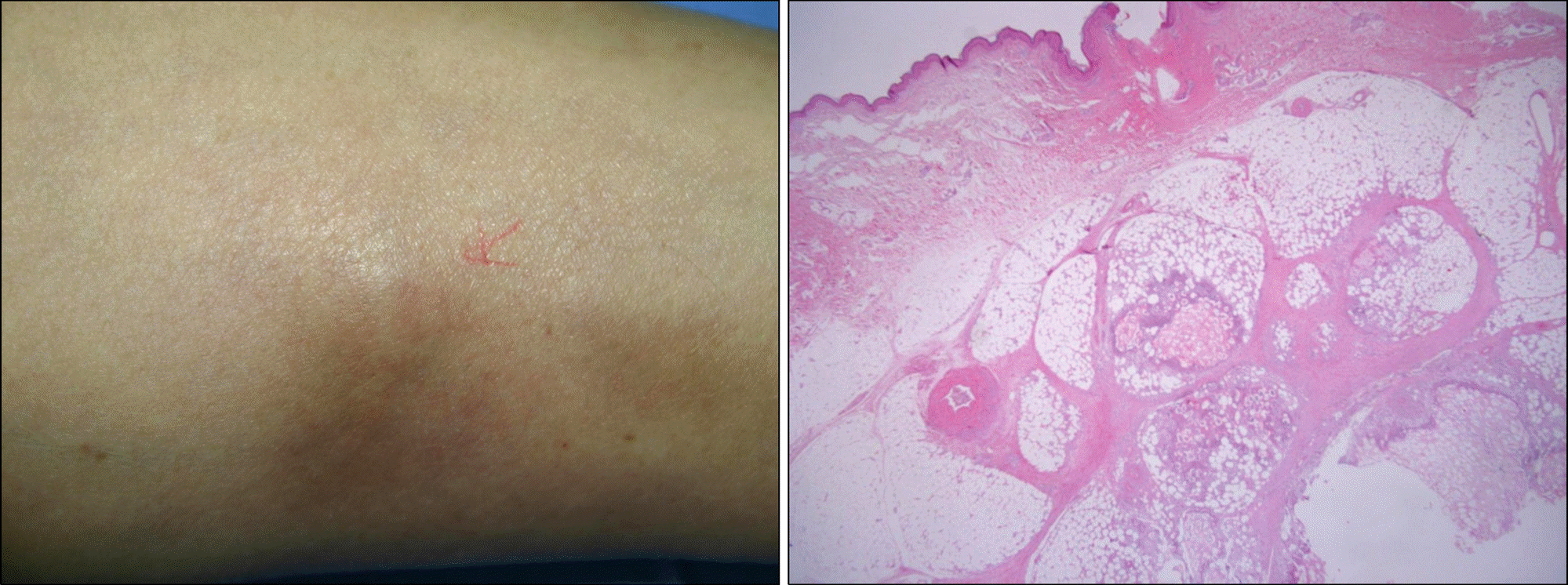
Fig. 1.
Subcutaneous nodule finding. (A) 1-2 cm sized erythematous to brown colored tender subcutaneous nodule was seen on the left leg. (B) It showed fat necrosis with chronic active inflammation and granulomatous reaction (H&E, ×100).

Fig. 2.
Abdominal computed tomography finding. It showed diffuse enlargement and heteroge-nicity of pancreas with multiple low density lesions in parenchy-me.

Fig. 3.
Endoscopic ultrasonography finding. It showed diffuse parenchymal enlargement of pancreas. Multiple strictures and di-latations of main pancreatic duct were noted. About 6.4×6.6 cm sized isoechogenic mass like lesion was noted in body of pancreas.

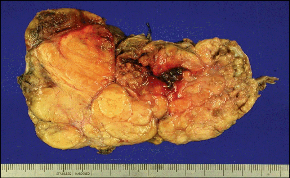
Fig. 4.
Tumor was lobulated and had yellowish cut surface with hemorrhage and necrosis. Tumor mass measured 13×9×4 cm.

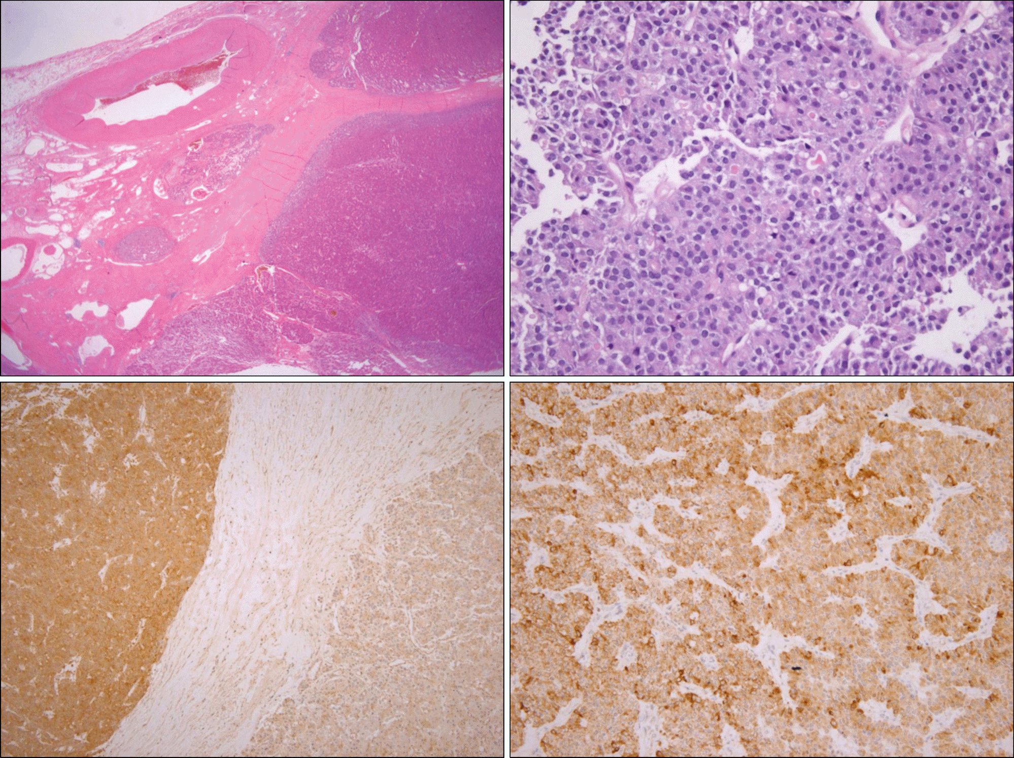
Fig. 5.
Microscopic finding. (A) Infiltrating solid nests of tumor cells with surrounding fibrosis were observed within the pancreatic tissue (H&E, ×100). (B) Acinar pattern with pleomorphic oval nucleoli and eosinophilic cytoplasm were observed (H&E, ×400). (C) The tumor cells were weak positive for synaptophysin (×100) and (D) positive for cytokeratin (×200).


 PDF
PDF ePub
ePub Citation
 Print
Print


 XML Download