Abstract
Spontaneous regression of hepatocellular carcinoma (HCC) is extremely rare. We report a case of 67-year-old man having HBV-associated HCC with multiple lung metastases which regressed spontaneously. The patient had single liver mass and received surgical resection. The mass was confirmed as HCC histopathologically. Nine years after surgical resection, a 3.3 cm sized recurred HCC was detected on the resection margin in CT scan. Transarterial chemoembolization (TACE) was performed 3 times, and lung metastases developed thereafter. The patient received 2 more sessions of TACE, however, metastatic lung nodules were in progress very rapidly. We decided to stop TACE and followed the patient regularly without any anti-cancer treatment. Nine months after development of lung metastasis, the size and number of metastatic lung nodules decreased and were not detected anymore after 14 months. Serum alphafetoprotein levels also decreased to normal range and no viable tumor was noted in the liver. The patient is still alive 12 years after the first diagnosis of HCC and 16 months after lung metastasis developed.
Go to : 
REFERENCES
1. El-Serag HB. Hepatocellular carcinoma: an epidemiologic view. J Clin Gastroenterol. 2002; 35:S72–78.
2. Yuen MF, Hou JL, Chutaputti A. Hepatocellular carcinoma in the asia pacific region. J Gastroenterol Hepatol. 2009; 24:346–353.

3. Oquiñ ena S, Iñ arrairaegui M, Vila JJ, Alegre F, Zozaya JM, Sangro B. Spontaneous regression of hepatocellular carcinoma: three case reports and a categorized review of the literature. Dig Dis Sci. 2009; 54:1147–1153.

5. Ikeda M, Okada S, Ueno H, Okusaka T, Kuriyama H. Spontaneous regression of hepatocellular carcinoma with multiple lung metastases: a case report. Jpn J Clin Oncol. 2001; 31:454–458.

6. Kondo S, Okusaka T, Ueno H, Ikeda M, Morizane C. Spontaneous regression of a hepatocellular carcinoma. Int J Clin Oncol. 2006; 11:407–411.
7. Gomez Sanz R, Moreno Gonzalez E, Colina Ruiz-Delgado F, Garcia-Munoz H, Ochando Cerdan F, Gonzalez-Pinto I. Spontaneous regression of a recurrent hepatocellular carcinoma. Dig Dis Sci. 1998; 43:323–328.
8. Tocci G, Conte A, Guarascio P, Visco G. Spontaneous remission of hepatocellular carcinoma after massive gastrointestinal haemorrhage. BMJ. 1990; 300:641–642.

9. Suzuki M, Okazaki N, Yoshino M, Yoshida T. Spontaneous regression of a hepatocellular carcinoma-a case report. Hepatogastroenterology. 1989; 36:160–163.
10. Sato Y, Fujiwara K, Nakagawa S, et al. A case of spontaneous regression of hepatocellular carcinoma with bone metastasis. Cancer. 1985; 56:667–671.

11. Markovic S, Ferlan-Marolt V, Hlebanja Z. Spontaneous regression of hepatocellular carcinoma. Am J Gastroenterol. 1996; 91:392–393.
12. McCaughan GW, Bilous MJ, Gallagher ND. Longterm survival with tumor regression in androgen induced liver tumors. Cancer. 1985; 56:2622–2626.
13. Gottfried EB, Steller R, Paronetto F, Lieber CS. Spontaneous regression of hepatocellular carcinoma. Gastroenterology. 1982; 82:770–774.

14. Takeda Y, Togashi H, Shinzawa H, et al. Spontaneous regression of hepatocellular carcinoma and review of literature. J Gastroenterol Hepatol. 2000; 15:1079–1086.

15. Heianna J, Miyauchi T, Suzuki T, Ishida H, Hashimoto M, Watarai J. Spontaneous regression of multiple lung metastases following regression of hepatocellular carcinoma after trans-catheter arterial embolization. A case report. Hepatogastroenterology. 2007; 54:1560–1562.
16. Iiai T, Sato Y, Nabatame N, Yamamoto S, Makino S, Hata-keyama K. Spontaneous complete regression of hepatocellular carcinoma with portal vein tumor thrombus. Hepatogastroenterology. 2003; 50:1628–1630.
17. Uenishi T, Hirohashi K, Tanaka H, Ikebe T, Kinoshita H. Spontaneous regression of a large hepatocellular carcinoma with portal vein tumor thrombi: report of a case. Surg Today. 2000; 30:82–85.

18. Abiru S, Kato Y, Hamasaki K, et al. Spontaneous regression of hepatocellular carcinoma associated with elevated levels of interleukin 18. Am J Gastroenterol. 2002; 97:774–775.

19. O'Beirne JP, Harrison PM. The role of the immune system in the control of hepatocellular carcinoma. Eur J Gastroenterol Hepatol. 2004; 16:1257–1260.
Go to : 
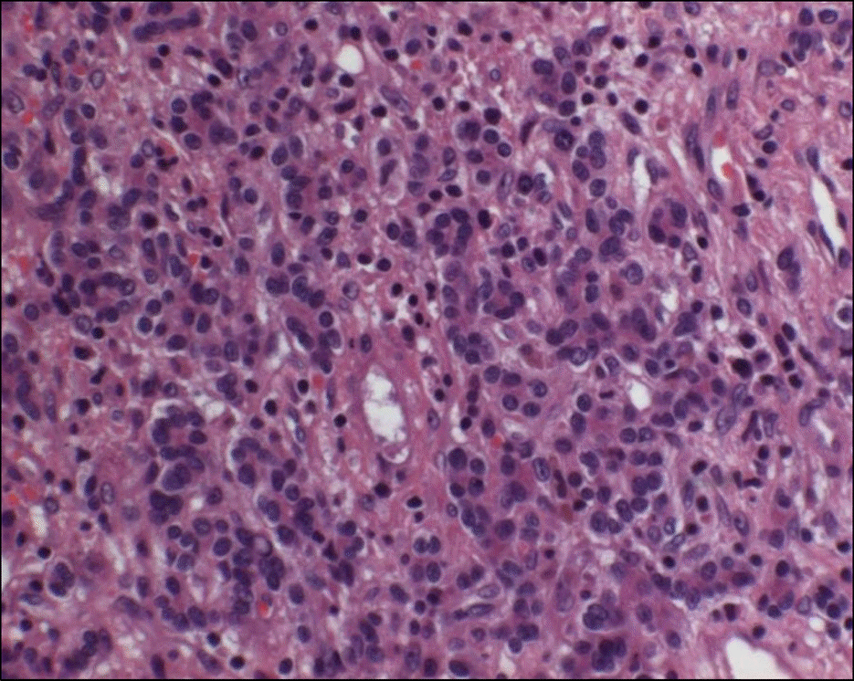
 | Fig. 1.Histological finding of surgically resected tumor. Pleomorphic tumor cells were compatible with hepatocellular carcinoma with trabecular growth pattern and Edmondson-Steiner grade III differentiation (H&E, ×200). |
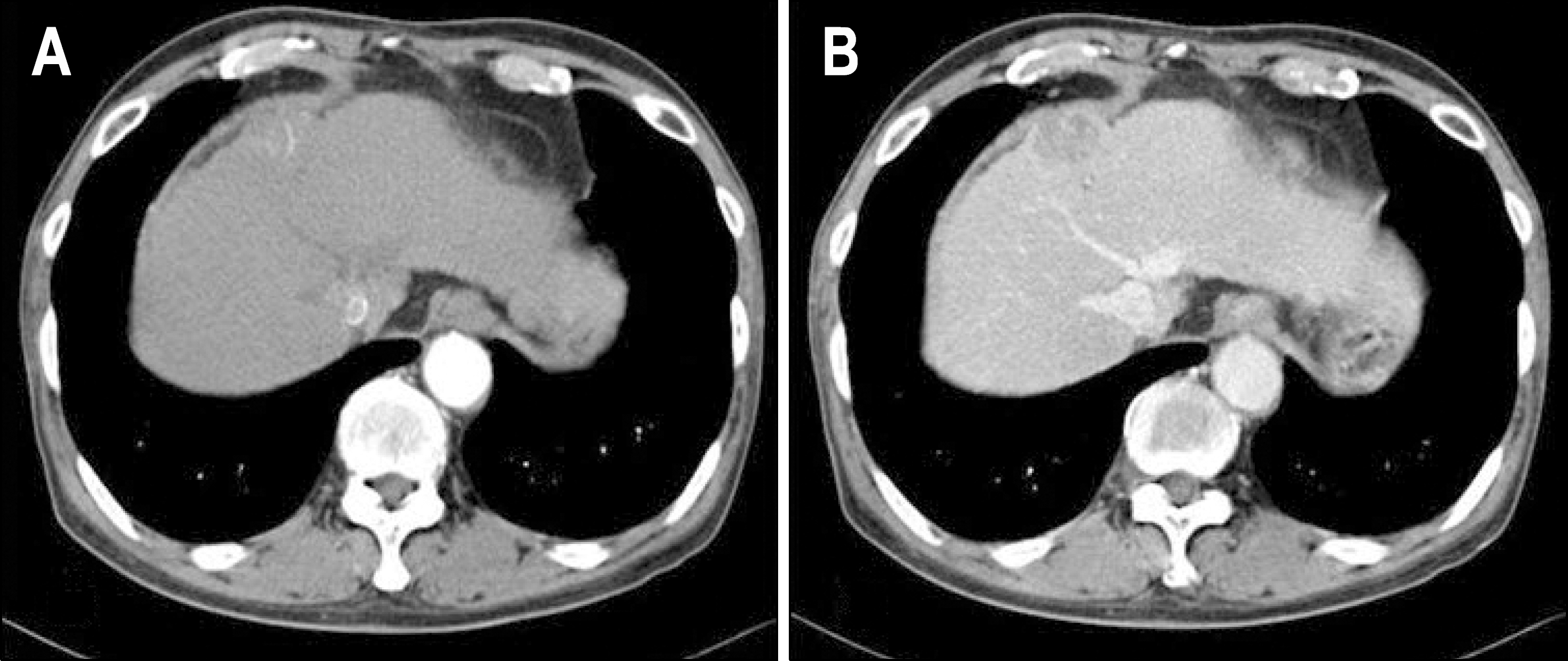
 | Fig. 2.Abdominal computed tomographic findings of recurred HCC. Contrast-enhanced abdominal computed tomography showed a 3.3 cm sized tumor in the liver. The tumor was enhanced on arterial phase (A) and wash-ed out on delayed phase (B). |
 | Fig. 3.Serial changes of chest X-ray and serum alphafetoprotein levels. A round nodule was detected on right upper lobe of lung in March 2008 (A). Lung nodules increased in number on right lobe and spread to left lobe (B, C). Metastatic lung nodules increased in size and number strikingly throughout the whole lung field (D). Lung nodules started to decrease without any anti-cancer treatment since December 2008 (E), and disappeared in May 2009 (F). Serum alphafetoprotein levels increased to 10,704 ng/mL as metastatic lung nodules progressed, and decreased to normal range as these regressed. |
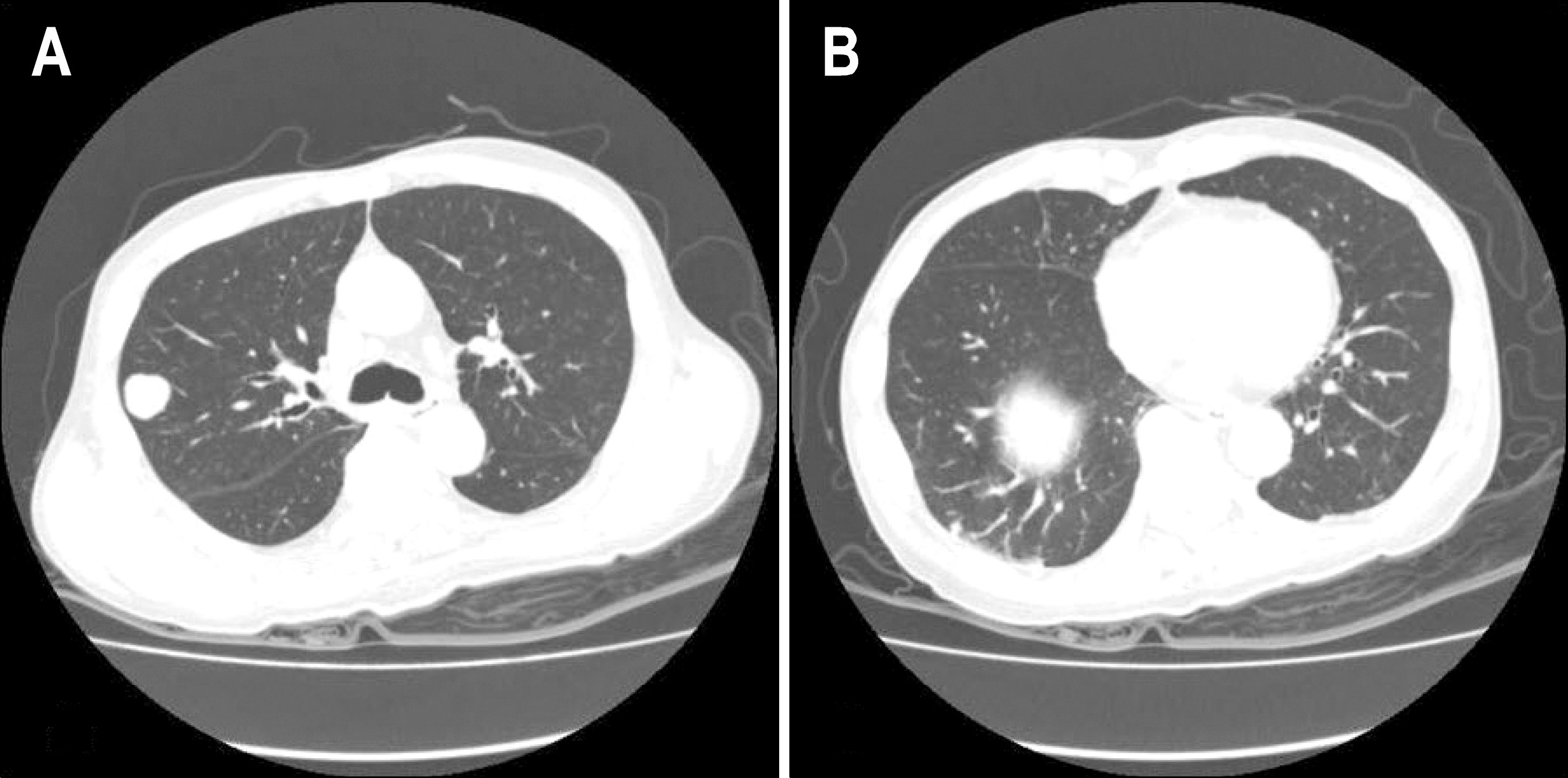
 | Fig. 4.Chest computed tomographic findings of metastatic lung nodules. Metastatic lung nodules were noted on right upper lobe (A) and right lower lobe (B). |
 | Fig. 5.Serial changes of abdominal computed tomography. After 4th session of TACE, viable tumor still remained in liver in April 2008 (A). After one more session of TACE, viable tumor around lipiodol laden mass increased further in size and invaded into diaphragm in July 2008 (B). However, without any anti-cancer treatment, viable tumor decreased spontaneously in December 2008 (C) and was no longer observed on abdominal CT in May 2009 (D). |


 PDF
PDF ePub
ePub Citation
 Print
Print


 XML Download