Abstract
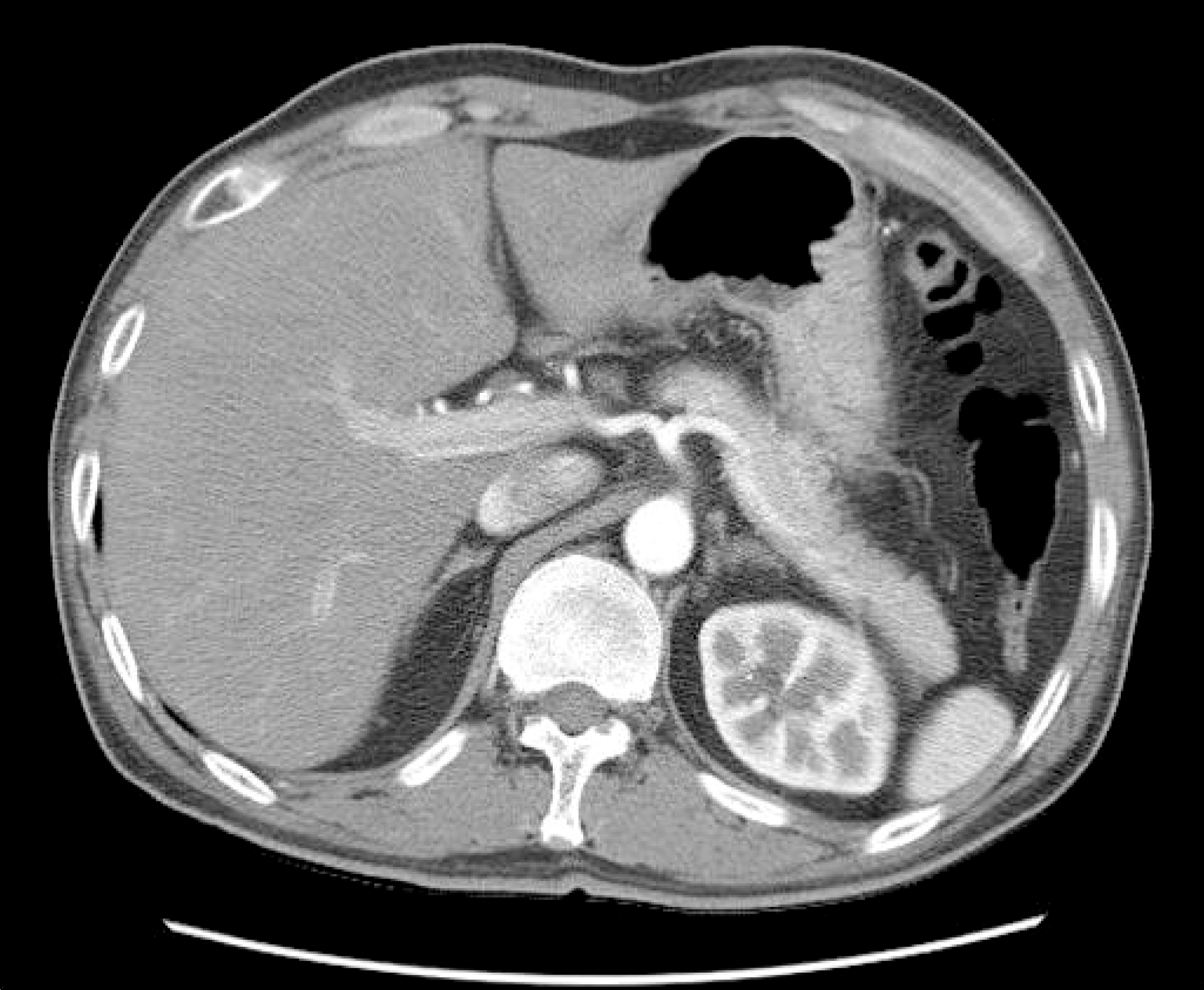
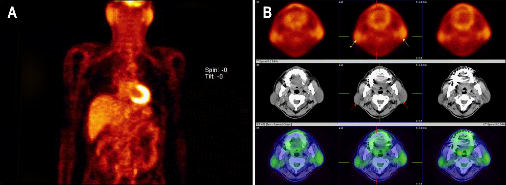
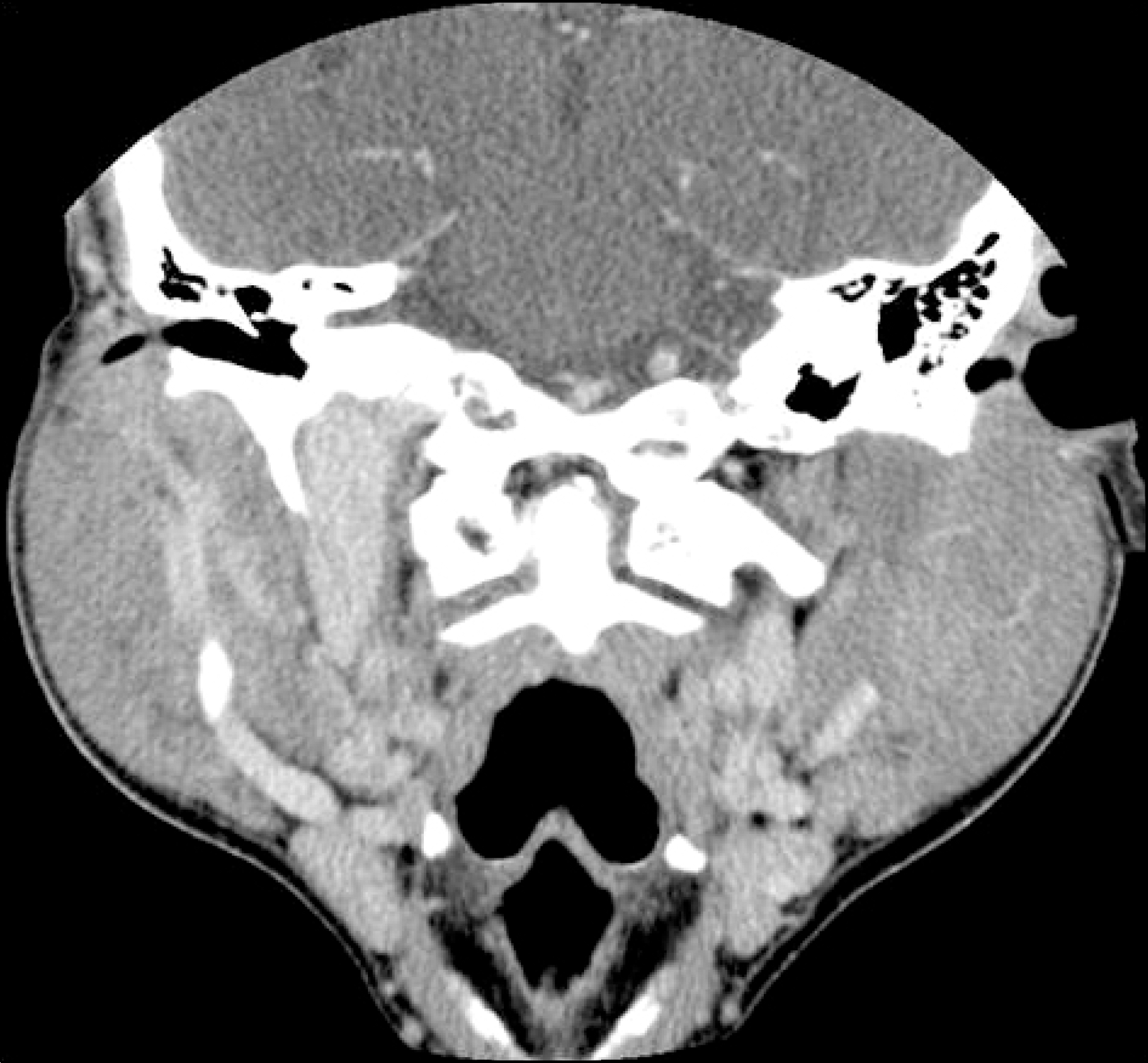
Sialadenosis is a unique form of non-inflammatory, non-neoplastic bilateral salivary gland disorder characterized by recurrent painless swelling which usually occurs in parotid glands. Alcoholism is one of the main causes of sialadenosis along with diabetes, bulimia, and other idiopathic causes. The prognosis is verified according to the degree of liver function. We present a case of a 46 year-old man who had alcoholic fatty liver disease diagnosed as alcoholic sialadenosis based on clinical points of recurrent bilateral parotid swelling after heavy alcohol drinking, computed tomography, and fine-needle aspiration biopsy. After stopping alcohol drinking and treated with conservative treatment, he got improved without specific sequela.
Go to : 
REFERENCES
1. Chilla R. Sialadenosis of the salivary glands of the head. Studies on the physiology and pathophysiology of parotid secretion. Adv Otorhinolaryngol. 1981; 26:1–38.
3. Henry-Stanley MJ, Beneke J, Bardales RH, Stanley MW. Fine-needle aspiration of normal tissue from enlarged salivary glands: sialosis or missed target? Diagn Cytopathol. 1995; 13:300–303.
4. Donath K, Seifert G. Ultrastructural studies of the parotid glands in sialadenosis. Virchows Arch A Pathol Anat Histol. 1975; 365:119–135.

5. Chilla R, Arglebe C. Function of salivary glands and sia-lochemistry in sialadenosis. Acta Otorhinolaryngol Belg. 1983; 37:158–164.
6. Kelly SA, Black MJ, Soames JV. Unilateral enlargement of the parotid gland in a patient with sialosis and contralateral parotid aplasia. Br J Oral Maxillofac Surg. 1990; 28:409–412.

7. Pape SA, MacLeod RI, McLean NR, Soames JV. Sialadenosis of the salivary glands. Br J Plast Surg. 1995; 48:419–422.

8. Santolaria F, Castilla A, Gonzalez-Reimers E, et al. Alcohol intake in a rural village: physical signs and biological markers predicting excessive consumption in apparently healthy people. Alcohol. 1997; 14:9–19.

9. Maier H, Born IA, Mall G. Effect of chronic ethanol and nic-otine consumption on the function and morphology of the salivary glands. Klin Wochenschr. 1988; 66(suppl 11):140–150.
10. Abelson DC, Mandel ID, Karmiol M. Salivary studies in alcoholic cirrhosis. Oral Surg Oral Med Oral Pathol. 1976; 41:188–192.

11. Dutta SK, Dukehart M, Narang A, Latham PS. Functional and structural changes in parotid glands of alcoholic cirrhotic patients. Gastroenterology. 1989; 96:510–518.

12. Barnett JL, Wilson JA. Alcoholic pancreatitis and parotitis: utility of lipase and urinary amylase clearance determinations. South Med J. 1986; 79:832–835.
13. Mandel L, Vakkas J, Saqi A. Alcoholic (beer) sialosis. J Oral Maxillofac Surg. 2005; 63:402–405.

14. Kastin B, Mandel L. Alcoholic sialosis. N Y State Dent J. 2000; 66:22–24.
16. Carranza M, Ferraris ME, Galizzi M. Structural and morpho-metrical study in glandular parenchyma from alcoholic sialosis. J Oral Pathol Med. 2005; 34:374–379.

17. Ogren FP, Huerter JV, Pearson PH, Antonson CW, Moore GF. Transient salivary gland hypertrophy in bulimics. Laryn-goscope. 1987; 97:951–953.

18. Ino C, Matsuyama K, Ino M, Yamashita T, Kumazawa T. Approach to the diagnosis of sialadenosis using sialography. Acta Otolaryngol Suppl. 1993; 500:121–125.

Go to : 


 PDF
PDF ePub
ePub Citation
 Print
Print


 XML Download