

 PDF
PDF ePub
ePub Citation
 Print
Print


Abstract
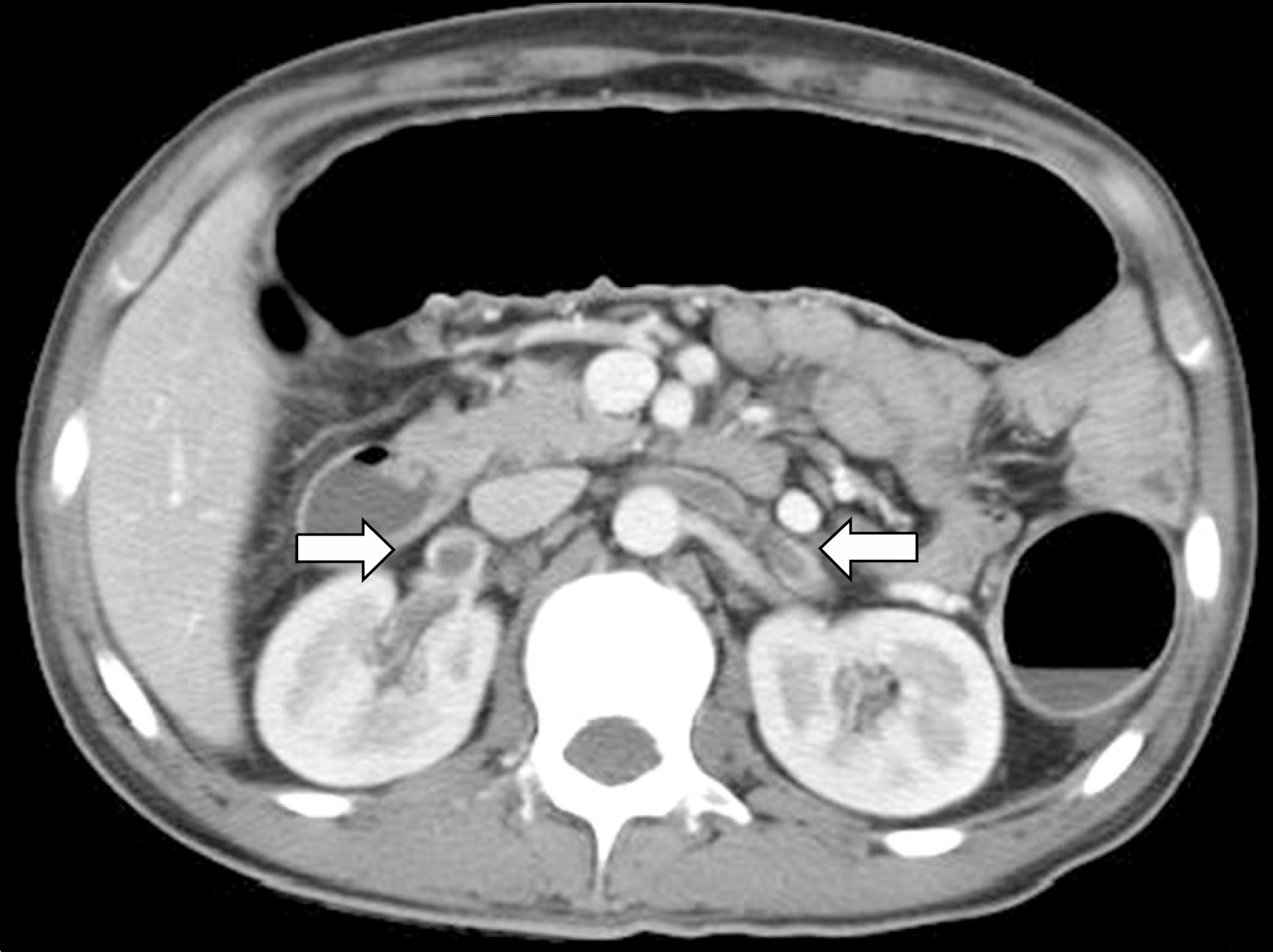
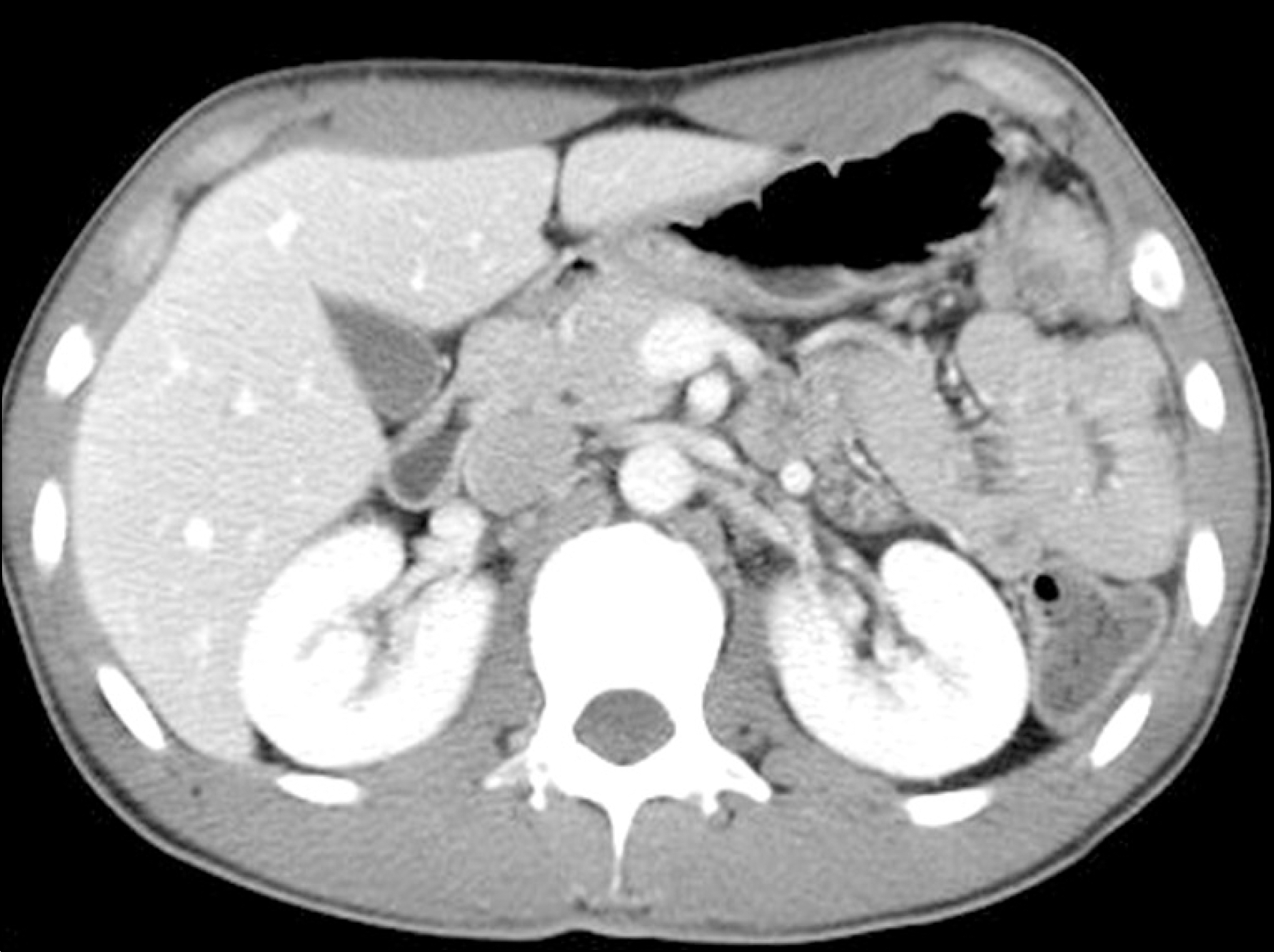
Venous thrombosis and thromboembolism appear to occur more often in patient with inflammatory bowel disease (IBD). The cause of thrombotic complications in IBD is generally considered to be associated with hypercoagulable conditions. Its prevalence rate ranges from 1% to 8% in clinical studies and rises to 39% in autopsy, but the renal vein thrombosis is very rare complication in ulcerative colitis patient. A 24-year-old man presented with intermittent abdominal pain and hematochezia for 6 months and recently developed pitting edema for few weeks. He was diagnosed as severe ulcerative colitis involving whole colon combined with thrombosis in both renal veins by colonoscopy and computed tomography scan of abdomen. We used steroid for the treatment of ulcerative colitis and both intravenous lower molecular weight heparin and warfarin for renal vein thrombosis. His symptoms were improved after treatment and maintained with mesalazine and warfarin. Follow-up abdominal CT scan showed complete resolution of both renal vein thrombosis. Currently he has been followed up for 2 years with oral mesalazine.
Go to : 
REFERENCES
1. Bernstein CN, Blanchard JF, Houston DS, Wajda A. The incidence of deep venous thrombosis and pulmonary embolism among patients with inflammatory bowel disease: a population-based cohort study. Thromb Haemost. 2001; 85:430–434.

2. Danese S, Papa A, Saibeni S, Repici A, Malesci A, Vecchi M. Inflammation and coagulation in inflammatory bowel disease: the clot thickens. Am J Gastroenterol. 2007; 102:174–186.
3. Bargen. J, Barker N. Extensive arterial and venous thrombosis complication chronic ulcerative colitis. Arch Intern Med. 1936; 58:17–31.
4. Graef V, Baggenstoss AH, Sauer WG. Veonus thrombosis occuring in nonspecific ulcerative. Arch Intern Med. 1966; 117:377–382.
5. Talbot RW, Heppell J, Dozois RR, Beart RW Jr. Vascular complications of inflammatory bowel disease. Mayo Clin Proc. 1986; 61:140–145.
6. Nam SW, Yang SK, Jung HY, et al. A case of cerebral venous thrombosis in active ulcerative colitis. Korean J Gastroenterol. 1998; 32:396–401.
7. Hong SJ, Hahm KB, Moon YS, et al. A case of superior sagittal sinus thrombosis in a patient with ulcerative colits. Korean J Gastroenterol. 1996; 28:445–450.
8. Kim BH, Han DS, Cho YJ, et al. A case of ulcerative colitis associated with portal vein and superior mensenteric vein thrombosis. Korean J Gastroenterol. 1998; 32:412–416.
9. Park JH, Choi GS, Kim MS, et al. A case of pulmonary thromboembolism in active ulcerative colitis. Korean J Gastroenterol. 2005; 45:301–305.
10. Tabibian JH, Lada SJ, Tabibian N. Combined inferior vena cava & renal vein thromboses: case and synopsis of thromboembolism in inflammatory bowel disease. Medscape J Med. 2008; 10:6.
11. Nachnani JS, Bhat R, Allen MJ. Renal vein thrombosis in inflammatory bowel disease. J Clin Gastroenterol. 2006; 40:651.
12. Freeman HJ. Venous thromboembolism with inflammatory bowel disease. World J Gastroenterol. 2008; 14:991–993.
13. Liebman HA, Kashani N, Sutherland D, McGehee W, Kam AL. The factor V Leiden mutation increases the risk of venous thrombosis in patients with inflammatory bowel disease. Gastroenterology. 1998; 115:830–834.
14. Frosst P, Blom HJ, Milos R, et al. A candidate genetic risk factor for vascular disease: a common mutation in methylenetetrahydrofolate reductase. Nat Genet. 1995; 10:111–113.
15. Asghar M, Ahmed K, Shah SS, Siddique MK, Dasgupta P, Khan MS. Renal vein thrombosis. Eur J Vasc Endovasc Surg. 2007; 34:217–223.
16. Kim HS, Fine DM, Atta MG. Catheter-directed thrombectomy and thrombolysis for acute renal vein thrombosis. J Vasc Interv Radiol. 2006; 17:815–822.
17. Lam KK, Lui CC. Successful treatment of acute inferior vena cava and unilateral renal vein thrombosis by local infusion of recombinant tissue plasminogen activator. Am J Kidney Dis. 1998; 32:1075–1079.
18. Laville M, Aguilera D, Maillet PJ, Labeeuw M, Madonna O, Zech P. The prognosis of renal vein thrombosis: a re-evaluation of 27 cases. Nephrol Dial Transplant. 1988; 3:247–256.
Go to :
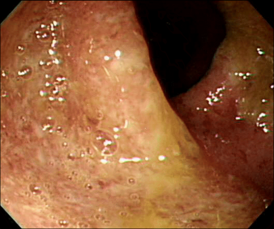 | Fig. 1.Colonoscopic finding showed diffuse edema and fine ulceration with friability from rectum to cecal base compatible with ulcerative colitis. |
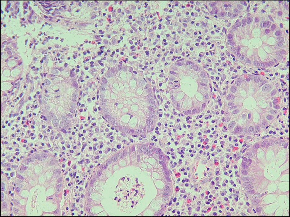 | Fig. 2.Microscopic finding revealed chronic active inflammation with crypt abscess in the colon (H&E stain, ×400). |
 XML Download