

 PDF
PDF ePub
ePub Citation
 Print
Print


Abstract
Background/Aims
This study was conducted to analyze the prognostic factors in patients with intrahepatic cholangiocarcinoma (ICC) who did not receive surgery.
Methods
Between August 1997 and November 2007, the medical records of 175 patients (mean age; 66 years, male/female 126/49), who were diagnosed as ICC, were reviewed retrospectively.
Results
Clonorchiasis and hepatolithiasis was found in 14.9%, and 6.3% of all patients, and no risk factors were identified in 77.8% of them. Surgical resection was performed in 29.1% (51 patients), chemotherapy +/− radiotherapy in 12.6% (22 patients), and palliative therapy in 58.3% (102 patients). The proportion of patients with stage I was 23.4% (41 patients). The prognostic factors in patients who did not receive surgery were alkaline phosphatase (ALP) and bilirubin levels by univariate and multivariate analysis. The median survival of patients with normal ALP and bilirubin levels was six months, whereas only one month in patients with elevated ALP and bilirubin levels (p<0.001). Tumor characteristics of patients with elevated bilirubin and ALP levels were infiltrative tumor, bile duct involvement, and very huge tumor.
Go to : 
REFERENCES
1. Malhi H, Gores GJ. Cholangiocarcinoma: modern advances in understanding a deadly old disease. J Hepatol. 2006; 45:856–867.

2. Suh BS, Lee SG, Min PC. A clinical review of peripheral cholangiocellular carcinoma. Korean J Gastroenterol. 1994; 26:987–994.
3. Nakeeb A, Pitt HA, Sohn TA, et al. Cholangiocarcinoma. A spectrum of intrahepatic, perihilar, and distal tumors. Ann Surg. 1996; 224:463–473.
4. Tang W. Multi-disciplinary treatment for cholangiocellular carcinoma. World J Gastroenterol. 2007; 13:1500–1504.
5. Anderson CD, Pinson CW, Berlin J, Chari RS. Diagnosis and treatment of cholangiocarcinoma. Oncologist. 2004; 9:43–57.
6. Khan SA, Thomas HC, Davidson BR, Taylor-Robinson SD. Cholangiocardinoma. Lancet. 2005; 366:1303–1314.
7. Lee WJ, Park JW. Review: analysis of survival rate and prognostic factors of intrahepatic cholangiocarcinoma: 318 cases in single institue. Korean J Hepatol. 2007; 13:125–128.
8. Chen MF, Jan YY, Jeng LB, et al. Experience of cholangiocarcinoma in Taiwan. J Hep Bil Pancr Surg. 1999; 6:136–141.
9. Song GW, Lee SG, Lee YJ, et al. Analysis of survival and factors of intrahepatic cholangiocarcinoma: 318 cases in single institue. Korean J Hepatol. 2007; 13:208–221.
10. Ryu KH, Lee KT, Lee JK, et al. Progostic factors in resectable peripheral cholangiocarcinoma. Korean J Gastroenterol. 2000; 36:686–694.
11. Kim HY, Choi CS, Choi YK. Clinicopathologic features and prognosis in peripheral cholangiocarcinoma. Korean J Gastroenterol. 1999; 33:268–275.
12. Chen MF. Peripheral cholangiocarcinoma (cholangiocellular carcinoma): clinical features, diagnosis and treatment. J Gastroenterol Hepatol. 1999; 14:1144–1149.
13. Kang KS, Kim KH, Suh KS, Kim SW, Lee KU. Clinical analysis of intrahepatic cholangiocarcinomas. J Korean Surg Soc. 1998; 54:396–404.
14. Lee HS, You JH, Nah YW. Peripheral cholangiocarcinoma. J Korean Surg Soc. 1997; 52:363–370.
15. Kim HJ, Yun SS, Jung KH, Kwun WH, Choi JH. Intrahepatic cholangiocarcinoma in Korea. J Hepatobiliary Pancreat Surg. 1999; 6:142–148.
16. Cherqui D, Tantawi B, Alon R, et al. Intrahepatic cholangiocarcinoma, results of aggressive surgical management. Arch Surg. 1995; 130:1073–1078.
17. Jarnagin WR, Shoup M. Surgical management of cholangiocarcinoma. Semin Liver Dis. 2004; 24:189–199.
18. Guo ZJ, Li Q, He K. Diagnosis and treatment of hepatic cholangiocarcinoma: report of 52 cases. Hepatobiliary Pancreat Dis Int. 2003; 2:62–65.
19. Chen MF, Jan YY, Wang CS, Jeng LB, Hwang TL. Clinical experience in 20 hepatic resection for peripheral cholangiocarcinoma. Cancer. 1989; 64:2226–2232.
20. Schlinkert RT, Nargoney DM, Van Heerden JA. Intrahepatic cholangiocarcinoma: clinical aspect, pathology and treatment. HBP Surg. 1992; 5:95–101.
21. Okabayashi T, Yamamoto J, Kosuge T, et al. A new staging system for mass-forming intrahepatic cholangiocarcinoma: analysis of preoperative and postoperative variables. Cancer. 2001; 92:2374–2383.
22. Park KW, Park JW, Cho SH, et al. Survival analysis for patients with hepatocellular carcinoma according to stage, liver Function and treatment modalities. Korean J Hepatol. 2006; 12:41–54.
23. Park JW. Practice guideline for diagnosis and treatment of hepatocellular carcinoma. Korean J Hepatol. 2004; 10:88–98.
24. Pang RW, Poon RT. From molecular biology to targeted therapies for hepatocellular carcinoma: the future is now. Oncology. 2007; 72(suppl 1):S30–S44.
25. http://www.cancer.go.kr
Go to :
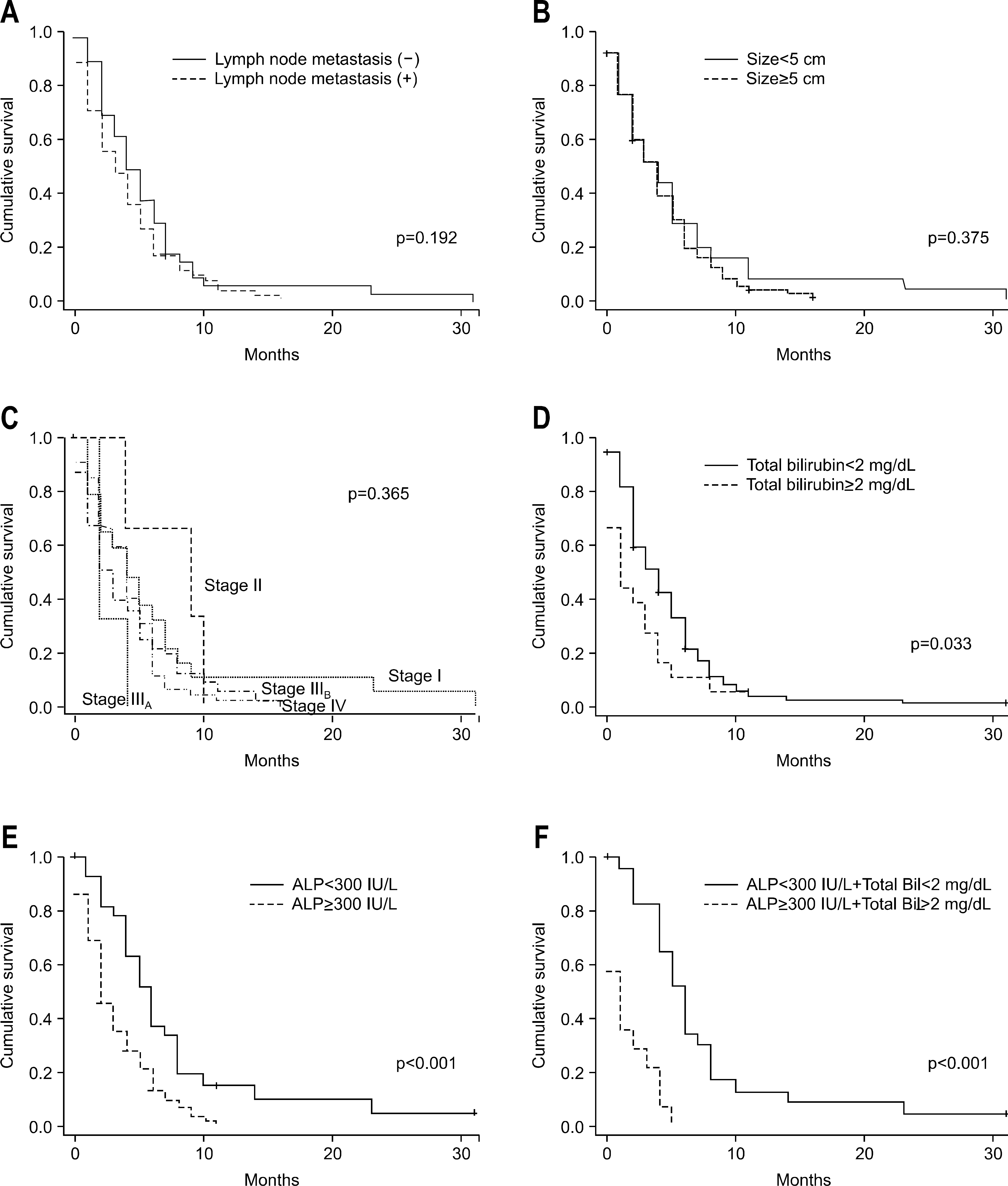 | Fig. 1.Comparison of survival rate according to clinical variables; lymph node metastasis (A), tumor size (B), stages (C), bilirubin level (D), ALP level (E), and bilirubin plus ALP levels (F). |
Table 1.
Baseline Clinical Characteristics
| Clinical factors | Surgical treatment (n=51) | Non-surgical treatment (n=124) | p-value |
|---|---|---|---|
| Gender, male, n (%) | 31 (60.8) | 95 (76.6) | 0.034 |
| Age, years | |||
| ≤39, n (%) | 1 (2.0) | 0 | |
| 40-49, n (%) | 0 | 9 (7.3) | |
| 50-59, n (%) | 9 (17.6) | 21 (16.9) | 0.083 |
| 60-69, n (%) | 24 (47.1) | 45 (36.3) | |
| 70-79, n (%) | 16 (31.4) | 38 (30.6) | |
| ≥80, n (%) | 1 (2.0) | 11 (8.9) | |
| Age, years (range) | 64.5±7.9 (37-83) | 66.5±10.0 (44-86) | 0.024∗ |
| Risk factors, n (%) | |||
| Clonorchiasis | 11 (21.6) | 15 (12.1) | |
| IHD stone | 5 (9.8) | 6 (4.8) | |
| HCV | 0 | 3 (2.4) | NS |
| Smoking | 6 (11.8) | 17 (13.7) | |
| Papillomatosis | 2 (3.9) | 0 | |
| Symptoms, n (%) | |||
| Abdominal pain | 22 (43.1) | 69 (55.6) | |
| Jaundice | 2 (3.9) | 16 (1.3) | |
| Palpable mass | 1 (1.7) | 5 (4.0) | NS |
| Weight loss | 1 (1.7) | 7 (5.6) | |
| Dyspepsia or nausea | 4 (7.8) | 18 (1.5) | |
| Asymptomatic | 22 (43.1) | 22 (1.8) | |
| Laboratory findings | |||
| Total bilirubin, mg/dL | 1.2±1.9 | 2.4±4.5 | NS∗ |
| ≥2.0, n (%) | 4 (7.8) | 20 (15.6) | NS |
| ALP, U/L | 340.6±193.6 | 567.0±450.2 | 0.001∗ |
| ≥300, n (%) | 21 (41.2) | 81 (65.3) | 0.005 |
| CA 19-9, u/mL | 1,314.0±3,052.1 | 3,879.4±15,282.7 | NS∗ |
| ≥37, n (%) | 26 (51.0) | 74 (59.7) | |
| <37 | 18 (35.3) | 24 (19.4) | NS |
| NA | 7 (13.7) | 26 (21.0) | |
| Diagnostic modalities, n (%) | |||
| CT | 51 (100) | 124 (100) | NS |
| MRI | 5 (9.8) | 9 (7.3) | NS |
| Angiography | 6 (11.8) | 11 (8.9) | NS |
| Biopsy | 17 (33.3) | 82 (66.1) | 0.001 |
| Median duration of F/U, mo (range) | 9.0 (0-122) | 3.5 (0-31) | <0.001∗ |
Table 2.
Tumor Characteristics
| Characteristics | Surgical treatment (n=51) | Non-surgical treatment (n=124) | p-value |
|---|---|---|---|
| Location, n (%) | |||
| Right | 23 (45.1) | 76 (61.3) | |
| Left | 25 (49.0) | 29 (23.4) | 0.003 |
| Both | 3 (5.9) | 19 (15.3) | |
| Number | |||
| One | 47 (92.2) | 98 (79.0) | |
| Two Three | 2 (3.9) 1 (2.0) | 5 (4.0) 16 (12.9) | 0.076 |
| Uncountable | 1 (2.0) | 5 (4.0) | |
| Size, cm, n (%) | |||
| ≤2.0 | 6 (11.8) | 3 (2.4) | |
| 2.1-5.0 | 20 (39.2) | 32 (25.8) | |
| 5.1-10.0 | 22 (43.1) | 69 (55.6) | 0.006 |
| ≥10.1 | 2 (3.9) | 17 (13.7) | |
| Unmeasurable | 1 (2.0) | 3 (2.4) | |
| (infiltrating tumor) | |||
| Size, cm (range) | 5.4±2.7 | 7.4±3.3 | <0.001∗ |
| Stage, n (%) | |||
| I | 20 (39.2) | 21 (16.9) | |
| II | 10 (19.6) | 3 (2.4) | |
| IIIA IIIB | 0 16 (31.4) | 3 (2.4) 55 (44.4) | <0.001 |
| IV | 5 (9.8) | 42 (33.9) | |
| LN metastasis, n (%) | 17 (33.3) | 86 (69.4) | <0.001 |
| Distant metastasis, n (%) | 5 (9.8) | 42 (33.9) | 0.001 |
| Lung | 1 | 14 | |
| Bone | 0 | 11 | |
| Peritoneal cavity | 1 | 11 | |
| Adrenal | 3 | 5 | |
| Brain | 0 | 2 | |
| Others | 1 | 4 |
Table 3.
Prognostic Factors in Patients Who Received Nonsurgical Treatment by Univariate Analysis
| Clinical factors | Survival (months) | p-value |
|---|---|---|
| Gender | ||
| Male (n=95) | 4.2±3.9 | 0.309 |
| Female (n=29) | 5.2±6.0 | |
| Age, years | ||
| <60 (n=30) | 3.9±3.4 | |
| ≥60 (n=94) | 4.6±4.8 | 0.434 |
| Stage | ||
| I (n=21) | 6.0±7.5 | |
| II (n=3) | 7.7±3.2 | |
| IIIA (n=3) | 2.7±1.2 | 0.272∗ |
| IIIB (n=55) | 4.0±3.9 | |
| IV (n=42) | 4.1±3.1 | |
| Size | ||
| <5 cm (n=25) | 5.8±7.2 | 0.109 |
| ≥5 cm (n=96) | 4.1±3.5 | |
| LN metastasis | ||
| Yes (n=87) | 4.0±3.7 | 0.114 |
| No (n=37) | 5.4±5.9 | |
| CA 19-9, U/mL | ||
| <37 (n=24) | 5.5±6.3 | 0.127 |
| ≥37 (n=74) | 4.0±3.4 | 0.127 |
| Total bilirubin, mg/dL | ||
| <2.0 (n=99) | 4.7±4.6 | 0.034 |
| ≥2.0 (n=20) | 2.4±3.0 | |
| ALP, U/L | ||
| <300 (n=38) | 6.7±5.9 | <0.001 |
| ≥300 (n=81) | 3.2±3.0 |
Table 4.
Prognostic Factors in Patients Who Received Nonsurgical Treatment by Multivariate Analysis
 XML Download