

 PDF
PDF ePub
ePub Citation
 Print
Print


Abstract
Several forms of colonic complications are rarely observed during the clinical course of acute pancreatitis, and potentially fatal in some cases. Colonic lesions associated with acute pancreatitis can be divided into several groups from a pathogenic point of view. Possible pathogenesis includes 1) spread of pancreatic enzymes through the retroperitoneum to mesocolon, causing pericolitis, 2) external inflammatory compression by mesocolic mass secondary to necrosis of fatty tissue, and 3) hypotension due to shock, and thrombosis of mesenteric arteries. These might lead to colonic infarction, fistula formation, perforation, and obstruction during follow-up. We report two cases of colonic obstruction following acute pancreatitis with possible different mechanisms and review Korean cases. One patient developed colonic obstruction due to severe necrotizing pancreatitis, possibly as a result of pericolitis, and the other developed stenosis as a result of ischemic colitis induced by acute pancreatitis.
REFERENCES
1. Whitcomb DC. Clinical practice. Acute pancreatitis. N Engl J Med. 2006; 354:2142–2150.
2. Kim YT. Medical management of acute pancreatitis and complications. Korean J Gastroenterol. 2005; 46:339–344.
3. Van Minnen LP, Besselink MG, Bosscha K, Van Leeuwen MS, Schipper ME, Gooszen HG. Colonic involvement in acute pancreatitis. A retrospective study of 16 patients. Dig Surg. 2004; 21:33–40.
4. Umeno Y, Otsuka J, Sasatomi E, Irie K. Development of colonic necrosis following severe acute pancreatitis. Intern Med. 2000; 39:305–308.

5. Kim YK, Dong SH, Kim HJ, Kim BH, Chang YW, Chung R. A case of pancreatico-colonic fistula after acute pancreatitis. Korean J Gastroenterol. 1992; 24:1186–1191.
6. Cho HG, Chung JP, Yum JS, et al. Spontaneous bowel perforation during the course of acute pancreatitis-a case report. Yonsei Med J. 1996; 37:158–164.
7. Cho YD, Hong SJ, Moon JH, et al. Stenosis of the colon due to chronic pancreatitis mimicking colon cancer. Korean J Gastrointest Endosc. 1998; 18:605–610.
8. Han YS, Dong SH, Kim HS, et al. A case of bowel infarction accompanied by acute necrotizing pancreatitis. Korean J Gastroenterol. 2002; 39:446–449.
9. Sohn BS, Jung JH, Song YT. Colonic complication of acute nectrotizing pancreatitis-a case report. J Korean Asso Pediatr Surg. 2003; 9:113–116.
10. Chung NS, Kim YS, Park CH, et al. A case of colon obstruction developed during the recovery period of acute pancreatitis. Korean J Gastroenterol. 2005; 45:206–209.
11. Yoo SS, Choi SK, Lee DH, et al. A case of colon obstruction developed as a complication of acute pancreatitis. Korean J Gastroenterol. 2008; 51:255–258.
12. Mohamed SR, Siriwardena AK. Understanding the colonic complications of pancreatitis. Pancreatology. 2008; 8:153–158.
13. Gardner A, Gardner G, Feller E. Severe colonic complications of pancreatic disease. J Clin Gastroenterol. 2003; 37:258–262.
14. Small AJ, Young-Fadok TM, Baron TH. Expandable metal stent placement for benign colorectal obstruction: outcomes for 23 cases. Surg Endosc. 2008; 22:454–462.
15. Baron TH. Colonic stenting: technique, technology, and outcomes for malignant and benign disease. Gastrointest Endosc Clin N Am. 2005; 15:757–771.
Fig. 1.
Case 1. Abdominal CT findings on admission. (A) Extensive amount of peripancreatic fluid collection extending to pararenal space was observed. (B) Distal ascending colon obstruction with proximal bowel dilatation was seen.
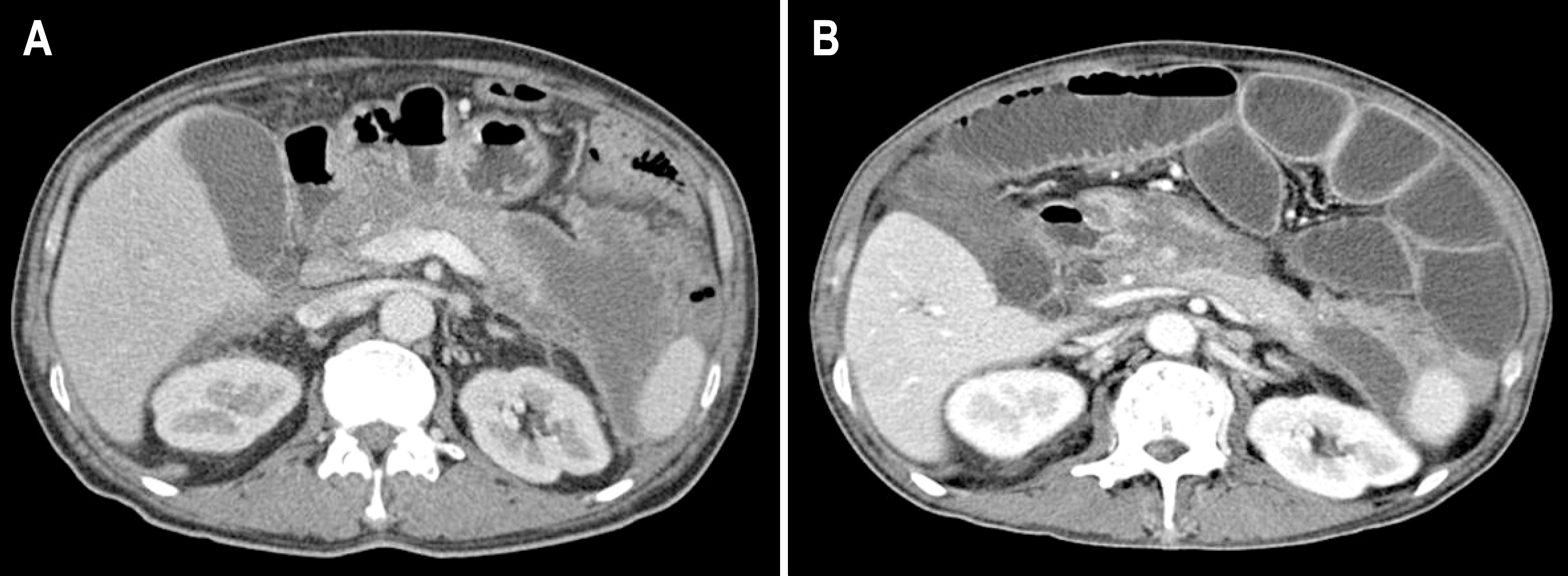
Fig. 2.
Case 1. Colonoscopic findings on 59th hospital day. In the distal ascending colon, luminal narrowing with mucosal edema and hyperemic changes were observed. Self-expanding metallic stent was inserted.

Fig. 3.
Case 2. Colonoscopic findings in the descending colon during initial hospital course of pancreatitis (A) and late obstruction event (B). (A) Longitudinal ulcer with edema and bleeding was seen, which was compatible with ischemic colitis. (B) Stricture, mucosal nodularity, and hyperemic changes were noted.

Fig. 4.
Case 2. Abdominal CT findings on admission. Stenosis of the descending colon with proximal bowel dilatation was noted. Arrow indicates stenosed descending colon.
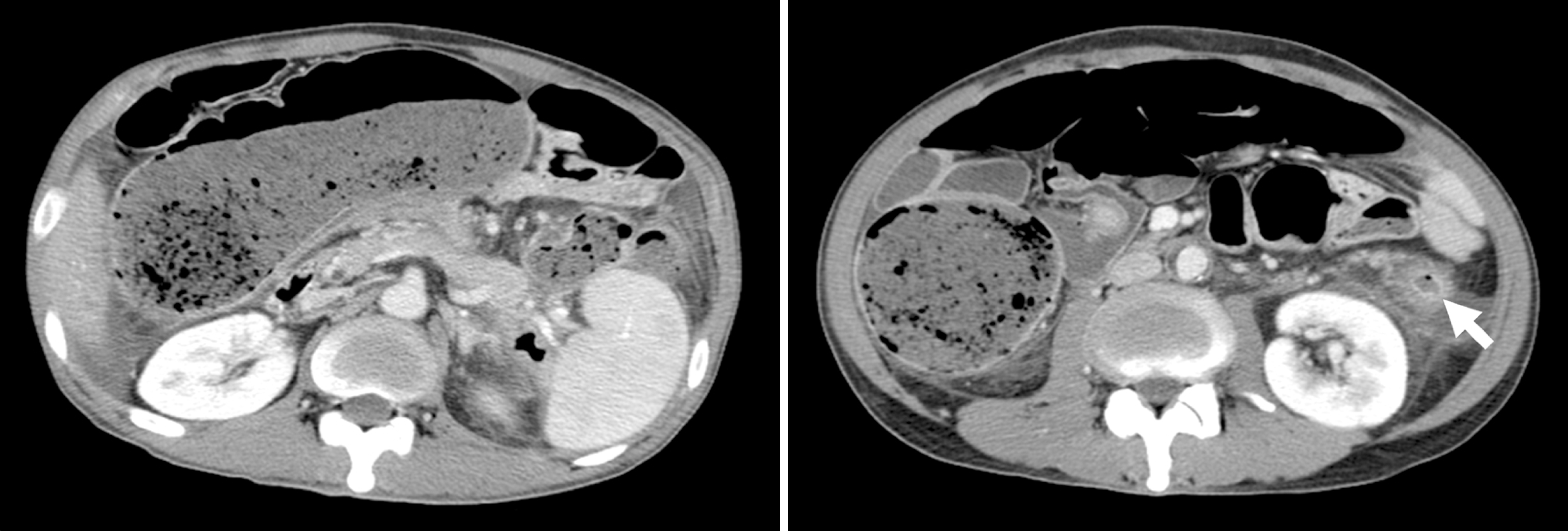
Table 1.
Clinical Features of Colonic Complications after Pancreatitis
| Age Sex Etiology | Interval after pancreatitis | CT grade | Colon complication | Colon Segment Involved | Colonoscopy Findings | Treatment | Outcome | |
|---|---|---|---|---|---|---|---|---|
| Case 1 | 49 M Alcoholic | 46 days | E | Obstruction | A∗ | Edema | Ileostomy | Recovery |
| Case 2 | 45 M Alcoholic | 27 days | E | Obstruction | SF†, D‡ | Stenosis, hyperemia | Bypass | Recovery |
| Yoo11 | 43 M Alcoholic | 42 days | E | Obstruction | D | Resection | Recovery | |
| Chung10 | 46 M Alcoholic | 17 days | E | Obstruction | SF | Edema, hemorrhage | Ileostomy | Death |
| Sohn9 | 10 M Unknown | 10 days | E | Obstruction | D | Normal mucosa | Resection | Recovery |
| Han8 | 41 F CRF | 21 days | E | Infarction | A | Resection | Death | |
| Cho7 | 56 M Alcoholic | - | Obstruction | SF, D | Edema, erosion | Resection | Recovery | |
| Cho6 | 63 M Alcoholic | 29 days | D | Perforation | T§ | Hyperemia | Resection | Recovery |
| Kim5 | 53 M Alcoholic | 60 days | E | Fistula | T | Drainage | Recovery |
 XML Download