

 PDF
PDF ePub
ePub Citation
 Print
Print


Abstract
Transcatheter arterial chemoembolization (TACE) is the mainstay of treatment for unresectable hepatocellular carcinoma (HCC). Although various complications of TACE have been reported, cerebral lipiodol embolism after TACE is rare. We report a 67-year-old man, who had patent foramen ovale and developed cerebral lipiodol embolism after TACE via the inferior phrenic artery. At 20 months after third TACE of 3 cm sized HCC in the left hepatic lobe, computed tomography (CT) revealed about 1.6 cm newly developed HCC in the anterior superior segment of right hepatic lobe. The angiogram revealed the HCC was supplied from the right inferior phrenic artery. Toward the end of TACE, there were accumulations of the iodized oil in the pulmonary vasculature. Immediately after TACE, he complained of weakness in right upper and lower limbs and sensory decrease in right limbs and right hemitrunk. Magnetic resonance imaging revealed a cerebral lipiodol embolism. Transesophageal echocardiography revealed no visible thrombi but contrast-echocardiography using hand agitated saline revealed an intracardiac right to left shunt consistent with patent foramen ovale. Motor weakness and sensory decrease were gradually improved, and all neurological symptoms disappeared over 4 weeks.
Go to : 
REFERENCES
1. Llovet JM, Burroughs A, Bruix J. Hepatocellular carcinoma. Lancet. 2003; 362:1907–1917.

2. Chen MS, Li JQ, Zhang YQ, et al. High-dose iodized oil transcatheter arterial chemoembolization for patients with large hepatocellular carcinoma. World J Gastroenterol. 2002; 8:74–78.
3. Tang ZY. Hepatocellular carcinoma–cause, treatment and metastasis. World J Gastroenterol. 2001; 7:445–454.
4. Yip D, Findlay M, Boyer M, Tattersall MH. Hepatocellular carcinoma in central Sydney: a 10-year review of patients seen in a medical oncology department. World J Gastroenterol. 1999; 5:483–487.
5. Ramsey DE, Kernagis LY, Soulen MC, Geschwind JF. Chemoembolization of hepatocellular carcinoma. J Vasc Interv Radiol. 2002; 13(suppl):S211–S221.
6. Sakamoto I, Aso N, Nagaoki K, et al. Complications associated with transcatheter arterial embolization for hepatic tumors. Radiographics. 1998; 18:605–619.
7. Wu RH, Tzeng WS, Chang CM. Iodized oil embolization to brain following transcatheter arterial embolization of liver. J Gastroenterol Hepatol. 2005; 20:1465–1467.
8. Yoo KM, Yoo BG, Kim KS, Lee SU, Han BH. Cerebral lipiodol embolism during transcatheter arterial chemoembolization. Neurology. 2004; 63:181–183.
9. Takao H, Makita K, Doi I, Watanabe T. Cerebral lipiodol embolism after transcatheter arterial chemoembolization of hepatocellular carcinoma. J Comput Assist Tomogr. 2005; 29:680–682.
10. Tajima T, Honda H, Kuroiwa T, et al. Pulmonary complications after hepatic artery chemoembolization or infusion via the inferior phrenic artery for primary liver cancer. J Vasc Interv Radiol. 2002; 13:893–900.
11. Cho SH, Kim JH, Kim BS, Jang J. A case of acute lung injury complicated by transcatheter arterial chemoembolization for hepatocellular carcinoma. Tuberc Respir Dis. 1995; 42:781–786.
12. Chung JW, Park JH, Im JG, Han JK, Han MC. Pulmonary oil embolism after transcatheter oily chemoembolization of hepatocellular carcinoma. Radiology. 1993; 187:689–693.
13. Kan Z, Sato M, Ivancev K, et al. Distribution and effect of iodized poppyseed oil in the liver after hepatic artery embolization: experimental study in several animal species. Radiology. 1993; 186:861–866.
14. Matsumoto K, Nojiri J, Takase Y, et al. Cerebral lipiodol embolism: a complication of transcatheter arterial chemoembolization for hepatocellular carcinoma. Cardiovasc Intervent Radiol. 2007; 30:512–514.
Go to :
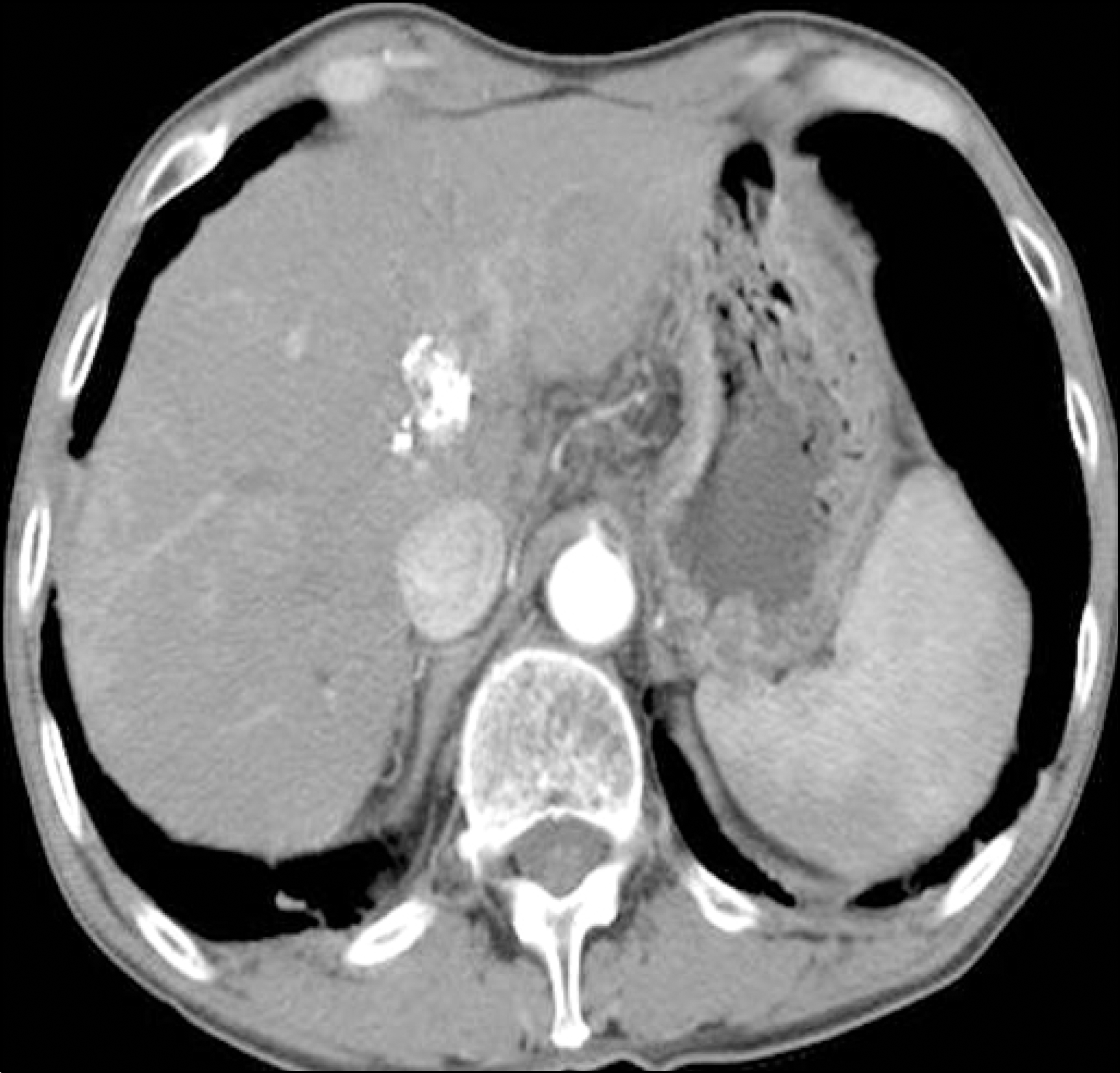 | Fig. 1.Computed tomography (CT) revealed previous lipiodol uptake in the medial segment of the left hepatic lobe and a newly developed HCC in the anterior superior segment of the right hepatic lobe, approximately 1.6 cm in size. |
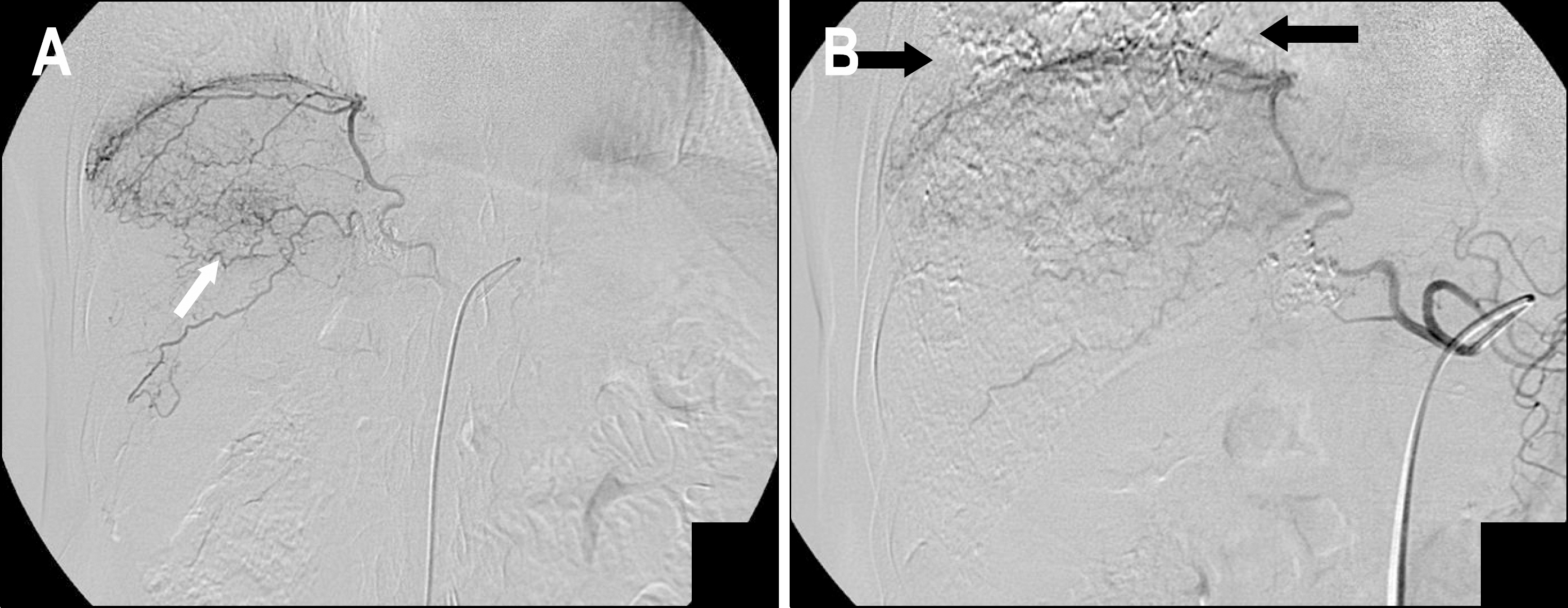 | Fig. 2.(A) Right inferior phrenic angiogram showed a hypervascular tumor in the right lobe of the liver (arrow). (B) Transar-terial infusion of adriamycin emulsified in iodized oil was infused into the right inferior phrenic artery. Delayed angiogram showed a lipiodol uptake in the right lower lung field (arrow). |
 | Fig. 3.(A) Magnetic resonance images of the brain revealed multiple, discrete, high signal intensities in the frontal lobe, parietal lobe, occipital lobe, and cerebellum on T2-weighted and diffusion-weighted imaging. (B) A non-contrast-enhanced CT scan of the brain obtained 7 days after the procedure revealed disseminated hyperattenuation along the gyri of both frontal lobe and subcortical white matter of the parietal lobe, suggesting deposition of iodized oil. |
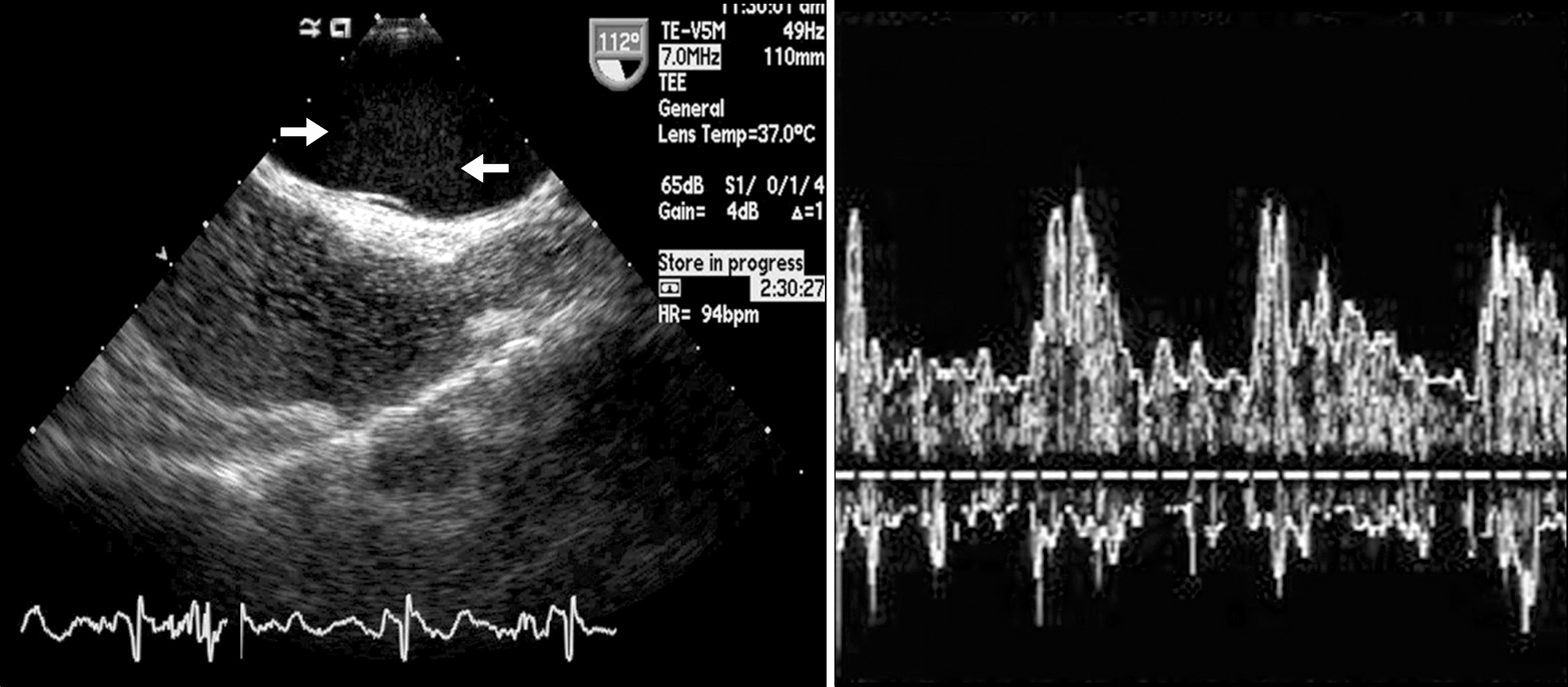 | Fig. 4.Agitated saline injections were performed at rest and during maximal Valsalva maneuver. Transesophageal echocardiogram showed visualization of an injected saline microbubble in the left atrium within three cardiac cycles of right atrial opacification, consistent with the presence of an intracardiac shunt (left). Transcranial Doppler through the temporal bone showed agitated saline bubbles passing through the middle cerebral artery (right). |
Table 1.
Cases of Cerebral Lipiodol Embolism after TACE in the Literature
 XML Download