

 PDF
PDF ePub
ePub Citation
 Print
Print


Abstract
The gastrointestinal (GI) tract is the most frequently involved site of mucosa-associated lymphoid tissue (MALT) lymphoma. Stomach is the most common site of involvement among the GI tract. However, MALT lymphoma of the large intestine is rare. A diagnosis is established by pathological examination of the surgical or endoscopic specimens. A 72-year-old man with low abdominal pain was diagnosed as a sigmoid MALT lymphoma, which was noted as an obstructing mass in a colonoscopic examination. A left hemicolectomy was performed, and the patient has had no recurrence postoperatively without any chemotherapy.
REFERENCES
1. Isaacson P, Wright DH. Malignant lymphoma of mucosa-associated lymphoid tissue. A distinctive type of B-cell lymphoma. Cancer. 1983; 52:1410–1416.

2. Dragosics B, Bauer P, Radaszkiewicz T. Primary gastrointestinal non-Hodgkin's lymphoma. A retrospective clinicopathologic study of 150 cases. Cancer. 1985; 55:1060–1073.
3. Isaacson PG. Gastric MALT lymphoma: from concept to cure. Ann Oncol. 1999; 10:637–645.
4. Mun HS, Park HJ, Lee KB, et al. A case of primary rectal MALT lymphoma presented as multiple submucosal tumors. Korean J Gastrointest Endosc. 2007; 35:272–276.
5. Matsuo S, Mizuta Y, Hayashi T, et al. Mucosa-associated lymphoid tissue lymphoma of the transverse colon: a case report. World J Gastroenterol. 2006; 14:5573–5576.
6. Kim JH, Moon YS, Lee SH, et al. A case of primary B cell mucosa-associated lymphoid tissue lymphoma presenting as a solitary rectal mass. Korean J Gastrointest Endosc. 2008; 36:102–106.
7. Harris NL, Jaffe ES, Stein H, et al. A revised European-American classification of lymphoid neoplasms: a proposal from the International Lymphoma Study Group. Blood. 1994; 84:1361–1392.
8. Jaffe ES, Harris NL, Diebold J, Muller-Hermelink HK. World Health Organization classification of neoplastic disease of the hematopoietic and lymphoid tissues. A progress report. Am J Clin Pathol. 1999; 111(suppl):S8–S12.
9. Shepherd NA, Hall PA, Coates PJ, Levison DA. Primary malignant lymphoma of the colon and rectum: a histopathological and immunohistochemical analysis of 45 cases with clinicopathological correlations. Histopathology. 1988; 12:235–252.
10. Jinnai D, Iwasa Z, Watanuki T. Malignant lymphoma of the large intestine: operative results in Japan. Jpn J Surg. 1983; 13:331–336.
11. Park JS, Kim WH, Baek HJ, et al. A case of mucosa-associated lymphoid tissue lymphoma of the large intestine diagnosed by sigmoidscopy. Korean J Gastrointest Endosc. 1999; 19(suppl):S71–S75.
12. Sasaki M, Iwafuchi M, Watanabe H. Clinopathologic study on primary intestinal lymphoma. I to Cho (Stomach and Intestine). 1998; 23:1315–1322.
13. Thieblemont C, Berger F, Coiffier B. Mucosa-associated lymphoid tissue lymphomas. Curr Opin Oncol. 1995; 7:415–420.
14. Grunberger B, Wohrer S, Streubel B, et al. Antibiotic treatment is not effective in patients infected with Helicobacter pylori suffering from extragastric MALT lymphoma. J Clin Oncol. 2006; 24:1370–1375.
Fig. 1.
Colonoscopic view of a large polypoid tumor causing nearly total obstruction in the sigmoid colon. The surface of this tumor was ulcerative and covered with exudates.
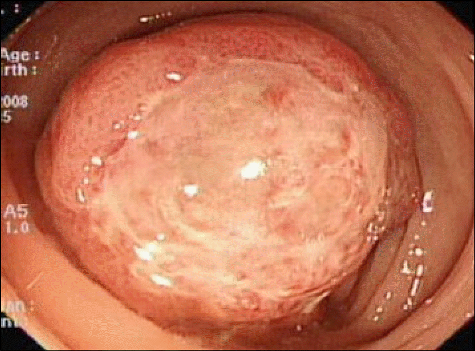
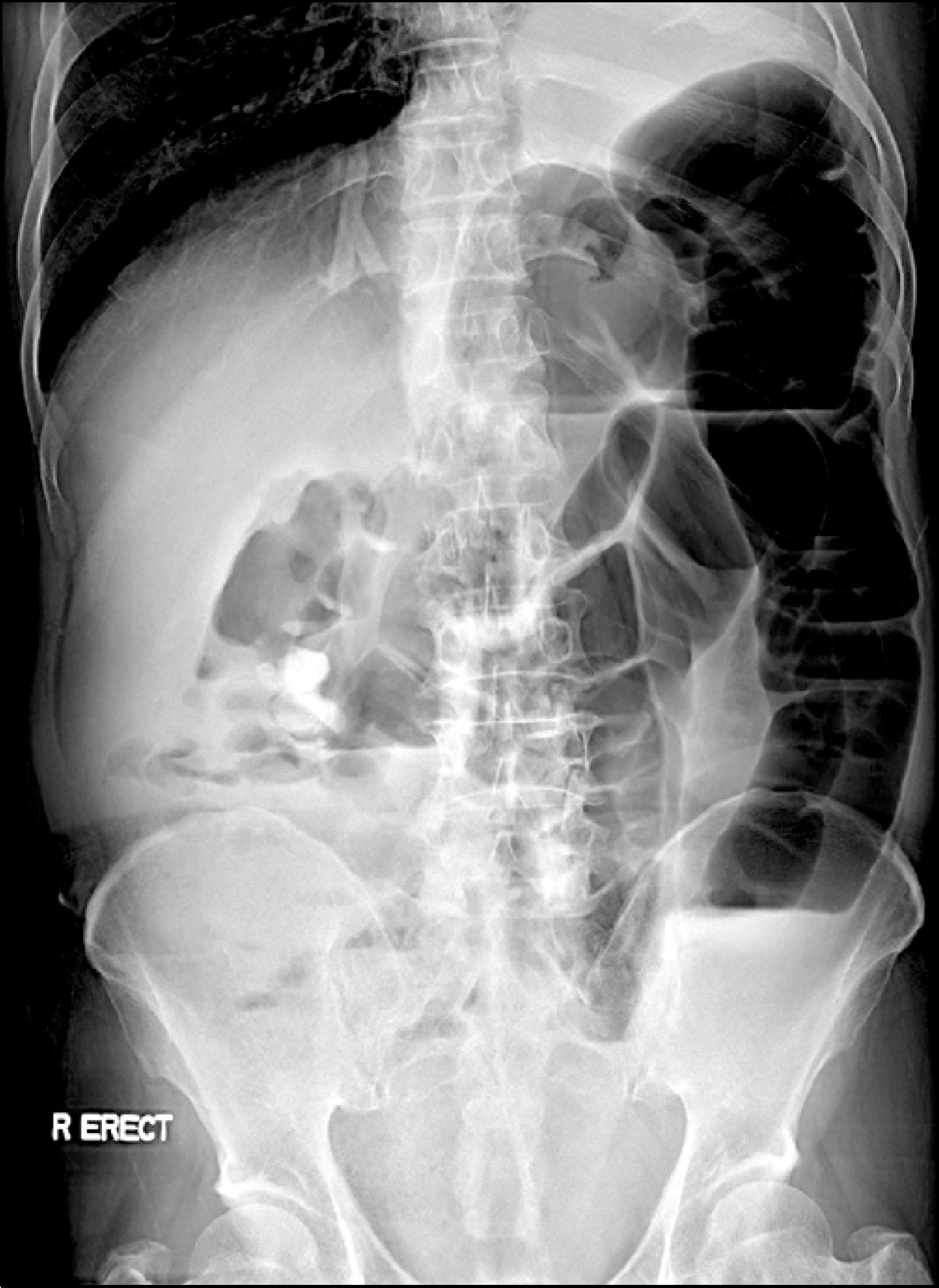
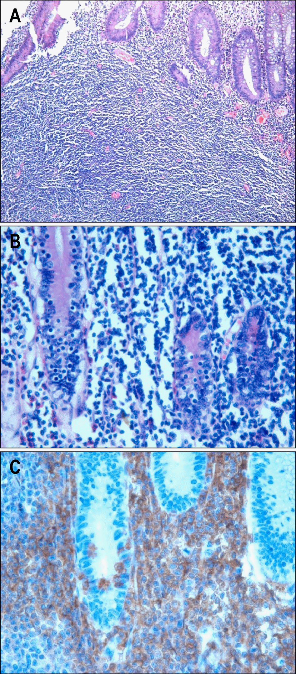
Fig. 3.
Microscopic findings. (A) Surgical specimens histologically showed diffuse proliferation of atypical small lymphocytes in the mucosa and submucosa layer (H&E stain, ×100). (B) In high power view, infiltrative lymphocytes were seen within the glandular epithelium (lymphoepithelial lesion) and the gland structures were destructed (H&E stain, ×200). (C) Immunohistochemical stain revealed infiltrative lymphocytes with immunoreactive for CD20 (CD20, ×200).
 XML Download