

 PDF
PDF ePub
ePub Citation
 Print
Print


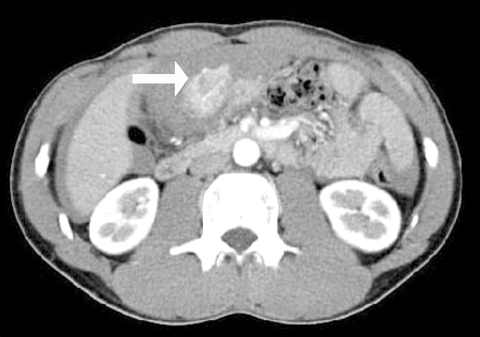
Abstract
The stomach is the most frequent site of gastrointestinal stromal tumor (GIST). The common clinical manifestation of GIST are melena and hematochezia caused by gastointestinal bleeding. However, hemoperitoneum due to GIST rupture is a very rare condition. We describe a 33-year-old man with gastric GIST causing hemoperitoneum. A preoperative CT scan demonstrated large amount of fluid collection and extraluminal mass lesion in gastric antral area. He underwent an emergent laparotomy. The antral mass was polypoid shaped and showed ruptured focus. We performed a distal gastrectomy. The tumor was revealed as GIST with intermediate malignant risk by pathologic examination. The patient had an uneventful postoperative course and remains well.
Go to : 
REFERENCES
1. Lucey BC, Varghese JC, Anderson SW, Soto JA. Spontaneous hemoperitoneum: a bloody mess. Emerg Radiol. 2007; 14:65–75.

2. Lucey BC, Varghese JC, Soto JA. Spontaneous hemoperitoneum: causes and significance. Curr Probl Diagn Radiol. 2005; 34:182–195.
3. Gupta P, Tewari M, Shukla HS. Gastrointestinal stromal tumor. Surg Oncol. 2008; 17:129–138.
4. Cichoz-Lach H, Kasztelan-Szczerbińska B, Słomka M. Gastrointestinal stromal tumors: epidemiology, clinical picture, diagnosis, prognosis and treatment. Pol Arch Med Wewn. 2008; 118:216–221.
5. Hirasaki S, Fujita K, Matsubara M, et al. A ruptured large extraluminal ileal gastrointestinal stromal tumor causing hemoperitoneum. World J Gastroenterol. 2008; 14:2928–2931.
6. Cegarra-Navarro MF, de la Calle MA, Girela-Baena E, García-Santos JM, Lloret-Estañ F, de Andrés EP. Ruptured gastrointestinal stromal tumors: radiologic findings in six cases. Abdom Imaging. 2005; 30:535–542.
7. Bucher P, Poletti PA, Myit S, Morel P. Spontaneous rupture of a gastrointestinal stromal tumour associated with life-threatening nontraumatic hemoperitoneum. Can J Surg. 2008; 51:38–39.
8. Gold JS, Dematteo RP. Combined surgical and molecular therapy: the gastrointestinal stromal tumor model. Ann Surg. 2006; 244:176–184.
9. Van der Zwan SM, Dematteo RP. Gastrointestinal stromal tumor: 5 years later. Cancer. 2005; 104:1781–1788.
Go to :

 XML Download