

 PDF
PDF ePub
ePub Citation
 Print
Print


Abstract
Objective
The aim of this study was to evaluate generational accumulation of murine fetal ovarian genes following prenatal exposure to 1.765-GHz microwave radiation.
Methods
A 1.765-GHz microwave generator was used. Twenty pregnant ICR mice were divided into two groups: the microwave-exposed experimental (irradiated) group, and the sham-exposed (sham) group. On the fifth day post-mating, dam mice were exposed to microwave irradiation in the insulated cage for 8 hours each day. The remaining mice were treated in the same way. Second generation mice were raised for 8 weeks then classified into four groups for examination. We removed the neonatal ovaries on the seventh day after the third delivery. We investigated the expression of six genes in the ovaries: Tnfaip 8, TNFsf 12, Cfd, CCL 11, Zfp 74, and Brd 3. Real time reverse transcription-polymerase chain reaction was performed using total RNA extracted from the removed ovaries.
Results
In the third-generation offspring, we detected some differences in ovarian gene expression between the first group and the fourth. Expression of CCL 11, and TNFsf 12 was decreased in the first group compared to the fourth group. Expression of Tnfaip 8, brd 3, Cfd, and Zfp 74 was higher in the first group than in the fourth group. We found differing results when we compared ovarian gene expression in mice of the second generation with those of the third.
Figures and Tables
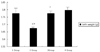 | Figure 1The comparison of birth weight in pregnant mice of the second generation. The birth weight of the first group is not different from that in the fourth group. However, that of the second group is decreased compared with other groups. aP<0.05 between 4 group and other groups, bP<0.05 between 1 group and other groups, cP<0.05 between 2 group and other groups.
|
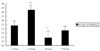 | Figure 2The comparison of number of offspring per pregnant mice of the second generation. The number of offspring in the first group is not different from that in the fourth group. However, the number of offspring in the second group is significantly increased compared with that in other groups. aP<0.05 between 4 group and other groups, bP<0.05 between 1 group and other groups, cP<0.05 between 2 group and other groups.
|
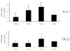 | Figure 3The results of TNFaip 8 and CCL gene expression by real time reverse transcription-polymerase chain reaction. Expression of CCL 11, and TNFsf 12 was decreased in the first group compared to the fourth group. aP<0.05 between 4 group and other groups, bP<0.05 between 1 group and other groups, cP<0.05 between 2 group and other groups.
|
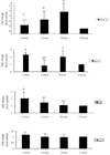 | Figure 4The results of Tnfsf 12, Zfp 74, Cfd, and Brd 3 gene expression by real time reverse transcription-polymerase chain reaction. Expression of Tnfaip 8, brd 3, Cfd, and Zfp 74 was higher in the first group than in the fourth group. aP<0.05 between 4 group and other groups, bP<0.05 between 1 group and other groups, cP<0.05 between 2 group and other groups.
|


References
1. Heynick LN, Johnston SA, Mason PA. Radio frequency electromagnetic fields: cancer, mutagenesis, and genotoxicity. Bioelectromagnetics. 2003. Suppl 6. 74–100.

2. Wertheimer N, Leeper E. Electrical wiring configurations and childhood cancer. Am J Epidemiol. 1979. 109:273–284.
3. Merritt CR, Kremkau FW, Hobbins JC. Diagnostic ultrasound: bioeffects and safety. Ultrasound Obstet Gynecol. 1992. 2:366–374.
4. Robert E. Intrauterine effects of electromagnetic fields (low frequency, mid-frequency RF, and microwave): review of epidemiologic studies. Teratology. 1999. 59:292–298.
5. Ahlbom A, Green A, Kheifets L, Savitz D, Swerdlow A. Epidemiology of health effects of radiofrequency exposure. Environ Health Perspect. 2004. 112:1741–1754.
6. Hwang JY, Na SH, Lee HA, Lee DH, Lee HJ, Kim SI, et al. Microarray analysis of gene expression in mice ovaries exposed to a 1.765 GHz microwave in utero. Korean J Obstet Gynecol. 2009. 52:602.
7. Ortner MJ, Galvin MJ, McRee DI. Studies on acute in vivo exposure of rats to 2450-MHz microwave radiation. Radiat Res. 1981. 86:580–588.
8. Bergonie J, Tribondeau L. Interpretation of some results from radiotherapy and an attempt to determine a rational treatment technique. 1906. Yale J Biol Med. 2003. 76:181–182.
9. Michaelson SM. Health implications of exposure to radiofrequency/microwave energies. Br J Ind Med. 1982. 39:105–119.
10. Ritenour ER. Health effects of low level radiation: carcinogenesis, teratogenesis, and mutagenesis. Semin Nucl Med. 1986. 16:106–117.
11. Wertheimer N, Savitz DA, Leeper E. Childhood cancer in relation to indicators of magnetic fields from ground current sources. Bioelectromagnetics. 1995. 16:86–96.
12. Sigler AT, Lilienfield AM, Cohen BH, Westake IE. Radiation exposure in parents of children with mongolism (Down's syndrome). Bull Johns Hopk Hosp. 1965. 117:374–399.
13. Cohen BH, Lilienfield AM, Kramer S, Hyman LC. Hook EB, Porter IH, editors. Parental factors in Down's syndrome: results of the second 'Baltimore case-control study.'. Population cytogenetics studies in humans. 1977. New York: Academic Press;301–352.
14. Larsen AI, Olsen J, Svane O. Gender-specific reproductive outcome and exposure to high-frequency electromagnetic radiation among physiotherapists. Scand J Work Environ Health. 1991. 17:324–329.
15. Guberan E, Campana A, Faval P, Guberan M, Sweetnam PM, Tuyn JW, et al. Gender ratio of offspring and exposure to shortwave radiation among female physiotherapists. Scand J Work Environ Health. 1994. 20:345–348.
16. Larsen AI. Congenital malformations and exposure to high-frequency electromagnetic radiation among Danish physiotherapists. Scand J Work Environ Health. 1991. 17:318–323.
17. Lindbohm ML, Hietanen M, Kyyronen P, Sallmen M, von Nandelstadh P, Taskinen H, et al. Magnetic fields of video display terminals and spontaneous abortion. Am J Epidemiol. 1992. 136:1041–1051.
18. Berman E, Carter HB. Decreased body weight in fetal rats after irradiation with 2450-MHz (CW) microwaves. Health Phys. 1984. 46:537–542.
19. Nawrot PS, McRee DI, Staples RE. Effects of 2.45 GHz CW microwave radiation on embryofetal development in mice. Teratology. 1981. 24:303–314.
20. Heynick LN, Merritt JH. Radiofrequency fields and teratogenesis. Bioelectromagnetics. 2003. Suppl 6. S174–S186.
21. Nakamura H, Matsuzaki I, Hatta K, Nobukuni Y, Kambayashi Y, Ogino K. Nonthermal effects of mobile-phone frequency microwaves on uteroplacental functions in pregnant rats. Reprod Toxicol. 2003. 17:321–326.
22. Ono T, Saito Y, Komura J, Ikehata H, Tarusawa Y, Nojima T, et al. Absence of mutagenic effects of 2.45 GHz radiofrequency exposure in spleen, liver, brain, and testis of lacZ-transgenic mouse exposed in utero. Tohoku J Exp Med. 2004. 202:93–103.
23. Diem E, Schwarz C, Adlkofer F, Jahn O, Rudiger H. Non-thermal DNA breakage by mobile-phone radiation (1800 MHz) in human fibroblasts and in transformed GFSH-R17 rat granulosa cells in vitro. Mutat Res. 2005. 583:178–183.
 XML Download