

 PDF
PDF ePub
ePub Citation
 Print
Print


Abstract
Objective
We investigated the usefulness of shortening of the fetal femur length (FL) and humeral length (HL) to predict Down syndrome at the middle gestation of pregnancy in Korean subjects.
Methods
This retrospective study involved 41 fetuses with Down syndrome and 328 fetuses with normal chromosome between 14+0 and 28+6 weeks of gestation. The expected FL and HL for any biparietal diameter (BPD) was calculated based on the control group data. The odds ratios for measure to expected FL and HL in comparison between normal fetuses and Down syndrome fetuses were calculated. The sensitivities of short FL and HL to predict Down syndrome were analyzed at a fixed false positive rate of 5%.
Results
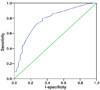
The lengths of femur and humerus long bone for any BPD in Down syndrome group were significantly shorter than the lengths in control group (P<0.001). A low ratio of measured to expected FL and HL increased the risk of fetal Down syndrome (P<0.001). At a fixed false positive rate of 5%, the sensitivities were 21.3% (95% confidence interval [CI] 0.698~0.852, P<0.001) in FL and 29.9% (95% CI 0.773~0.914, P<0.001) in HL.
Figures and Tables
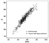 | Figure 1Regression lines of femur lengths (FL) with respect to biparietal diameter (BPD) in the study group and control group. FL=-6.150+0.754*BPD in study group. FL=-6.470+0.812*BPD in control group.
|
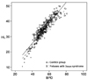
 | Figure 2Regression lines of humeral length (HL) with respect to biparietal diameter (BPD) in the study group and control group. HL=-1.893+0.625*BPD in study group. HL=-0.940+0.672*BPD in control group.
|
 | Figure 3Receiver operating characteristiccurve for a Down syndrome prediction, based on the ratio of measured to expected femur length (95% confidence interval 0.698~0.852).
|
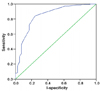 | Figure 4Receiver operating characteristic curve for a Down syndrome prediction, based on the ratio of measured to expected humeral length (95% confidence interval 0.773~0.914).
|
Table 1
Odds ratio for Mefl2 and Mehl2 derived in comparison between normal fetuses and Down syndrome fetuses in the same gestational age

![]()


References
1. Nicolaides KH. Screening for chromosomal defects. Ultrasound Obstet Gynecol. 2003. 21:313–321.

2. DeVore GR, Romero R. Genetic sonography: an option for women of advanced maternal age with negative triple-marker maternal serum screening results. J Ultrasound Med. 2003. 22:1191–1199.
3. Yeo L, Vintzileos AM. The use of genetic sonography to reduce the need for amniocentesis in women at high-risk for Down syndrome. Semin Perinatol. 2003. 27:152–159.
4. Smith-Bindman R, Hosmer W, Feldstein VA, Deeks JJ, Goldberg JD. Second-trimester ultrasound to detect fetuses with Down syndrome: a meta-analysis. JAMA. 2001. 285:1044–1055.
5. Bethune M. Literature review and suggested protocol for managing ultrasound soft markers for Down syndrome: thickened nuchal fold, echogenic bowel, shortened femur, shortened humerus, pyelectasis and absent or hypoplastic nasal bone. Australas Radiol. 2007. 51:218–225.
6. Smith-Bindman R, Chu P, Goldberg JD. Second trimester prenatal ultrasound for the detection of pregnancies at increased risk of Down syndrome. Prenat Diagn. 2007. 27:535–544.
7. Benacerraf BR. The role of the second trimester genetic sonogram in screening for fetal Down syndrome. Semin Perinatol. 2005. 29:386–394.
8. Harper LM, Gray D, Dicke J, Stamilio DM, Macones GA, Odibo AO. Do race-specific definitions of short long bones improve the detection of down syndrome on second-trimester genetic sonograms? J Ultrasound Med. 2010. 29:231–235.
9. Borgida AF, Zelop C, Deroche M, Bolnick A, Egan JF. Down syndrome screening using race-specific femur length. Am J Obstet Gynecol. 2003. 189:977–979.
10. Kovac CM, Brown JA, Apodaca CC, Napolitano PG, Pierce B, Patience T, et al. Maternal ethnicity and variation of fetal femur length calculations when screening for Down syndrome. J Ultrasound Med. 2002. 21:719–722.
11. Benacerraf BR, Gelman R, Frigoletto FD. Sonographic identification of second-trimester fetuses with Down's syndrome. N Engl J Med. 1987. 317:1371–1376.
12. Benacerraf BR, Neuberg D, Bromley B, Frigoletto FD. Sonographic scoring index for prenatal detection of chromosomal abnormalities. J Ultrasound Med. 1992. 11:449–458.
13. Perrella R, Duerinckx AJ, Grant EG, Tessler F, Tabsh K, Crandall BF. Second-trimester sonographic diagnosis of Down syndrome: role of femur-length shortening and nuchal-fold thickening. AJR Am J Roentgenol. 1988. 151:981–985.
14. Tannirandorn Y, Manotaya S, Uerpairojkit B, Tanawattanacharoen S, Wacharaprechanont T, Charoenvidhya D. Evaluation of fetal femur length to detect Down syndrome in a Thai population. Int J Gynaecol Obstet. 2001. 73:117–123.
15. Krantz DA, Hallahan TW, Macri VJ, Macri JN. Genetic sonography after first-trimester Down syndrome screening. Ultrasound Obstet Gynecol. 2007. 29:666–670.
16. Rodis JF, Vintzileos AM, Fleming AD, Ciarleglio L, Nardi DA, Feeney L, et al. Comparison of humerus length with femur length in fetuses with Down syndrome. Am J Obstet Gynecol. 1991. 165:1051–1056.
17. Benacerraf BR, Neuberg D, Frigoletto FD. Humeral shortening in second-trimester fetuses with Down syndrome. Obstet Gynecol. 1991. 77:223–227.
18. Vintzileos AM, Egan JF, Smulian JC, Campbell WA, Guzman ER, Rodis JF. Adjusting the risk for trisomy 21 by a simple ultrasound method using fetal longbone biometry. Obstet Gynecol. 1996. 87:953–958.
19. Zelop CM, Borgida AF, Egan JF. Variation of fetal humeral length in second-trimester fetuses according to race and ethnicity. J Ultrasound Med. 2003. 22:691–693.
20. Mastrobattista JM, Pschirrer ER, Hamrick MA, Glaser AM, Schumacher V, Shirkey BA, et al. Humerus length evaluation in different ethnic groups. J Ultrasound Med. 2004. 23:227–231.
21. Ogasawara KK. Variation in fetal ultrasound biometry based on differences in fetal ethnicity. Am J Obstet Gynecol. 2009. 200:676e1–e4.
22. Snijders RJ, Platt LD, Greene N, Carlson D, Krakow D, Gregory K, et al. Femur length and trisomy 21: impact of gestational age on screening efficiency. Ultrasound Obstet Gynecol. 2000. 16:142–145.
23. Longo D, DeFigueiredo D, Cicero S, Sacchini C, Nicolaides KH. Femur and humerus length in trisomy 21 fetuses at 11-14 weeks of gestation. Ultrasound Obstet Gynecol. 2004. 23:143–147.
24. Leung TN, Pang MW, Daljit SS, Leung TY, Poon CF, Wong SM, et al. Fetal biometry in ethnic Chinese: biparietal diameter, head circumference, abdominal circumference and femur length. Ultrasound Obstet Gynecol. 2008. 31:321–327.
25. Saksiriwuttho P, Ratanasiri T, Komwilaisak R. Fetal biometry charts for normal pregnant women in northeastern Thailand. J Med Assoc Thai. 2007. 90:1963–1969.
26. Jung SI, Lee YH, Moon MH, Song MJ, Min JY, Kim JA, et al. Reference charts and equations of Korean fetal biometry. Prenat Diagn. 2007. 27:545–551.
 XML Download