

 PDF
PDF ePub
ePub Citation
 Print
Print


Cervical cancer is the second most common cancer among women worldwide and is the primary cause of cancer-related deaths in women in developing countries. Of the estimated 500,000 annual incident cases worldwide and the 250,000 deaths attributed to cervical cancer, over 80% occur in low-resource countries.1,2 In developing countries, where cervical cancer accounts for 15% of female cancers, with a risk before age 65 of 1.5%, while in developed countries it accounts for only 3.6% of new cancers, with a cumulative risk (age, 0~64) of 0.8%.3 The highest incidence rates are observed in sub-Saharan Africa, Melanesia, Latin America and the Caribbean, South-Central Asia, and South East Asia.4
Traditional cervical screening methods are conventional Papanicolaou smear cytology and the pelvic examination. Since Dr. George Papanicolaou introduced this test for cervical cancer in 1939, the mortality rate for cervical squamous cell carcinoma in the United States decreased by 70~75% between 1955 and 1992. But unfortunately, this method contains numerous inherent opportunities for error leading to an acceptably high false negative rate. 53~90% of total false negative errors are due to sampling and preparation rather than interpretation errors.5 In February 1999, a report from the Agency for Health Care Policy and Research in the US admitted that estimates of the sensitivity of conventional Papanicolau (Pap) screening are not as high as previously reported.6 The report mentioned that, based on the few studies that avoided severe biases, conventional Pap smear cytology showed sensitivity of 51% and specificity of 98%. Furthermore, the report stated that the conventional Pap test is more accurate when an high-grade squamous intraepithelial lesion (HSIL) threshold is used, with the goal of detecting a high-grade lesion, than when lower thresholds, such as a low-grade squamous intraepithelial lesion (LSIL) or atypical squamous cells of undetermined significance (ASCUS), are used, with the goal of detecting low or high-grade dysplasia. Dunton7 said that the future of the Pap smear may best be summed up by this statement: "The 'new paradigm' of cervical cancer screening could be a combination of the new technology enhancements-liquid-based sampling, computer-assisted screening, and reflex molecular testing."
The major risk factors for developing cervical cancer include early age of marriage or sexual exposure, multiplicity of sexual partners, low socio-economic status, smoking, oral contraceptives and multiparity.8 Sexual behavior has been linked to cervical neoplasia. Recent epidemiological studies showed that cervical cancer is the result of the process that is initiated by infection of the genital tract with specific types of high risk human papillomaviruses (HR-HPVs). Genital HPV types are divided into those of low and high oncogenic risk. The most representative of these low risk is HPV 6 and 11 and the most representative of the high risk types are HPV 16, 18, 31, and 45. Low risk HPVs cause benign genital warts and have no oncogenic potential. By contrast, high risk HPVs are the causative agents of cervical cancer and its intraepithelial precursors. HPVs 16, 18, 31, 35, 39, 45, 51, 52, 56, 58, 59, and 68 account for about 93% of infections in high grade cervical intraepithelial neoplasias (CINs) and cancer. HPV 16 and HPV 18 are the HPV types most commonly identified in neoplasias.9-11
More than 99% of cervical cancers contain one or more of the approximately 15 HPV genotypes that have been associated with the development of cervical cancer. As approximately 50~60% of these cancers contain HPV 16, and another 10~20% contain HPV 18.12 Persistent infection with high-risk HPV types, especially 16 and 18, is strongly predictive of cervical neoplasia and cancer.
The purpose of this study was to evaluate the detection rate of HPV DNA Test (type 16 and 18) and to investigate the clinical significance of HPV DNA test in patients with cervical neoplasia.
Materials and Methods
1. Study design
This university hospital-based clinical study was performed in the outpatient clinic of the Department of Obstetrics and Gynecology at Dankook University Medical Center from January 1, 2002 to December 31, 2004. Of the 708 patients aged 21~83 years who had undergone both conventional Papanicolaou cervical cytologic test and HPV DNA test by polymerase chain reaction (PCR), 383 cases underwent histologic diagnoses. Clinicopathological characteristics were obtained from the patient's clinical records. Specific exclusion criteria consisted of patients under 18 years of age, pregnant women, women less than 6 weeks post-partum, patients who had active menstruation or active vaginal bleeding, and patients without cervix.
2. Specimen collection for conventional Papanicolaou smear cytology
The cervical sample was taken in the usual way by the physician using a combination of plastic spatula and endocervical brush (cytobrush). Following collection according to the guidelines, the collection device was spread on the surface of the slide as evenly and thinly as possible. Immediately the slide was fixed in 95% ethyl alcohol solution. The slide and a case report form containing the patient's identification number, initials and medical history were sent to the pathology laboratory.
3. Papanicolaou cytology slide preparation and evaluation
The slides were prepared with the laboratory's routine Papanicolaou staining method, and evaluated according to The Bethesda System (TBS 2001) criteria. In brief, specimens that are not within normal limits can contain changes deemed either as benign cellular changes, or as cells that are consistent with an epithelial cell abnormality. The latter category includes both squamous cell and glandular cell abnormalities. Squamous cell abnormalities are consistent with atypical squamous cells (ASC), LSIL, HSIL, and squamous cell carcinoma (SCC). ASC refers to cytologic changes suggestive of SIL, which are qualitatively or quantitatively insufficient for a definitive interpretation. All interpretations of ASC should be qualified as "Of undetermined significance" (ASC-US) or "cannot exclude HSIL" (ASC-H). Glandular cell abnormalities are consistent with atypical glandular cells (AGC), endocervical adenocarcinoma in situ (AIS), and adenocarcinoma (ACC). Pap slides were initially screened by cytotechnologists, and finally confirmed by a pathologist.
4. Histology
Histologic diagnoses were made with biopsy specimens by various kinds of method: colposcopy-directed exocerevical biopsies using punch or LEEP biopsy, endocervical or endometrial curettage, conization, and hysterectomy.
The biopsy specimen was fixed in 10% buffered formalin, paraffin embedded, and prepared as conventional histopathologic slides stained with hematoxylin and eosin. The biopsies were interpreted and diagnosed using a combined reporting system, including TBS and CIN designations by a pathologist. Atypical category were defined as histology that were atypical but could not be categorized as intraepithelial lesion.
5. Specimen collection for HPV DNA test
The specimens were collected by scraping the uterine cervical canal with a small cytobrush and then the brush was put into a 15 ml centrifuge tube containing phosphate buffered saline (PBS).
6. DNA extraction and HPV PCR
DNA extraction and HPV PCR were performed with Bio-Core HPV 16/18 PCR kit (Biocore, Seoul, Korea) according to manufacturer's recommendations, after careful and thorough Quality Control by the Department of Medical Science of Diagnostic Test. Briefly, the specimen in 15 mL centrifuge tube was centrifuged at 3,000 rpm for 15 minutes and the supernatant was removed. The collected pellet was washed twice with 1 mL of PBS and treated with 50 µL of HPV 16/18 extraction buffer for 20 minutes at 100℃. After centrifugation at 13,000 rpm for 3 minutes, the isolated DNA from supernatant was stored at -20℃ until use.
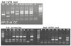
HPV DNA amplication was performed using consensus primers for HPV16 and HPV18 in the Bio-Core HPV 16/18 PCR kit. The PCR was performed mixing 4.5 µL of the isolated DNA with 15.5 µL of HPV 16/18 PCR mixture included in the kit using a PTC200 Thermo cycler (MJ Research, Waltham, MA, USA) under the following conditions: initial denaturation at 95℃ for 5 minutes, followed by 35 cycles of denaturation at 94℃ for 45 seconds, annealing at 60℃ for 45 seconds, extension at 72℃ for 45 seconds, and a final cycle of 72℃ for 5 minutes. Included in each run were a positive control containing of β-globin DNA primers and distilled water as a negative control to monitor the performance of the PCR methods.
PCR products were separated on a 2% agarose gel stained with 0.5 µg/mL ethidium bromide and analyzed by UV transilluminator (Fig. 1).
7. Data analysis
The cytologic diagnoses of conventional Pap smear test and the HPV DNA test were compared with the histologic diagnoses. Comparisons were performed using standard contingency table analyses. The statistical analysis was performed using SPSS version 10.0. The Chi-square test was used to know whether the significant difference between cervical cytology and HPV DNA test in the screening of the cervical lesion is present. P<0.05 was considered statistically significant.
Results
The 708 women had undergone conventional Papanicolaou cervical cytologic test and HPV DNA test. The mean age was 45.6 years ranging from 21 to 83 years. The mean gravidity was 4.0 (from 0 to 15) and the mean parity was 2.0 (from 0 to 9). Of the 708 women, histologic diagnoses were performed in 383 cases.
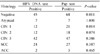
Of the 708 cytologic diagnoses, there were 11 (73.3%) positive HPV DNA test diagnoses in SCC, 41 (61.2%) in HSIL, 20 (54.1%) in LSIL, 41 (36.9%) in ASC, and 86 (18.3%) in negative cytology (Table 1). Of the 383 histologic diagnoses, there were 24 (72.7%) positive HPV DNA test diagnoses in SCC, 42 (59.2%) in CIN 3, 12 (50.0%) in CIN 2, 12 (42.9%) in CIN 1, 7 (38.9%) in atypical change, and 45 (22.0%) in negative histology (Table 2). Of the 383 histologic diagnoses, there were 16 (48.5%) positive HPV 16 DNA test diagnoses in SCC, 2 (50.0%) in ACC, 24 (33.8%) in CIN 3, 2 (8.3%) in CIN 2, 2 (7.1%) in CIN 1, 1 (5.6%) in atypical change, and 22 (10.7%) in negative histology. Of the 383 histologic diagnoses, there were 11 (33.3%) positive HPV 18 DNA test diagnoses in SCC, 2 (50.0%) in ACC, 17 (23.9%) in CIN 3, 2 (8.3%) in CIN 2, 3 (10.7%) in CIN 1, 3 (16.7%) in atypical change, and 8 (3.9%) in negative histology. There was no significant difference between cervical cytology and HPV DNA test for the screening of the cervical lesion except for negative and the CIN 1 lesion (P<0.05) (Table 2).
Of the 708 cases, distribution of HPV DNA test outcomes according to the age showed 24 (45.3%) positive HPV DNA test diagnoses under the age of 30, 36 (21.4%) in the thirties, 77 (29.4%) in the forties, 36 (23.5%) in the fifties, 17 (35.4%) in the sixties, and 12 (54.5%) in the seventies.
Of the 239 patients with negative HPV DNA test, 68 (28.5%) cases showed histologic diagnoses of CIN 1 or worse lesion including SCC and ACC (Table 3). Of the 46 patients with negative cytology and positive HPV DNA test, 23 (50%) cases showed histologic diagnoses of CIN 1 or worse lesion including SCC (Table 4).
When ASC/AGC or worse lesions were defined as positive in the cytology and CIN 1 or worse lesions were defined as positive in the histology, Pap smear cytology showed 148 true-negative, 44 false-negative, 75 false-positive, and 116 true-positive, in the histology comparison study. Diagnostic accuracy of Pap smear cytology showed sensitivity of 72.5%, specificity of 66.4%, false-positive rate of 33.6%, false-negative rate of 27.5%, positive predictive value of 60.7%, and negative predictive value of 77.1%.
When other types of HPV were included in the HPV DNA test and CIN 1 or worse lesions were defined as positive in the histology, HPV DNA test showed 171 true-negative, 68 false-negative, 52 false-positive, and 92 true-positive, in the histology comparison study. Diagnostic accuracy of HPV DNA test showed sensitivity of 57.5%, specificity of 76.7%, false-positive rate of 23.3%, false-negative rate of 42.5%, positive predictive value of 63.9%, and negative predictive value of 71.5%.
Combined test (Pap cytology and HPV DNA test) showed sensitivity of 86.9%, specificity of 56.1%, false-positive rate of 43.9%, false-negative rate of 13.1%, positive predictive value of 58.6%, and negative predictive value of 85.6% (Table 5).
Discussion
Because cervical cancer affects relatively young women, it is an important cause of lost years of life in the developing world.4 Yang et al13 found that it was responsible for 2.7 million (age-weighted) years of life lost worldwide in 2000 and it is the biggest single cause of years of life lost from cancer in developing world. But uniquely, cervical cancer remains eminently preventable. The key to prevention is the timely identification and management of precancerous lesions through accessible and affordable screening programs.
There is little doubt that well organized cytology based screening programs are effective in saving lives. However, a programme based on solely conventional cytology has important limitations with sensitivity and specificity and an ideal screening test should be performed infrequently and be capable of detecting precursor or early easily treatable lesions with great accuracy. Recently, the accumulated molecular and clinical evidences have left no doubt that HPV directly influences the pathogenesis for cervical neoplasia. It is evident that most cervical neoplasias are causally related to HPV.14
HPV is an 8 kb circular double-stranded DNA virus. To date, over 120 distinctive types of HPV have been identified. Each HPV type has a predilection for infecting certain areas of the body. Over 30 types of HPV are considered genital types. These viruses can be divided into three groups: high-risk types (16, 18, 31, 33, 35, 39, 45, 51, 52, 56, 58, 59, 68, 82); low-risk types (6, 11, 40, 42, 43, 44, 53, 54, 61, 72, 73, 81); and intermediate- risk types (possible high-risk types 26, 66, 73).15,16
The HPV screening method offers the possibility of greater sensitivity, reduced follow up of low grade cytological abnormalities and treated lesions, increased screening intervals and overall cost reductions. A large number of other studies conducted in different parts of the world suggest HPV infection to be a common phenomenon in cervical cancers and premalignant lesions with HPV presence detected in 48~100% of cervical cancers. Among the high risk group, HPV 16 and 18 has been most commonly associated in cervical cancers, with HPV 16 constituting the most prevalent type, ranging from 44 to 84%, followed by HPV 18 ranging from 2 to 39%.17
The most common of the high-risk types is HPV 16. HPV 16 is found in approximately half of all women with cervical cancers. HPV 16 is also the single most commonly identified HPV type in high-grade cervical intraepithelial neoplasia (CIN 2, 3), as well as among women in the general population. The data of this study are consistent with results of other studies.14,16,17 The second most common high-risk virus is HPV 18. HPV 18 is common not only in squamous lesions but also is especially common in adenocarcinoma and adenocarcinoma in situ (AIS) lesions of the cervix. Adjusted for age, the odds ratio for cervical cancer was 435 for infection with HPV 16 and was 248 for infection with HPV 18.15,16 To put this dramatic increase in risk into perspective, most studies of cigarette smoking in lung cancer have reported odds ratios of less than 20.15,16
Abnormal Paps develop within 5 years in 17~36% of HPV DNA positive women. 3~10% of high-risk HPV DNA positive women will develop CIN 2, 3. Women with persistent high-risk HPV DNA infections are at greatest risk for CIN 2, 3, and cancer.15,16,18
High-risk, oncogenic HPV types 16 and 18 are responsible for approximately 70% of cervical cancer cases, where low-risk types 6 and 11 cause approximately 90% of genital warts cases, abnormal cytology, and recurrent respiratory papillomatosis. In 2 large prospective trials, 5 to 27% of women who tested positive for HPV 16 or 18 developed CIN 3 or cervical cancer within a 3-year period.19
Among young women with a prevalent HPV infection, 60 to 75% become HPV negative after 30 months, and the median duration of HPV infection is estimated at 8 months. The risk of HSIL development is estimated to 14 times higher among women who test positive at least 3 times for high-risk HPV types compared with HPV-negative women. Prospective studies estimate 15 to 30% of women who test positive for high-risk HPV types will develop HSILs within a 4-year period. Persistent infection with high-risk HPV types (16 and 18) is strongly predictive of cervical neoplasias and cancer.19,20
Of the 383 histologic diagnoses, there were 24 (72.7%) positive HPV DNA test diagnoses in SCC, 42 (59.2%) in CIN 3, 12 (50.0%) in CIN 2, 12 (42.9%) in CIN 1, 7 (38.9%) in atypical change, and 45 (22.0%) in negative histology. Of the 383 histologic diagnoses, positive HPV 16 DNA test diagnoses showed 48.5% (16/33) in SCC, 50.0% (2/4) in ACC, 33.8% (24/71) in CIN 3, 8.3% (2/24) in CIN 2, 7.1% (2/28) in CIN 1, 5.6% (1/18) in atypical change, and 10.7% (22/205) in negative histology. Of the 383 histologic diagnoses, positive HPV 18 DNA test diagnoses showed 33.3% (11/33) in SCC, 50.0% (2/4) in ACC, 23.9% (17/71) in CIN 3, 8.3% (2/24) in CIN 2, 10.7% (3/28) in CIN 1, 16.7% (3/18) in atypical change, and 3.9% (8/205) in negative histology. These data show that the more advanced high-grade lesions have more frequent HPV infections including HPV 16 and/or 18 infection. There was no significant difference between Pap cytology and HPV DNA test for detecting cervical histology except for negative and CIN 1 lesion (P<0.05).
Published literature indicates that 35% of untreated CIN 2 lesions will persist and 22% will progress to carcinoma in situ or invasive cervical cancer. For comparison, 56% of untreated CIN 3 lesions will persist and 14% will progress.21 Of the 111 patients with cytologic diagnosis of ASC, there were 70 (63.1%) negative HPV DNA test diagnoses and 41 (36.9%) positive diagnoses. Of the 88 patients with cytologic diagnosis of ASC, there were 45 (51.1%) histologic diagnoses of negative, 4 (4.5%) of atypical, 12 (13.6%) of CIN 1, 6 (6.8%) of CIN 2, 14 (15.9%) of CIN 3, and 7 (8.0%) of SCC. Overall, 30.7% (27/88) of patients with cytologic diagnosis of ASC had CIN 2 or worse lesion.
Cervical cancer is largely preventable through screening programmes designed to diagnose and treat cervical lesions that may progress to invasive cancer. Early detection of LSIL and HSIL is very important because some lesions have a high-risk potential of developing into more severe lesions, including invasive cancer. The issue of false negative Pap smears was drawn to public attention in 1987 following a series of invetigative reports in the Wall Street Journal (November 2, 1987:1, 20; December 29, 1987: 17). Linda and Zahniser summarized that the consequences of this and subsequent reports include the implementation of the Clinical Laboratory Improvement Amendments of 1988, an erosion in Public confidence in the Pap smear, an unprecedented liability crisis for those who practice cervical cytology, a dramatic increase in the percentage of cases designated 'atypical squamous cells of undetermined significance (ASCUS)' as cytopathologists attempt to protect themselves from potential liability, overuse of colposcopy and cervical biopsy in treating women with ASCUS Pap, and the application of new technology to Pap testing.22 Our data confirmed that the physician must be cautious in managing the patient with the cytologic diagnosis of ASC.
Of the 239 patients with negative HPV DNA test, 28.5% (68/239) showed histologic diagnoses of CIN 1 or worse lesion. Of the 93 patients with negative HPV DNA test and ASC or worse cytology, 37.6% (35/93) revealed histologic diagnoses of CIN 2 or worse lesion. Our data for HPV DNA test might be incorrect because our institution does not seem to have highly trained specialized pathologist and technicians. Some variation may represent regional or population-based differences or different laboratory practice patterns. Anyway the physician should not overrate HPV DNA test in managing the patient with cervical neoplasia.
Approximately 5% of HPV DNA positive, cytology negative women will have CIN 2, 3.15,18 Despite low specificity for low-grade cervical lesions, HPV DNA testing significantly increases the sensitivity of predicting HSIL and carcinoma: a prospective study demonstrated that women with negative cervical cytologic screens and positive HPV DNA test results were at much higher risk compared with women who were HPV DNA negative (assessed at 1 to 2 year, intervals during a 5-year period).23 Jeon et al24 reported that frequent exams at less than 12 months intervals is unnecessary for women with positive for high risk HPV and negative cytology.
Of the 46 patients with positive HPV DNA test and negative cytology, 50.0% (23/46) showed histologic diagnoses of CIN 1 or worse lesion. Of the 29 patients with positive HPV 16 and/or 18 and negative cytology, 41.4% (12/29) revealed histologic diagnoses of CIN 2 or worse lesion. These data confirmed HPV DNA test is an important diagnostic method for cervical neoplasia.
If the goal of cervical screening is to identify patients with cervical lesions, sensitivity is more important than specificity. As is well known, sensitivity and specificity are interdependent; both vary according to the test threshold used, but in opposite directions. When ASC/AGC or worse lesions were defined as positive in the cytology and CIN 1 or worse lesions were defined as positive in the histology, diagnostic accuracy of Pap smear cytology showed sensitivity of 72.5%, specificity of 66.4%, false-positive rate of 33.6%, false-negative rate of 27.5%, positive predictive value of 60.7%, and negative predictive value of 77.1%. When other types of HPV were included in the HPV DNA test and CIN 1 or worse lesions were defined as positive in the histology, diagnostic accuracy of HPV DNA test showed sensitivity of 57.5%, specificity of 76.7%, false-positive rate of 23.3%, false-negative rate of 42.5%, positive predictive value of 63.9%, and negative predictive value of 71.5%. Combined test (Pap cytology and HPV DNA test) showed sensitivity of 86.9%, specificity of 56.1%, false-positive rate of 43.9%, false-negative rate of 13.1%, positive predictive value of 58.6%, and negative predictive value of 85.6% (Table 5). The data in this study showed combined test are superior to Pap smear cytology or HPV DNA test.
HPV DNA testing should not be used for screening in women under 30 years of age due to the very high rate of transient HPV infection in this age group.15 Even when use as a screening test is restricted to women 30 years and older, 5 to 15% of all women screened will be high-risk HPV DNA positive. For comparison, the rate of CIN 2, 3, and cervical cancer is approxinately 0.5 to 1% in this age group. Negativity for both cytology and HPV DNA test has a very high negative predictive value in CIN 2+. And so, women negative by both cytology and HPV DNA testing should not be rescreened before 3 years.15 The data in this study showed 45.3% (24/53) positive HPV DNA test diagnoses under the age of 30.
Effective strategies for comprehensive cervical cancer prevention include primary, secondary, and tertiary prevention. Primary prevention are health promotion and prophylactic HPV vaccination. Health promotion are dissemination of information for awareness- raising activities and advice on healthy sexual behavior, for example, delaying sexual debut, limiting the number of sexual partners, using condoms correctly and consistently, and avoiding tobacco use. HPV vaccines have been developed for the prevention of HPV infection. Recently, the quadrivalent HPV 6, 11, 16, 18 vaccine of Merk and the bivalent HPV 16, 18 vaccine of GSK, are shown to be effective to prevent persistent HPV infection and cytologic abnormality. Secondary prevention is cervical cancer screening-visul inspection with acetic acid or Lugol's iodine, conventional or liquid-based Pap cytology, cervicography, and HPV DNA test. Detection of high-grade lesions plays a central role in the screening and detection of pre-invasive and invasive disease! In the U.S., the central purpose of cervical cancer screening is the detection and management of high-grade disease, particularly CIN 3 (severe dysplasia and carcinoma in situ). Tertiary prevention is effective treatment for precancerous cervical lesions, which is a critical component of a successful cervical cancer prevention programme.2,5,15,19
This study confirmed that the primary standard Pap cytology and HPV DNA test are adjunctive. Also this study showed that physicians always should not overrate Pap cytology or HPV DNA test in managing the patient with cervical neoplasia. HPV DNA test combined with Pap cytology is a very effective diagnostic method for detecting cervical neoplasia.


 XML Download