Ectopic pregnancy after hysterectomy is very rare. It was first described by Wendeler in 1895, and only 56 cases of ectopic pregnancy have been reported in the world's literature since.1,2
Although post-hysterectomy ectopic pregnancy is rare, it can occur, and the delayed diagnosis has disastrous complications. Any woman, who presents with an acute abdominal pain should be screened for pregnancy despite a history of hysterectomy.
We present the case of an ectopic pregnancy in a 37-year-old woman after total abdominal hysterectomy.
Case Report
A 37-year-old woman with no medical history, G3P2 visited our emergency department with lower abdominal pain. The patient had developed an acute onset of pain 3 hours earlier. The pain was associated with nausea and several episodes of bilious vomiting. She had no symptoms such as fever, chills, constipation, diarrhea, and vaginal bleeding.
The patient had a surgical history of total abdominal hysterectomy that was performed 31 days before, for uterine myoma. She had no episodes of vaginal bleeding or pelvic pain after hysterectomy. She reported having intercourse a few days before surgery and denied any history of using oral contraception or barrier contraception.
On physical examination, vital sign was as follows: blood pressure 100/60 mm Hg, pulse rate 74 beats/min, respiratory rate 20 breaths/min, and body temperature 36.2℃. Abdominal examination revealed a soft and flat abdomen, with a healed Pfannenstiel incision scar. The abdomen was tender in both the lower quadrants, with rebound tenderness. Pelvic examination showed an intact vaginal cuff. No bleeding or fluctuation was noted at the suture line.
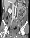
The results of the laboratory investigations were as follows: white blood cell count, 12,680/mm3; hemoglobin, 13.2 g/dL; hematocrit, 37.8%; and platelets, 208,000/mm3. Urine tested positive for the human chorionic gonadotropin (hCG) hormone, and serum hCG level was 16,313 mIU/mL. Transvaginal sonography revealed a 2.85×1.88 cm sized right adnexal mass containing small cystic lesions (Fig. 1). Computed tomography scan showed a 3.6×2.3 cm sized ovoid cystic lesion with peripheral hypervascularity in the right adnexa (Fig. 2). A large amount of high attenuation fluid was observed in the pelvic cavity. Differential diagnosis included a ruptured ectopic pregnancy versus ruptured ovarian cyst.
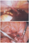
During laparoscopy, we observed a right-sided hyperemic pelvic complex containing a ruptured fallopian tube. There was active bleeding from the ruptured site and the hemoperitoneum (Fig. 3). The right fallopian tube was resected. On the second postoperative day, the hCG titer decreased to 4,255 mIU/mL. The patient was discharged home on the following day.
Discussion
Ectopic pregnancy after hysterectomy is quite rare, and this could be a reason for the delay in diagnosis when such a patient presents with abdominal pain. Since the first case reported in 1895, there have been 56 cases described in literature, but no such case has been reported in Korea till date.1,2
Post-hysterectomy ectopic pregnancies are divided into 2 subtypes-early and late-on the basis of the amount of time elapsed between the hysterectomy and the pregnancy.3 In "early" ectopic pregnancy, conception occurs immediately before hysterectomy, and the subsequent conceptus migrates through the fallopian tube at the time of hysterectomy, precluding the development of a normal intrauterine pregnancy. The time interval between hysterectomy and the symptoms for such an occurrence has been reported to range from 29 to 96 days.1 In contrast, pregnancies where a significant amount of time has elapsed between hysterectomy and fertilization are described as "late." The time interval for such cases has been reported to range from 7 months to 11 years.4 These pregnancies most likely result from fertilization enabled by a fistulous tract between the vagina and the peritoneal cavity or the fallopian tubes. Sperms have been reported to be recovered from the peritoneal cavity after coitus.5
As in our case, the majority of reported cases are early ectopic pregnancies that occurred due to inadequate preoperative contraception along with an inability to diagnose early pregnancy at the time of surgery.3
In our case, on pathologic examination after total hysterectomy, a secretory endometrium was found indicating that ovulation had occurred with no evidence of pregnancy. When a fertilized ovum is present, the endometrium responds with a decidual reaction. Since this reaction develops by the fifth postovulatory day, we postulate that fertilization must have occurred just before hysterectomy. When the uterus was removed and the fallopian tube was ligated, the fertilized ovum was sequestered.6
Cases of ectopic pregnancies after hysterectomy remain a diagnostic dilemma because of the absence of a history of vaginal bleeding or amenorrhea, along with a low index of suspicion.7 The classic triad of pelvic pain, amenorrhea, and an adnexal mass supports the diagnosis of an ectopic pregnancy. However, women presenting to the emergency department may not have all of these findings. The diagnosis of an ectopic pregnancy is often delayed because symptoms and signs such as protracted abdominal pain, pelvic hematoma formation, vaginal cuff infection, and vaginal bleeding are attributed to common immediate post-hysterectomy complications, until additional imaging or a repeat operation confirms it. Thus, a serum pregnancy test and pelvic ultrasonography should be performed to conform to a high index of clinical suspicion and prevent potentially fatal delays in patient management.2,8
Ectopic pregnancy after hysterectomy is a rare but potentially fatal complication. A delay in diagnosis and treatment may be catastrophic. Therefore, the diagnosis of ectopic pregnancy must not be excluded in any woman of reproductive age who presents with abdominal pain, vaginal bleeding, or a pelvic mass after hysterectomy.7,9 Definitive therapy consists of timely recognition and early laparotomy to minimize maternal morbidity and mortality.
The standard procedure during hysterectomy with conservation of ovaries has been the preservation of tubes with the clamps placed as close to the uterine corpus as possible. This is suggested to decrease interference with the vascular structures in the mesosalpinx and the mesovarium. However, it is not clear whether tubal conservation at the time of hysterectomy has any impact on ovarian blood flow, which has a dual blood supply both from the terminal ascending branch of the uterine arteries and the corresponding ovarian arteries.10 The complete removal of fallopian tubes during hysterectomy is only considered for the prevention of ectopic pregnancy after hysterectomy.


 PDF
PDF ePub
ePub Citation
 Print
Print


 XML Download