

 PDF
PDF ePub
ePub Citation
 Print
Print


Abstract
Objective
The aim of this study was to evaluate whether the Bishop score, length, volume and gray-scale histogram of the cervix has a predictive value of assessing the rate of success in trial of induction.
Methods
Forty-one nulliparous patients with its Bishop score six or less were enrolled for this prospective study. All were on prostaglandin E2 (PGE2, Propess®; Controlled Therapeutics Ltd) pessary. Three-dimensional transvaginal ultrasound scans of the cervix were performed on the ACCUVIX XQ (Medison) to measure length, volume, and gray-scale histogram. Bishop score was determined by digital examination. The successful induction was defined as the ability to achieve the active phase of labor corresponding to a cervical dilatation of ≥4 cm within 12 hours of removing the PGE2 pessary. The receiver operating characteristics (ROC) curves were also used to estimate an optimal cutoff point for the Bishop score, length, volume, and gray-scale histogram of the cervix. Logistic regression analysis was used for statistical analyses.
Results
The overall successful rate of labor induction was 73.2% (30/41). Multiple logistic regression analyses demonstrated that the value of anterior lip histogram was significantly associated with the successful labor induction. ROC curve for anterior lip histogram value in predicting success of induction indicated a significant relationship with successful induction. The best cutoff value was 69.4.
Figures and Tables
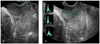 | Fig. 1Ultrasonographic assessment of the uterine cervix. (A) Cervix length, (B) Histogram (anterior lip).
|
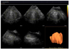 | Fig. 2Cervix volume by 3-D sonography using extended imaging virtual organ computer-aided analysis (XI VOCAL) technique.
|
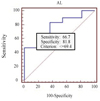 | Fig. 3Receiver operating characteristic curve for anterior lip histogram value in predicting success of induction. The cutoff value is 69.4 (Area under the curve 0.785; Standard error 0.0757; z=3.765; P<0.01). AL: anterior lip.
|


References
1. Andersen HF. Transvaginal and transabdominal ultrasonography of the uterine cervix during pregnancy. J Clin Ultrasound. 1991. 19:77–83.
2. Maitra N, Sharma D, Agarwal S. Transvaginal measurement of cervical length in the prediction of successful induction of labour. J Obstet Gynaecol. 2009. 29:388–391.
3. Verhoeven CJ, Oudenaarden A, Hermus MA, Porath MM, Oei SG, Mol BW. Validation of models that predict Cesarean section after induction of labor. Ultrasound Obstet Gynecol. 2009. 34:316–321.
4. Tan PC, Vallikkannu N, Suguna S, Quek KF, Hassan J. Transvaginal sonographic measurement of cervical length vs. Bishop score in labor induction at term: tolerability and prediction of Cesarean delivery. Ultrasound Obstet Gynecol. 2007. 29:568–573.
5. Bueno B, San-Frutos L, Perez-Medina T, Barbancho C, Troyano J, Bajo J. The labor induction: integrated clinical and sonographic variables that predict the outcome. J Perinatol. 2007. 27:4–8.
6. Severi FM, Bocchi C, Florio P, Picciolini E, D'Aniello G, Petraglia F. Comparison of two-dimensional and three-dimensional ultrasound in the assessment of the cervix to predict preterm delivery. Ultrasound Med Biol. 2003. 29:1261–1265.
7. Rovas L, Sladkevicius P, Strobel E, Valentin L. Three-dimensional power Doppler ultrasound assessment of the cervix for the prediction of successful induction of labor with prostaglandin in prolonged pregnancy. J Ultrasound Med. 2005. 24:933–939.
8. Maeda K, Utsu M, Yamamoto N, Serizawa M. Echogenicity of fetal lung and liver quantified by the grey-level histogram width. Ultrasound Med Biol. 1999. 25:201–208.
9. Maeda K, Utsu M, Kihaile PE. Quantification of sonographic echogenicity with grey-level histogram width: a clinical tissue characterization. Ultrasound Med Biol. 1998. 24:225–234.
10. Tekesin I, Hellmeyer L, Heller G, Romer A, Kuhnert M, Schmidt S. Evaluation of quantitative ultrasound tissue characterization of the cervix and cervical length in the prediction of premature delivery for patients with spontaneous preterm labor. Am J Obstet Gynecol. 2003. 189:532–539.
11. Chandra S, Crane JM, Hutchens D, Young DC. Transvaginal ultrasound and digital examination in predicting successful labor induction. Obstet Gynecol. 2001. 98:2–6.
12. Rozenberg P, Chevret S, Chastang C, Ville Y. Comparison of digital and ultrasonographic examination of the cervix in predicting time interval from induction to delivery in women with a low Bishop score. BJOG. 2005. 112:192–196.
13. Park KH. Transvaginal ultrasonographic cervical measurement in predicting failed labor induction and cesarean delivery for failure to progress in nulliparous women. J Korean Med Sci. 2007. 22:722–727.
14. Pandis GK, Papageorghiou AT, Ramanathan VG, Thompson MO, Nicolaides KH. Preinduction sonographic measurement of cervical length in the prediction of successful induction of labor. Ultrasound Obstet Gynecol. 2001. 18:623–628.
15. Ware V, Raynor BD. Transvaginal ultrasonographic cervical measurement as a predictor of successful labor induction. Am J Obstet Gynecol. 2000. 182:1030–1032.
16. Gabriel R, Darnaud T, Chalot F, Gonzalez N, Leymarie F, Quereux C. Transvaginal sonography of the uterine cervix prior to labor induction. Ultrasound Obstet Gynecol. 2002. 19:254–257.
17. Berghella V, Tolosa JE, Kuhlman K, Weiner S, Bolognese RJ, Wapner RJ. Cervical ultrasonography compared with manual examination as a predictor of preterm delivery. Am J Obstet Gynecol. 1997. 177:723–730.
18. Guzman ER, Walters C, Ananth CV, O'Reilly-Green C, Benito CW, Palermo A, et al. A comparison of sonographic cervical parameters in predicting spontaneous preterm birth in high-risk singleton gestations. Ultrasound Obstet Gynecol. 2001. 18:204–210.
19. Heath VC, Southall TR, Souka AP, Novakov A, Nicolaides KH. Cervical length at 23 weeks of gestation: relation to demographic characteristics and previous obstetric history. Ultrasound Obstet Gynecol. 1998. 12:304–311.
20. Iams JD, Goldenberg RL, Mercer BM, Moawad A, Thom E, Meis PJ, et al. The Preterm Prediction Study: recurrence risk of spontaneous preterm birth. National Institute of Child Health and Human Development Maternal-Fetal Medicine Units Network. Am J Obstet Gynecol. 1998. 178:1035–1040.
21. Yu SY, Tozzi CA, Babiarz J, Leppert PC. Collagen changes in rat cervix in pregnancy--polarized light microscopic and electron microscopic studies. Proc Soc Exp Biol Med. 1995. 209:360–368.
22. Leppert PC, Yu SY. Apoptosis in the cervix of pregnant rats in association with cervical softening. Gynecol Obstet Invest. 1994. 37:150–154.
23. Aspden RM. Collagen organisation in the cervix and its relation to mechanical function. Coll Relat Res. 1988. 8:103–112.
 XML Download