

 PDF
PDF ePub
ePub Citation
 Print
Print


Abstract
Cervical cancer of gynecologic cancer is considered a preventable disease because it has a long preventive state, cervical cytology screening programs are currently available, and treatment of precancerous lesions is effective. Proper management of precancerous disease is important because improper management of precancerous disease can increase risk of invasive cancer on the one hand and can result in complications from overtreatment on the other. The decision as to which therapeutic option to use in an individual patient depends on considerations such as patient age, parity, desire for future childbearing, preferences, prior cytology and treatment history, and history of default from follow-up, operator experience, and nonvisualization of the transformation zone.
Figures and Tables
Figure 1
Management of women with a histological diagnosis of cervical intraepithelial neoplasia grade 1 (CIN 1) preceded by atypical squamous cells (ASC)-US, ASC-H, low-grade squamous intraepithelial lesion (LSIL) cytology.
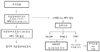
Figure 2
Management of women with a histological diagnosis of cervical intraepithelial neoplasia grade 1 (CIN 1) preceded by high-grade squamous intraepithelial lesion (HSIL) or atypical glandular cells not otherwise specified (AGC-NOS) cytology.
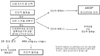
Figure 3
Management of adolescent women (20 years and younger) with a histological diagnosis of cervical intraepithelial neoplasia grade (CIN) 1.
HSIL: high-grade squamous intraepithelial lesion, ASC: atypical squamous cells.
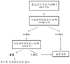
Figure 4
Management of women with a histological diagnosis of cervical intraepithelial neoplasia grade (CIN) 2, 3.
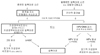
Figure 5
Management of adolescent and young women with a histological diagnosis of cervical intraepithelial neoplasia grade (CIN) 2, 3.
References
1. Cancer incidence in five continents. Volume VIII. IARC Sci Publ. 2002. (155):1–781.
2. Global cancer facts and figures 2007: estimated number of new cancer cases by world area. American Cancer Society. 2007. cited 2008 Jan 26. Atlanta (GA): American Cancer Society;Available from: URL:
http://www.cancer.org/downloads/STT/Global_Facts_and_Figures_2007_rev2.pdf.
5. Melnikow J, Nuovo J, Willan AR, Chan BK, Howell LP. Natural history of cervical squamous intraepithelial lesions: a meta-analysis. Obstet Gynecol. 1998. 92:727–735.
6. Ostor AG. Natural history of cervical intraepithelial neoplasia: a critical review. Int J Gynecol Pathol. 1993. 12:186–192.
7. Mitchell MF, Tortolero-Luna G, Wright T, Sarkar A, Richards-Kortum R, Hong WK, et al. Cervical human papillomavirus infection and intraepithelial neoplasia: a review. J Natl Cancer Inst Monogr. 1996. (21):17–25.
8. Wright TC Jr, Massad LS, Dunton CJ, Spitzer M, Wilkinson EJ, Solomon D. 2006 American Society for Colposcopy and Cervical Pathology-sponsored Consensus Conference. 2006 consensus guidelines for the management of women with cervical intraepithelial neoplasia or adenocarcinoma in situ. J Low Genit Tract Dis. 2007. 11:223–239.
9. Addis IB, Hatch KD, Berek JS. Berek JS, editor. Intraepithelial disease of the cervix, vagina, and vulva. Berek & Novak's gynecology. 2007. 14th ed. Philadelphia: Lippincott Williams & Wilkins;561–600.
10. El-Bastawissi AY, Becker TM, Daling JR. Effect of cervical carcinoma in situ and its management on pregnancy outcome. Obstet Gynecol. 1999. 93:207–212.

11. Kyrgiou M, Tsoumpou I, Vrekoussis T, Martin-Hirsch P, Arbyn M, Prendiville W, et al. The up-to-date evidence on colposcopy practice and treatment of cervical intraepithelial neoplasia: the Cochrane colposcopy & cervical cytopathology collaborative group (C5 group) approach. Cancer Treat Rev. 2006. 32:516–523.
12. Martin-Hirsch PL, Paraskevaidis E, Kitchener H. Surgery for cervical intraepithelial neoplasia. Cochrane Database Syst Rev. 2000. (2):CD001318.
13. Nuovo J, Melnikow J, Willan AR, Chan BK. Treatment outcomes for squamous intraepithelial lesions. Int J Gynaecol Obstet. 2000. 68:25–33.
14. Kalliala I, Nieminen P, Dyba T, Pukkala E, Anttila A. Cancer free survival after CIN treatment: comparisons of treatment methods and histology. Gynecol Oncol. 2007. 105:228–233.
15. Soutter WP, Sasieni P, Panoskaltsis T. Long-term risk of invasive cervical cancer after treatment of squamous cervical intraepithelial neoplasia. Int J Cancer. 2006. 118:2048–2055.
16. Kyrgiou M, Koliopoulos G, Martin-Hirsch P, Arbyn M, Prendiville W, Paraskevaidis E. Obstetric outcomes after conservative treatment for intraepithelial or early invasive cervical lesions: systematic review and meta-analysis. Lancet. 2006. 367:489–498.
17. Samson SL, Bentley JR, Fahey TJ, McKay DJ, Gill GH. The effect of loop electrosurgical excision procedure on future pregnancy outcome. Obstet Gynecol. 2005. 105:325–332.
18. Sadler L, Saftlas A, Wang W, Exeter M, Whittaker J, McCowan L. Treatment for cervical intraepithelial neoplasia and risk of preterm delivery. JAMA. 2004. 291:2100–2106.
19. Bruinsma F, Lumley J, Tan J, Quinn M. Precancerous changes in the cervix and risk of subsequent preterm birth. BJOG. 2007. 114:70–80.
20. Jakobsson M, Gissler M, Sainio S, Paavonen J, Tapper AM. Preterm delivery after surgical treatment for cervical intraepithelial neoplasia. Obstet Gynecol. 2007. 109:309–313.
21. Bell MC, Alvarez RD. Chemoprevention and vaccines: a review of the nonsurgical options for the treatment of cervical dysplasia. Int J Gynecol Cancer. 2005. 15:4–12.
22. Stern PL. Immune control of human papillomavirus (HPV) associated anogenital disease and potential for vaccination. J Clin Virol. 2005. 32:Suppl 1. S72–S81.
23. Persad VL, Pierotic MA, Guijon FB. Management of cervical neoplasia: a 13-year experience with cryotherapy and laser. J Low Genit Tract Dis. 2001. 5:199–203.
24. Ueda M, Ueki K, Kanemura M, Izuma S, Yamaguchi H, Nishiyama K, et al. Diagnostic and therapeutic laser conization for cervical intraepithelial neoplasia. Gynecol Oncol. 2006. 101:143–146.
25. van Hamont D, van Ham MA, Struik-van der Zanden PH, Keijser KG, Bulten J, Melchers WJ, et al. Long-term follow-up after large-loop excision of the transformation zone: evaluation of 22 years treatment of high-grade cervical intraepithelial neoplasia. Int J Gynecol Cancer. 2006. 16:615–619.
26. Wang SS, Sherman ME, Hildesheim A, Lacey JV Jr, Devesa S. Cervical adenocarcinoma and squamous cell carcinoma incidence trends among white women and black women in the United States for 1976-2000. Cancer. 2004. 100:1035–1044.
27. Paraskevaidis E, Arbyn M, Sotiriadis A, Diakomanolis E, Martin-Hirsch P, Koliopoulos G, et al. The role of HPV DNA testing in the follow-up period after treatment for CIN: a systematic review of the literature. Cancer Treat Rev. 2004. 30:205–211.
28. Zielinski GD, Bais AG, Helmerhorst TJ, Verheijen RH, de Schipper FA, Snijders PJ, et al. HPV testing and monitoring of women after treatment of CIN 3: review of the literature and meta-analysis. Obstet Gynecol Surv. 2004. 59:543–553.
29. Insinga RP, Glass AG, Rush BB. Diagnoses and outcomes in cervical cancer screening: a population-based study. Am J Obstet Gynecol. 2004. 191:105–113.
30. SEER cancer statistics review 1975-2006. National Cancer Institue. 2009. cited 2009 May 4. Atlanta (GA): National Cancer Institue;Available from: URL:
http://seer.cancer.gov/csr/1975_2006/results_merged/sect_05_cervix_uteri.pdf.
31. Moscicki AB, Shiboski S, Hills NK, Powell KJ, Jay N, Hanson EN, et al. Regression of low-grade squamous intra-epithelial lesions in young women. Lancet. 2004. 364:1678–1683.
32. Economos K, Perez Veridiano N, Delke I, Collado ML, Tancer ML. Abnormal cervical cytology in pregnancy: a 17-year experience. Obstet Gynecol. 1993. 81:915–918.
33. Yost NP, Santoso JT, McIntire DD, Iliya FA. Postpartum regression rates of antepartum cervical intraepithelial neoplasia II and III lesions. Obstet Gynecol. 1999. 93:359–362.
34. Robinson WR, Webb S, Tirpack J, Degefu S, O'Quinn AG. Management of cervical intraepithelial neoplasia during pregnancy with LOOP excision. Gynecol Oncol. 1997. 64:153–155.
35. Connor JP. Noninvasive cervical cancer complicating pregnancy. Obstet Gynecol Clin North Am. 1998. 25:331–342.
36. Paraskevaidis E, Koliopoulos G, Kalantaridou S, Pappa L, Navrozoglou I, Zikopoulos K, et al. Management and evolution of cervical intraepithelial neoplasia during pregnancy and postpartum. Eur J Obstet Gynecol Reprod Biol. 2002. 104:67–69.
37. Wright TC Jr, Cox JT, Massad LS, Carlson J, Twiggs LB, Wilkinson EJ. American Society for Colposcopy and Cervical Pathology. 2001 consensus guidelines for the management of women with cervical intraepithelial neoplasia. Am J Obstet Gynecol. 2003. 189:295–304.
38. Stoler MH, Schiffman M. Atypical Squamous Cells of Undetermined Significance-Low-grade Squamous Intraepithelial Lesion Triage Study (ALTS) Group. Interobserver reproducibility of cervical cytologic and histologic interpretations: realistic estimates from the ASCUS-LSIL Triage Study. JAMA. 2001. 285:1500–1505.
39. Clifford GM, Rana RK, Franceschi S, Smith JS, Gough G, Pimenta JM. Human papillomavirus genotype distribution in low-grade cervical lesions: comparison by geographic region and with cervical cancer. Cancer Epidemiol Biomarkers Prev. 2005. 14:1157–1164.
40. Schlecht NF, Platt RW, Duarte-Franco E, Costa MC, Sobrinho JP, Prado JC, et al. Human papillomavirus infection and time to progression and regression of cervical intraepithelial neoplasia. J Natl Cancer Inst. 2003. 95:1336–1343.
41. Nobbenhuis MA, Helmerhorst TJ, van den Brule AJ, Rozendaal L, Voorhorst FJ, Bezemer PD, et al. Cytological regression and clearance of high-risk human papillomavirus in women with an abnormal cervical smear. Lancet. 2001. 358:1782–1783.
42. Numnum TM, Kirby TO, Leath CA 3rd, Huh WK, Alvarez RD, Straughn JM Jr. A prospective evaluation of "see and treat" in women with HSIL Pap smear results: is this an appropriate strategy? J Low Genit Tract Dis. 2005. 9:2–6.
43. Dunn TS, Burke M, Shwayder J. A "see and treat" management for high-grade squamous intraepithelial lesion pap smears. J Low Genit Tract Dis. 2003. 7:104–106.
44. Massad LS, Collins YC, Meyer PM. Biopsy correlates of abnormal cervical cytology classified using the Bethesda system. Gynecol Oncol. 2001. 82:516–522.
45. Robertson AJ, Anderson JM, Beck JS, Burnett RA, Howatson SR, Lee FD, et al. Observer variability in histopathological reporting of cervical biopsy specimens. J Clin Pathol. 1989. 42:231–238.
46. Castle PE, Stoler MH, Solomon D, Schiffman M. The relationship of community biopsy-diagnosed cervical intraepithelial neoplasia grade 2 to the quality control pathology-reviewed diagnoses: an ALTS report. Am J Clin Pathol. 2007. 127:805–815.
47. Wright TC Jr. CHAPTER 3 Pathology of HPV infection at the cytologic and histologic levels: basis for a 2-tiered morphologic classification system. Int J Gynaecol Obstet. 2006. 94:Suppl 1. S22–S31.
48. Park TW, Richart RM, Sun XW, Wright TC Jr. Association between human papillomavirus type and clonal status of cervical squamous intraepithelial lesions. J Natl Cancer Inst. 1996. 88:355–358.
49. Wright TC Jr, Kurman RJ, Ferenczy AF. Kurman RJ, editor. Precancerous lesions of the cervix. Blaustein's pathology of the pemale genital tract. 2002. 5th ed. New York: Springer-Verlag;253–324.
50. Smith JS, Lindsay L, Hoots B, Keys J, Franceschi S, Winer R, et al. Human papillomavirus type distribution in invasive cervical cancer and high-grade cervical lesions: a meta-analysis update. Int J Cancer. 2007. 121:621–632.
51. Fine BA, Feinstein GI, Sabella V. The pre- and postoperative value of endocervical curettage in the detection of cervical intraepithelial neoplasia and invasive cervical cancer. Gynecol Oncol. 1998. 71:46–49.
52. Duggan BD, Felix JC, Muderspach LI, Gebhardt JA, Groshen S, Morrow CP, et al. Cold-knife conization versus conization by the loop electrosurgical excision procedure: a randomized, prospective study. Am J Obstet Gynecol. 1999. 180:276–282.
53. Vedel P, Jakobsen H, Kryger-Baggesen N, Rank F, Bostofte E. Five-year follow up of patients with cervical intra-epithelial neoplasia in the cone margins after conization. Eur J Obstet Gynecol Reprod Biol. 1993. 50:71–76.
54. Gardeil F, Barry-Walsh C, Prendiville W, Clinch J, Turner MJ. Persistent intraepithelial neoplasia after excision for cervical intraepithelial neoplasia grade III. Obstet Gynecol. 1997. 89:419–422.
55. Zaitoun AM, McKee G, Coppen MJ, Thomas SM, Wilson PO. Completeness of excision and follow up cytology in patients treated with loop excision biopsy. J Clin Pathol. 2000. 53:191–196.
56. Felix JC, Muderspach LI, Duggan BD, Roman LD. The significance of positive margins in loop electrosurgical cone biopsies. Obstet Gynecol. 1994. 84:996–1000.
57. Mohamed-Noor K, Quinn MA, Tan J. Outcomes after cervical cold knife conization with complete and incomplete excision of abnormal epithelium: a review of 699 cases. Gynecol Oncol. 1997. 67:34–38.
58. Paraskevaidis E, Kalantaridou SN, Paschopoulos M, Zikopoulos K, Diakomanolis E, Dalkalitsis N, et al. Factors affecting outcome after incomplete excision of cervical intraepithelial neoplasia. Eur J Gynaecol Oncol. 2003. 24:541–543.
59. Ørbo A, Arnesen T, Arnes M, Straume B. Resection margins in conization as prognostic marker for relapse in high-grade dysplasia of the uterine cervix in northern Norway: a retrospective long-term follow-up material. Gynecol Oncol. 2004. 93:479–483.
60. Reich O, Lahousen M, Pickel H, Tamussino K, Winter R. Cervical intraepithelial neoplasia III: long-term follow-up after cold-knife conization with involved margins. Obstet Gynecol. 2002. 99:193–196.
61. Lapaquette TK, Dinh TV, Hannigan EV, Doherty MG, Yandell RB, Buchanan VS. Management of patients with positive margins after cervical conization. Obstet Gynecol. 1993. 82:440–443.
62. Murdoch JB, Morgan PR, Lopes A, Monaghan JM. Histological incomplete excision of CIN after large loop excision of the transformation zone (LLETZ) merits careful follow up, not retreatment. Br J Obstet Gynaecol. 1992. 99:990–993.
63. Andersen ES, Nielsen K. Adenocarcinoma in situ of the cervix: a prospective study of conization as definitive treatment. Gynecol Oncol. 2002. 86:365–369.
64. Kennedy AW, Biscotti CV. Further study of the management of cervical adenocarcinoma in situ. Gynecol Oncol. 2002. 86:361–364.
65. Krivak TC, Rose GS, McBroom JW, Carlson JW, Winter WE 3rd, Kost ER. Cervical adenocarcinoma in situ: a systematic review of therapeutic options and predictors of persistent or recurrent disease. Obstet Gynecol Surv. 2001. 56:567–575.
66. Soutter WP, Haidopoulos D, Gornall RJ, McIndoe GA, Fox J, Mason WP, et al. Is conservative treatment for adenocarcinoma in situ of the cervix safe? BJOG. 2001. 108:1184–1189.
67. Azodi M, Chambers SK, Rutherford TJ, Kohorn EI, Schwartz PE, Chambers JT. Adenocarcinoma in situ of the cervix: management and outcome. Gynecol Oncol. 1999. 73:348–353.
68. Lea JS, Shin CH, Sheets EE, Coleman RL, Gehrig PA, Duska LR, et al. Endocervical curettage at conization to predict residual cervical adenocarcinoma in situ. Gynecol Oncol. 2002. 87:129–132.
69. Hwang DM, Lickrish GM, Chapman W, Colgan TJ. Long-term surveillance is required for all women treated for cervical adenocarcinoma in situ. J Low Genit Tract Dis. 2004. 8:125–131.
70. Shin CH, Schorge JO, Lee KR, Sheets EE. Conservative management of adenocarcinoma in situ of the cervix. Gynecol Oncol. 2000. 79:6–10.
71. McHale MT, Le TD, Burger RA, Gu M, Rutgers JL, Monk BJ. Fertility sparing treatment for in situ and early invasive adenocarcinoma of the cervix. Obstet Gynecol. 2001. 98:726–731.
72. Kennedy CM, Boardman LA. New approaches to external genital warts and vulvar intraepithelial neoplasia. Clin Obstet Gynecol. 2008. 51:518–526.
73. Howe HL, Wingo PA, Thun MJ, Ries LA, Rosenberg HM, Feigal EG, et al. Annual report to the nation on the status of cancer (1973 through 1998), featuring cancers with recent increasing trends. J Natl Cancer Inst. 2001. 93:824–842.
74. Holschneider CH, Berek JS. Berek JS, editor. Vulvar cancer. Berek & Novak's gynecology. 2007. 14th ed. Philadelphia: Lippincott Williams & Wilkins;1549–1580.
75. van Seters M, van Beurden M, de Craen AJ. Is the assumed natural history of vulvar intraepithelial neoplasia III based on enough evidence? A systematic review of 3322 published patients. Gynecol Oncol. 2005. 97:645–651.
76. Todd RW, Luesley DM. Medical management of vulvar intraepithelial neoplasia. J Low Genit Tract Dis. 2005. 9:206–212.
77. Wolcott HD, Gallup DG. Wide local excision in the treatment of vulvar carcinoma in situ: a reappraisal. Am J Obstet Gynecol. 1984. 150:695–698.
78. Modesitt SC, Waters AB, Walton L, Fowler WC Jr, Van Le L. Vulvar intraepithelial neoplasia III: occult cancer and the impact of margin status on recurrence. Obstet Gynecol. 1998. 92:962–966.
79. Stanley M. Chapter 17: Genital human papillomavirus infections--current and prospective therapies. J Natl Cancer Inst Monogr. 2003. 31:117–124.
80. Centers for Disease Control and Prevention. Workowski KA, Berman SM. Sexually transmitted diseases treatment guidelines, 2006. MMWR Recomm Rep. 2006. 55:1–94.
81. Edwards L, Ferenczy A, Eron L, Baker D, Owens ML, Fox TL, et al. HPV Study Group. Self-administered topical 5% imiquimod cream for external anogenital warts. Human PapillomaVirus. Arch Dermatol. 1998. 134:25–30.
82. Megyeri K, Au WC, Rosztoczy I, Raj NB, Miller RL, Tomai MA, et al. Stimulation of interferon and cytokine gene expression by imiquimod and stimulation by Sendai virus utilize similar signal transduction pathways. Mol Cell Biol. 1995. 15:2207–2218.
83. Mayeaux EJ Jr, Dunton C. Modern management of external genital warts. J Low Genit Tract Dis. 2008. 12:185–192.
84. van Seters M, van Beurden M, ten Kate FJ, Beckmann I, Ewing PC, Eijkemans MJ, et al. Treatment of vulvar intraepithelial neoplasia with topical imiquimod. N Engl J Med. 2008. 358:1465–1473.
85. Smith JS, Backes DM, Hoots BE, Kurman RJ, Pimenta JM. Human papillomavirus type: distribution in vulvar and vaginal cancers and their associated precursors. Obstet Gynecol. 2009. 113:917–924.
86. Robinson JB, Sun CC, Bodurka-Bevers D, Im DD, Rosenshein NB. Cavitational ultrasonic surgical aspiration for the treatment of vaginal intraepithelial neoplasia. Gynecol Oncol. 2000. 78:235–241.
87. Hoffman MS, Roberts WS, LaPolla JP, Fiorica JV, Cavanagh D. Laser vaporization of grade 3 vaginal intraepithelial neoplasia. Am J Obstet Gynecol. 1991. 165:1342–1344.
88. Kim HS, Park NH, Park IA, Park JH, Chung HH, Kim JW, et al. Risk factors for recurrence of vaginal intraepithelial neoplasia in the vaginal vault after laser vaporization. Lasers Surg Med. 2009. 41:196–202.
 XML Download