

 PDF
PDF ePub
ePub Citation
 Print
Print


Abstract
Sclerosing stromal tumor (SST) of the ovary is a rare, benign tumor. The most common clinical symptom is menstrual irregularity. Diagnosis of SST is often made by postoperative pathologic examination. The important differential diagnoses are other sex cord stromal tumors including fibroma, thecoma and etc. We present four cases of SST of the ovary during 10 years with a brief review of the literature.
Figures and Tables
Fig. 1
(A) Abdomen X-ray shows a 3.6×2.4 cm calcified mass. (B) Contrast-enhanced computed tomography scans shows a 12.6×7.4×13.3 cm mass with peripheral enhancement.
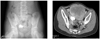
Fig. 2
(A) Magnetic resonance imaging on dynamic contrast enhanced image, the tumor reveal early peripheral enhancement with centripetal progression. (B) T2-weighted image shows low-intensity nodules set against high-intensity stroma.
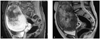
Fig. 3
This photograph is cut surface of the both ovarian masses of case 1. (A) The right ovary, measuring 13.5 cm in diameter, is totally replaced by whitish gray solid firm mass. (B) The wedge resected left ovary shows a cystic follicle in the upper portion and a 0.5 cm sized whitish firm mass in the lower portion.
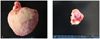
Fig. 4
This photograph is microscopic findings. (A) The right tumor of case 1 is composed of spindle cells and polyhedral cells with numerous vasculature (H&E stain, ×200). (B) The left tumor of case 1 consists of fibrotic stroma and occasional calcification (upper) alternating with the cellular area (H&E stain, ×100). (C) Immunohistochemistry of case 1 exhibit positive staining for smooth muscle actin in the perivascular polygonal cells (Avidin biotin complex for actin, ×200). (D) Low power view of case 3 demonstrates typical pseudolobular pattern of cellular area and loose edematous stromal portion (H&E stain, ×40).
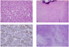
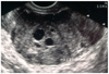
Fig. 5
Ultrasonography shows a 7.1×5.9 cm sized, mixed echogenic mass with multiple focal hypoechoic area.
References
1. Youm HS, Cha DS, Han KH, Park EY, Hyon NN, Chong Y. A case of huge sclerosing stromal tumor of the ovary weighing 10 kg in a 71-year-old postmenopausal woman. J Gynecol Oncol. 2008. 19:270–274.
2. Chalvardjian A, Scully RE. Sclerosing stromal tumors of the ovary. Cancer. 1973. 31:664–670.
3. Saitoh A, Tsutsumi Y, Osamura RY, Watanabe K. Sclerosing stromal tumor of the ovary. Immunohistochemical and electron-microscopic demonstration of smooth-muscle differentiation. Arch Pathol Lab Med. 1989. 113:372–376.
4. Kuscu E, Oktem M, Karahan H, Bilezikci B, Demirhan B. Sclerosing stromal tumor of the ovary: a case report. Eur J Gynaecol Oncol. 2003. 24:442–444.
5. Prat J. Pathology of the ovary. 2004. Philadelphia: Saunders.
6. Korczyinski J, Gottwald L, Pasz-Walczak G, Kubiak R, Bienkiewicz A. [Sclerosing stromal tumor of the ovary in a 30-year-old woman. A case report and review of the literature]. Ginekol Pol. 2005. 76:471–475.
7. Ihara N, Togashi K, Todo G, Nakai A, Kojima N, Ishigaki T, et al. Sclerosing stromal tumor of the ovary: MRI. J Comput Assist Tomogr. 1999. 23:555–557.
8. Lee MS, Cho HC, Lee YH, Hong SR. Ovarian sclerosing stromal tumors: gray scale and color Doppler sonographic findings. J Ultrasound Med. 2001. 20:413–417.
9. Yoon GS, Kim MS. A case of sclerosing stromal tumor of the ovary. Korean J Obstet Gynecol. 2003. 46:2052–2055.
10. Gee DC, Russell P. Sclerosing stromal tumours of the ovary. Histopathology. 1979. 3:367–376.
11. Fox H, Wells M. Haines and Taylor Obstetrical and Gynecological Pathology. 2002. 5th ed. New York: Cherchill Livingstone.
12. Terauchi F, Onodera T, Nagashima T, Kobayashi Y, Moritake T, Oharaseki T, et al. Sclerosing stromal tumor of the ovary with elevated CA125. J Obstet Gynaecol Res. 2005. 31:432–435.
13. Lee HC, Kim BR, Lee JS, Lim BI, Kim HG, Moon HB, et al. A case of ovarian sclerosing stromal tumor with massive ascites and elevated CA 125. Korean J Obstet Gynecol. 2009. 52:120–124.
 XML Download