

 PDF
PDF ePub
ePub Citation
 Print
Print


INTRODUCTION
Hardly any modern study nowadays does not claim that prostate cancer should be classified on the basis of molecular or genetic features. It could almost be said that progress in all areas of prostate cancer research is impossible until we determine the basic principles of this classification. Indeed, the number of fundamental research programs in prostate cancer molecular biology and genetics is overwhelming. It is obvious that the time has come for the translation of these data to the clinic. However, prostate cancer is characterized by prominent genetic heterogeneity, which could be a very difficult barrier to overcome [1]. How far we are now from the valuable clinical translation of the results of molecular genetic studies is the theme of this review.
GENE EXPRESSION STUDIES AND GENETIC SIGNATURES
Gene expression analysis (mRNA analysis) is an attractive method of tumor tissue analysis (for example, after biopsy or during the final pathology examination). Current technologies (DNA microarrays, quantitative polymerase chain reaction, RNAseq) provide the possibility for streamed analysis of thousands genes in a relatively small volume of extracted tissue. Key issues for gene expression analysis are the quality of the analyzed material (better with fresh-frozen tissues), the amount of tumor tissue in the probe (a certain amount of stromal tissue contaminates the material), and the necessity to use reference genes (as a rule, housekeeping genes, which are supposed to be expressed equally in all tissues) and normalization to account for differences in the RNA quantity in the probes. Recent studies have shown that formalin-fixed, paraffin-embedded (FFPE) tissues can also be used for analysis despite fixation-related RNA degradation if special extraction and preparation techniques are used [2]. In view of the large amount of generated data, certain demanding bioinformatics approaches are necessary for the analysis and to compensate for possible errors.
Gene expression studies in the area of prostate cancer have been carried out for more than 10 years. The principle idea of all these studies is to develop a gene expression signature, which could be useful for characterization of the tumor (mainly the aggressive, metastatic, lethal, or latent nature of the tumor) and for prognosis of outcomes or sensitivity to certain therapeutic modalities [3]. Different groups of scientists have undertaken efforts to extract these data from the analysis of gene expression [4,5,6,7,8,9,10,11,12,13,14]. Some signatures were developed in cooperation with genetic companies, some almost to the stage of being ready for clinical application [15,16,17,18,19,20,21]. Some of the signatures also used noncoding regions in the genome and not only protein-coding mRNAs with clear cellular functions [17,18].
Nevertheless, all these signatures have some common problems that hamper their rapid integration into clinical practice. First, they do not account for intra- and interfocal heterogeneity, because the sampling for investigation included only the highest grade tumors. Second, some interpretation errors are possible owing to the use of FFPE archive tissues (the quality of which is lower than that of fresh or fresh-frozen tissues, which are hard and expensive to handle). Third, the use of these genetic expression signatures with biopsy tissues is difficult owing to the undersampling issue and therefore entails possible bias due to characterization of less aggressive tumors. Fourth, the performance of these signatures is only slightly better than that of clinical variables and it is hard to estimate whether the prospective translation of these assays and their implementation for certain clinical cases will preserve this advantage. Fifth, the signatures were not compared to other important contemporary diagnostic modalities, e.g., multiparametric magnetic resonance tomography or some other biomarker [22] to see whether they will maintain their value.
Moreover, the method (gene expression analysis) itself is questionable for the purposes of high-resolution molecular characterization of prostate tumors. In the modern era, more than 60 studies of gene expression in prostate carcinoma have been published (e.g., in Oncomine Database, a publicly available database of stream gene expression data for various types of cancers). In almost all of these studies, thousands of genes were analyzed in a streamed fashion with regard to their mRNA expression. Two important conclusions stem from there. It was often found that the genes with altered expression in these studies did not correspond with the genes in other studies [6,10,11,12,16,17,21]. Second, the prognostic value in terms of clinical risks was never overwhelmingly high but rather low, with the primary endpoint of prostate-specific antigen recurrence not being a good surrogate of other prostate cancer outcomes [16,17,18,19,20,21]. The question arises of whether the expression of multiple genes is adequate for the aforementioned purposes. It seems that the gene expression analysis detects only virtually randomly mediated transcriptional reactions in the tissues, affecting thousands of genes, which are compensatory owing to changes in the cells and microenvironment but not a direct consequence of tumor growth. Behind this prominent transcriptional reaction lie some limited genetic oncogenic changes, which cannot be seen now because of this genetic chaos.
Nevertheless, selected signatures tested in real clinical practice showed some promising results in a postsurgical setting in patients with high risk of recurrence, which should be further prospectively evaluated [23]. However, the problem of valuable genetic characterization of a tumor at the time of biopsy (first minimally invasive contact with the tumor) is to date not solved.
GENETIC CHARACTERIZATION AND CLASSIFICATION OF PROSTATE TUMORS
A key finding in prostate tumor biology was the identification of the recurrent gene fusion (TMPRSS2: ERG, short abbreviation T2:ERG) in prostate cancer tumors [24]. This fusion is present in approximately 50% of all prostate adenocarcinomas [25] and is considered to be the early initial rearrangement, being also present in precancerous lesions (high-grade prostatic intraepithelial neoplasia) [26,27] and in a small percentage of benign prostatic hyperplasia tissues [28]. Several types of structural rearrangements leading to the T2:ERG fusion are known [29]. The principal result of the ERG and other ETS fusions is attachment of the coding region of powerful transcriptional factors from the ETS family (including ERG) to the strong promoter of the TMPRSS2 gene. This leads to the overexpression of ERG or other ETS genes, given that the TMPRSS2 gene is an androgen-regulated gene that is normally highly expressed in prostate tissue [30]. Other fusion partners from the ETS family of genes have also been identified, with ETV1, ETV4, ELK4, and ETV5 being most common [29]. Other genes with strong promoters may be also involved in fusion formation [29,31,32]. T2:ERG is the most common type of ETS-family-related fusion in prostate cancer, accounting for approximately 85% of all fusions [25,31,33].
The role of the aforementioned fusions for oncogenesis is currently understudied. Importantly, some functional studies show that overexpression of ERG as a single rearrangement is not enough for tumor formation, despite being obviously oncogenous, which suggests that additional mutations or rearrangements are necessary [34,35,36]. The results of attempts to link T2:ERG fusion to cancer aggressiveness and clinical outcomes have to date been unremarkable [37,38,39,40], especially in a postdefinitive therapy setting (for a review of studies, see reference [29]). This could, on one hand, be related to the retrospective nature of the performed studies. On the other hand, the multifocality and inter- and intratumoral heterogeneity of the tumors were not accounted for, which are issues that seem to be extremely important for prostate cancer.
From a logical point of view, T2:ERG fusion is a relatively early event and gives rise to the two big molecular branches of prostate cancer. Correspondingly, the T2:ERG fusion represents two ways of cancer evolution and should provide some differences in tumor phenotype at late stages. The resulting late genetic aberrations, which are important for tumor formation, in these ETS+ and ETS- tumors are common. It could also be that there are many unique sprouting pathways in both tumor groups, which could lead to both aggressive and unaggressive entities independent of ETS status. The evidence for ERG-dependent aberrations is emerging (see below), but to date there are not enough data to make any final conclusions. However, given the high prevalence of these fusions, they could already be used as potent diagnostic biomarkers alone or together with other assays [41].
With the introduction of next-generation sequencing technologies into prostate cancer research, it became possible to gain deep insight into tumor genetics. On the one hand, it became possible to obtain important information in recurrently mutated genes and to classify the tumors by some of these genes. On the other hand, the mechanisms underlying the genetic rearrangements in prostate cancer were elucidated.
Some investigations show that the rate of somatic mutations in prostate tumors is very low, with many gene dysfunctions being a result of gene rearrangements [42,43,44]. The prominent article by Baca et al. [42] indicates that these complex genetic rearrangements are caused by the phenomenon called "chromoplexy," which entails multiple breaks of DNA chains with newly arising interconnections and copy-number variations owing to inadequate reparation. Importantly, the breakpoint distribution and assembling are not random and involve adjacent fragments of the broken DNA chain. Chromoplexy is thereby responsible for the punctuated accumulation of genetic rearrangements. Nevertheless, the tempo of chromoplexy is not known. Thus, the pace of progression and time to clinically significant tumor formation remain obscure.
One remarkable success has been the identification of common recurrent genetic aberrations in prostate cancer. Many genes and altered pathways have been related to prostate cancer [42,43,44; for review see also reference 1]. The main genes recurrently affected in prostate tumors are ERG and the genes of the ETS family, TMPRSS2, Ki67, MYC, NKX3-1, PTEN, CHD1, Ras/Raf/MAPK pathway, PI3K pathway, NCOA2, SPINK1, EZH2, P53, RB1, HOXC6, CDKN2A, BMI1, SPOP, MED12, FOXA1, MLL2, CDKN1B, KDM6A, and MAGI2 [1,42,43,44]. Importantly, a single gene alteration in the pathway is enough to cause a pathway dysfunction [43]. This points at the importance of assessing genetic rearrangements with regard to pathways and their multiple interconnections and not in reference to selected genes [9].
A significant point is that localized and treatment-naïve prostate cancers carry a relatively small number of genetic rearrangements and mutations. Thereby, castration-refractory lethal cancers are highly mutated [42,43,44], indicating that hormonal therapy itself is a significant promoter of the mutational processes, which is a major object of contemporary research.
Information obtained from the aforementioned studies provides a first basis for a highly desired molecular classification of prostate tumors. It seems that, being an early event, T2:ERG fusion and other ETS fusions are major classification criteria that can be used to divide all tumors into ETS-positive or ETS-negative. Such division could in turn have an almost unique set of further associated mutations, which again proves that the mutational process is not random [42]. The genes common for ETS+/- tumors are discussed in detail elsewhere [1,45].
The main question is how to use this information on recurrent genetic aberrations for clinical purposes. The only way is to assess these aberrations in all patients in the clinic prospectively with strict respect for multifocality and intra- and interfocal heterogeneity. Such prospective evaluation will provide us with valuable information on the phenotypic properties of the tumors with regard to their genotype. Importantly, this genetic and molecular information from the primary tumor should be linked to outcomes and to the molecular genetic features of the metastatic lesions to understand the patterns of evolution to metastatic disease. This could give insight into which genetic alterations are responsible for invasion, metastasis, and progression and provide important data for clinical stratification of risks. Indeed, newly emerging prostate cancer-related genes and pathways should be included in this prospective model with time.
The first steps are already being taken in this direction. For example, PTEN loss and C-MYC gains are considered to be good tissue markers (fluorescence in situ hybridization analysis or immunohistochemistry) to rule out tumors with more aggressive phenotypes [46,47]. This is particularly important for Gleason stage 3 tumors, which could be considered more invasive and unfit for active surveillance in the presence of PTEN loss. Being a later and decisive genetic rearrangement by many prostate carcinomas [1,42,43,44,48,49], PTEN loss seems to have less prognostic significance in high Gleason score tumors.
Whereas copy number variations (CNVs) are the most common type of genetic rearrangement in prostate carcinoma, array comparative genomic hybridization (aCGH) can be used to detect the loci of CNVs and to cluster the cases in terms of aggressiveness and prognosis given that high-resolution arrays are available [43,47,50]. Overall, rough CNV burden estimation also seems to be a promising tool for identification of aggressive tumors [50,51]; nevertheless, the technique is still far from clinical application. Other successful examples of molecular subclassification have also been published recently [8,52,53,54,55,56,57,58]. The new examples will warrant development of a new molecular classification model (analogous to the Gleason score) in the next few years with possible applications at the biopsy stage and in the post-radical-therapy setting.
However, some tumors will probably always be outstanding. For example, some tumors, according to several studies [43,44], have no typical prostate cancer mutations, meaning that prostate carcinoma can be developing in ways other than genetic regulation and that some important genetic or epigenetic rearrangements, which could explain the oncogenesis in those tumors, are to date not in scope.
MULTIFOCALITY AND INTERFOCAL AND INTRAFOCAL HETEROGENEITY
Multifocality is a well-known feature of prostate cancer and is found in from 60% to 90% of prostate tumors [59]. Therefore, at the time of biopsy, tissue sampling may be inconclusive with regard to the index (dominant, most aggressive) lesion. Moreover, multifocal tumors within one prostate arise independently (interfocal heterogeneity), therefore having different sets of genetic rearrangements and representing separate issues with diverse behaviors [60,61]. In simple words, two tumors in one patient could be as different as two tumors in two different patients. This should always be accounted for in research and in the clinical setting.
The other emerging issue is intrafocal heterogeneity. This term represents two different conditions: intrafocal heterogeneity due to the merger of two independent tumor loci in the process of their growth and intrafocal heterogeneity due to clonality of the cell populations within one focus. The latter seems to be an understudied issue and could be a major obstacle for clinical translation of genetic information.
Emerging evidence [61,62,63,64] shows that substantial interfocal heterogeneity is present in the individual tumor foci with regard to TMRSS2:ERG fusion formation and its structural type, to PTEN loss, CNVs, and epigenetic alterations across the whole genome. In most cases, these genetically different tumors seem to be identical in terms of Gleason grade and visual appearance.
By contrast, certain intratumoral heterogeneity with regard to Gleason grade (presence of Gleason grade 3 and 4 tumors in one focus) often mirrors the clonality issue (with Gleason 3 being a predecessor of Gleason 4 tumors) with shared genetic rearrangements between these tumors [49,65]. This is an interesting yet understudied component of contemporary research outlining the evolution of low-grade cancers. When we compare mutations present in Gleason 3 and Gleason 4 tumors, which could be partly common and partly different, we can gain insight into which mutations the tumor progresses through to the next stage.
The main questions that arise with the reports of intrafocal heterogeneity and which may significantly influence the clinical application of genetic analyses are as follows: how many clones can reside within one tumor focus? Which spatial relations are typical for the clones (are the cells from different clones lying in layers, zones, or mixed)? Is the dominant clone advantage in terms of growth the biggest advantage (volume) in the tumor focus? How can we detect the most aggressive or the most important clones in terms of progression? These questions should be answered in the next few years.
EPIGENETICS AND FIELD EFFECT
Epigenetic alterations are typical for many cancers [66]. Three of the most important epigenetic regulators are DNA methylation, histone modifications, and microRNAs [67]. Methylation of DNA in the promoter region, which blocks the expression of the affected genes, is the most studied epigenetic alteration. The epigenetics of prostate cancer is a newly developing area of research with a limited amount information on the significance of epigenetic events for oncogenesis, progression, and important clinical issues (diagnosis, prognosis, treatment selection). Information is also lacking on the manifestation of these events in the natural course of prostate cancer. Nevertheless, the importance of some of these alterations for prostate tumors is confirmed by many relevant studies (for review, see reference [67]). A detailed review of the epigenetic events in prostate cancer was not the aim of this review; nevertheless, one issue with significant impact on everyday practice is worth discussing here. One of the most controversial issues in the genetics and epigenetics of prostate cancer is a field effect: the possibility of the cancer focus being associated with changes in the surrounding normal tissues, which appear visually as nonimpacted. The field effect is a cumulative concept consisting of several issues that must be clearly distinguished:
(1) Germline genetic or molecular changes in the tissues, which predispose to prostate cancer development owing to the altered functioning of intracellular pathways (for example, as the consequence of germline mutations or single-nucleotide polymorphisms in key genes).
(2) The field effect as a consequence of systemic actions of the prostate (viral/mycotic or bacterial infection, urine reflux, aging, etc.) that could lead to changes in the tissues that predispose to the development of prostate cancer.
(3) A real field effect associated with the presence of the tumor (as a hypothesis, the affected cells could be the primary precursor clones of tumor cells with a visual appearance indistinguishable from normal cells but already with certain genetic/epigenetic traits of the tumor or as a result of extracellular transport of genetic or epigenetic material to the normal cells with subsequent changes).
(4) A microenvironment response to the tumor, which is likely most important in the clinical setting, which is a real field effect with the only difference being that the changes in the cells may not predispose to the development of new tumors as they would with the real field effect.
The evidence of a field effect in prostate tumors stems from studies that investigated morphologically, genetically, proteomically, and epigenetically the tissues adjacent to the prostate cancer (for review of these studies, see reference [68]). The most promising data emerged from the assessment of the following epigenetic DNA methylation markers: GSTP1, APC, RASSF1A, and RARB [69,70,71,72]. Also, some studies showed that gene expression in benign tissues near the tumors could be altered [73]. These latter findings are hard to interpret, because modified genetic expression could be the result of the microenvironment response (the function of the affected genes is mainly understudied).
The main clinical application of the field effect concept is the prediction of prostate cancer in patients with an initial negative biopsy result via analysis of normally appearing prostate tissues in the obtained samples. Prominent results were achieved by Partin et al. [74] with the use of DNA methylation assessment for the GSTP1, APC, and RASSF1 genes. That study showed that the epigenetic assay could be readily implemented in clinical practice with a potentially high impact. The application of the epigenetic assay resulted in a negative predictive value of 88%. In simple words, when the assay does not detect methylation of three genes in the normal tissue from biopsy cores after an initial negative biopsy result, the probability that this patient has prostate cancer is as low as 12%. This is a prominent result and a ready solution for the clinical dilemma of whether to perform a repeat biopsy in a patient with an initial negative biopsy result. The disadvantage of the assay, although the study presented the clinical implications of the field effect concept, is that it is not clear which dimensions have this field effect. Important information, such as whether a tumor was detected in a repeat biopsy in the area of the previously identified epigenetic changes and whether any correlation between these issues persisted, was also lacking.
Another similar study by Truong et al. [75] investigated the methylation of the other gene set: EVX1, CAV1, and FGF1. Those authors reported that the combination of EVX1 and FGF1 had a negative predictive value of approximately 91%, which is a little bit more than in the study of Partin et al. [74]. These assays will obviously have a significant place in clinical practice in the next several years.
CONCLUSIONS AND FUTURE DIRECTIONS
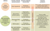
It is obvious that genetic characterization of prostate cancer is a mainstream component of contemporary translational prostate cancer research (Fig. 1). Nevertheless, the extremely heterogeneous nature of prostate tumors sets up substantial obstacles on the path to the clinical integration of the numerous findings.
Gene expression analysis is an interesting and simple implementation tool, but the contemporary evidence shows that the current state of this method does not reach the desired aim. Some obvious limitations are inevitably present.
The genetic characterization of prostate cancer with the use of contemporary molecular genetic methods is progressing unbelievably. There is nevertheless a substantial gap between the fundamental studies and the clinic. However, the emerging evidence shows that step by step, gene by gene, we are moving towards the clinically relevant genetic characterization of prostate cancer. The obvious limitations are the necessity of prospective evaluation of all findings and the outstanding interpatient and inter- and intrafocal heterogeneity of the prostate carcinoma. Given that the genetic characterization of the tumors by use of next-generation sequencing technologies is a labor-intensive task coupled with the analysis of huge amounts of data, international initiatives with division of tasks should play an important role in future progress (e.g., the International Cancer Genome Consortium, The Cancer Genome Atlas, projects similar to AURORA initiative for metastatic breast cancer [76]). One extremely important breakthrough has emerged from the epigenetic studies, which intended to solve the problem of repeat biopsies for most patients.
Importantly, a significant breach is now evident between the huge amount of studies of the genetic characterization of prostate cancer, which have limited translation to clinical practice or simply were not conceived to be so translated, and clinical practice. From a clinical point of view, this balance should be urgently shifted towards translation. Nevertheless, strict control of the significance of the new markers is necessary against the common clinical and pathological variables (e.g., Gleason score). This will guarantee protection from the enormous volume of insignificant data generated in the fundamental studies.
 XML Download