

 PDF
PDF ePub
ePub Citation
 Print
Print


INTRODUCTION
Peyronie's disease (PD) can be a debilitating psychosexual condition affecting up to 9% of adult men and is characterized by a constellation of penile symptoms and signs such as penile pain, deformity and plaque, with ensuing erectile dysfunction (ED) [1]. While no one truly knows the underlying pathophysiology of PD, it is generally agreed that PD is considered an abnormal wound healing disorder as a consequence of repetitive trauma to the erect penis resulting in fibrous plaque formation within the bi-layer of tunica albuginae in the genetically susceptible individuals [2]. This fibrotic, inelastic scar gives rise to penile curvature on the contralateral side when the penis is erect.
At present, PD process can be divided into 2 main phases, acute (inflammatory) and chronic (stable) stages. In the acute phase of PD, a patient usually describes a new onset of (less than 6 months) penile pain and penile curvature [2]. If left untreated, PD is a progressive sexual disorder in nearly 50% of men [3]. Since the disease process continues to evolve in the early phase, it is likely that the use of noninvasive therapy to halt and/or alter the disease progression may be effective. Low intensity extracorporeal shock wave therapy (LiESWT) has been used to treat men with PD since the late 1980s [4]. While the clinical outcomes of LiESWT in PD has been mixed, in recent years there has been a renewed interest in the use of LiESWT [1567].
We evaluate the efficacy, safety and patient satisfaction rate following LiESWT in Australian men with PD using a standardised protocol.
MATERIALS AND METHODS
1. Patient population
Following internal departmental ethics approval, patients with PD were prospectively enrolled from June 2013 to March 2014 in this open-label single arm study at the Department of Urology, Princess Alexandra Hospital, Brisbane, Australia. All patients received informed consent based on contemporary literature regarding LiESWT and were given the option to undergo surgery. Inclusion criteria included patient age ≥18 years, failed medical therapy, presence of palpable plaque and/or penile pain, and penile curvature. Exclusion criteria were complex curvature (more than 1 axis deviation or curvature >90 degrees), hourglass deformity, 2 or more palpable plaques, previous Peyronie's surgery (including penile prosthesis implant) and significant ED unresponsive to medical therapy.
2. Data collection
Penile curvature and plaque hardness, pain (on a visual analog scale ranging from 0 equals no pain to 10 equals strong pain), and International Index of Erectile Function (IIEF)-5 score were assessed at initial consultation, 6 weeks and 3 months following completion of LiESWT study. Assessment of penile curvature on erection was made based on digital photograph of the erect penis or in-room intracavernosal injection of 10-20 mcg of Alprostadil to induce maximal penile erection. Measurement of plaque size was performed by clinical palpation to document the length and width of the plaque. Patients were asked to score their overall satisfaction rate on a 5-point scale at the end of the treatment period. Adverse events such as penile pain, bruising and hematuria were recorded.
3. Procedure
The LiESWT was performed without local or systemic analgesia using Duolith SD1 ultra (Storz Medical AG, Tägerwilen, Switzerland) in the outpatient setting. Patients received instruction on the mechanics and procedure on LiESWT before informed consent was signed. Each patient stretched his penis fully by holding onto his glans penis with one hand while his other hand held the transducer and placed the probe directly onto the penile plaque. Ultrasound gel was used as a coupling solution. A clinical nurse will check on each patient at the start of treatment cycle to ensure that the patient adhered to protocol and that the probe was placed correctly over the Peyronie's plaque. It was explained to the patients that they would hear the generator noise during the course of LiESWT and that they will feel some minor discomfort along the penile shaft. Since no patient had undergone LiESWT before this study; therefore, no patient was aware of how this treatment felt like. Throughout each treatment session, a clinical nurse will periodically review the patient for any adverse event and to ensure correct positioning of probe.
All patients were treated twice weekly for 6 weeks, comprising of 3000 shock waves to the Peyronie's plaque per session with constant energy flow density of 0.25 mJ/mm2 and emission frequency of 3 Hz according to the manufacturer's guidelines. The shock wave generator implemented in our study has been used in the treatment of tenosynovitis and tendonitis.
4. Statistical analysis
Statistical analysis was performed with SAS 9.1.3 (SAS Institute Inc., Cary, NC, USA) computer software with comparison of pre- and posttreatment parameters at the 6 weeks and 3 months using paired t-test and McNemar chi-square test with p<0.05 considered statistical significant.
RESULTS
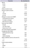
1. Patient demographics (Table 1)
Over the 10 months period, a total of 30 patients gave consent and were recruited in the study. The average age was 55 years (range, 42-68 years) and 27 patients (90%) reported the presence of penile curvature and/or deformity longer than 6 months (mean, 12.8 months; range, 6-24 months). Only 5 patients (17%) recalled a history of penile trauma. The range of penile curvature was 15 to 90 degrees and dorsal (12 patients, 40%) and left (10 patients, 33%) sided curvatures were most common. Penile plaque was palpable in all patients and 24 patients (80%) had palpable plaque size greater than 2 cm2×2 cm2. The presence of penile pain was reported in 6 patients (20%). Twenty of patients (66%) had received and failed medical therapy (10 patients received topical vitamin E, 5 patients had oral phentoxifylline, and 5 patients had oral colchicine therapies). Ten patients (33%) complained of concurrent mild to moderate ED but were responsive to oral phosphodiesterase (PDE) 5 inhibitors.
2. Efficacy, safety and patient satisfaction outcomes (Tables 1 and 2)
1) Efficacy
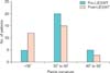
At the 6 weeks posttreatment assessment, there was an improvement in penile curvature (more than 15 degrees) observed in 10 patients (33%) with the range of improvement varying between 15 to 60 degrees. Two patients (7%) noticed worsening of penile curvature (15 and 20 degrees) while 10 patients (33%) did not noticed any change in penile curvature (Fig. 1). While the exact measurement of plaque volume was not feasible, no increase in penile plaque size was observed and 18 patients (60%) reported softening of the Peyronie's plaque on physical examination following LiESWT with a reduction in palpable penile plaque size by 2 cm2 observed in 8 patients (27%). These improvements remained similar at the 3-month follow-up visit. There was no significant difference found in the stretched penile length pre- and post-ESWL despite improvements in penile curvature and reduction in Peyronie's plaque size (p=0.48). Of the 6 patients who had penile pain, 4 patients (67%) reported resolution of pain during the course of therapy. Of the 10 patients who have ED, there was a moderate improvement in IIEF-5 score (>5 points) reported in 6 patients (60%) and 2 patients were able to resume sexual intercourse without oral PDE5 inhibitors.
2) Feasibility and safety
All patients completed the LiESWT course and there was no drop out in this study. Patients tolerated the treatment well and no analgesia was used during or after LiESWT. Most patients (90%) scored less than 2 out of 10 on the pain visual analog scale and none of the patients reported any persistent pain lasting more than 12 hours. No petechial bruising, dysuria or urethral bleeding was observed.
3) Patient satisfaction rate
The majority of patients (70%) were satisfied and rated 4 out of 5 in the overall satisfaction level. There is a strong positive correlation between changes in penile curvature and patient satisfaction outcome (p=0.03) and more than two-third of patients would recommend LiESWT to their friends or family members.
DISCUSSION
Published literature reports that the decrease in penile curvature varies between 21% and 74% among men who received LiESWT [5789]. However clinical outcomes in recent randomised controlled trials showed an actual change of less than 10° compared to control group [79]. In addition, Hatzichristodoulou et al. [7] reported an increase in penile deviation in 40% of patients following LiESWT although only 5 patients (10.9%) showed an increase in plaque size in this group. In our study, the improvement in penile curvature was observed in 33% of men and of these men, two-thirds reported a decrease in penile curvature from 30°-60° to <30°. Similarly our study showed a reduction in penile plaque by 2 cm2 in 8 of our patients (27%) at the completion of LiESWT, and the change in plaque size appears to correlate with the improvement in penile curvature. However it should be noted that our measurement of plaque size is two dimensional (i.e., not actual plaque volume) as plaque measurement is often be inaccurate and/or not feasible due to several factors such as operator dependent, configuration of the plaque and the use of appropriate ultrasound software. Furthermore it should be emphasized that our study lacks the control group and therefore we are not able to directly compare the natural progression and/or regression of PD against the success of LiESWT.
The improvement in penile curvature observed in our study in contrast to some of the published studies could be attributed to several factors. Firstly, we excluded all patients with complex PD such as the presence of more than 1 axis of penile curvature, curvature greater than 90°, presence of hour-glass deformity, and men with 2 or more palpable Peyronie's plaques. Secondly those men who reported improvement in penile curvature had PD history less than 12 months, indicating likely an active disease process which is more susceptible to mechanical effect. While the exact pathophysiology of PD remains largely unknown [12], the limited information available to date on the cellular basis of PD points to distinct alterations in wound healing and propagation of fibrotic process as the underlying cause. Therefore it is likely that treatment instituted during the active phase of PD will have the greatest impact and may alter the disease process. In our cohort, the 10 patients who reported improvement in penile curvature had mild plaque and duration of PD less than 12 months. Furthermore there was correspondingly softening and reduction in penile plaque size in this successful group of LiESWT men. It may be possible that LiESWT has a protective effect on PD progression by stabilizing the penile curvature and plaque progression [9] and actual underlying histological changes within the Peyronie's plaque [10].
In contrast to published literature supporting the role of LiESWT in men with ED [11], the reported changes in erectile function following LiESWT in PD has been mixed. While IIEF-5 score is frequently used to evaluate sexual function in men with PD, it has never been specifically validated for use in this disease state. Published meta-analysis in 2004 reported that the improvement in sexual function varies from 12% to 80% [5]. However more recent studies have found no significant difference between LiESWT and control group [78]. In our study, a small minority of patients reported an increase in erectile function and this improvement in IIEF score was seen in the 6 men who reported mild to moderate ED prior to LiESWT. We postulated that the improvement in the penile curvature with easier sexual penetration, and perhaps underlying neovascularization induced by LiESWT might play a role in the greater erectile function [11]. However any improvement in sexual function should be evaluated using formal penile hemodynamics study over a longer interval period.
A variety of contributing factors will likely influence the outcome of LiESWT for PD. Prolonged history of PD and presence of plaque calcification, as a marker of chronicity, indicate unlikely history of spontaneous regression. We acknowledge the various limitations in our study such as the lack of a sham (control) arm, objective penile hemodynamic measurements, small number of participants and short term follow-up study. In addition, detailed documentation of disease including sonographic changes such as tunical thickening, cavernosal calcification and fibrosis undertaken by an independent examiner will minimise certain biases [12]. Furthermore, our treatment protocol is based on manufacturer's guidelines [13] and is likely derived from previous orthopaedic literature.
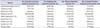
While published literature has largely failed to demonstrate significant benefit in the use of LiESWT to treat penile curvature [1578914151617181920] (Table 3), these outcomes should be interpreted with some caution due to underlying methodological flaws [789141516171819] and perhaps inappropriate use of shock wave energy flow density [715161718]. Interestingly, subgroup analysis of patients in the LiESWT group showed an overall better outcome in younger patients with a relatively milder degree of curvature [818]. Comparative studies between LiESWT with other treatment modality showed that LiESWT is not superior to other options [1510] and when used in combination with other therapeutic options such as intralesional injection or tadalafil for men with PD and ED, there were improvement in erectile function score and quality of life score while the plaque size and curvature were unchanged [1920].
We agree that the current literature on the use of LiESWT in PD population remains controversial. It may be possible that newer generation of shock wave lithotripter has an improved technology in disrupting the tunical plaque without inducing further plaque formation or injuring the underlying cavernosal tissue. While the exact therapeutic mechanism remains unclear, it is postulated that LiESWT may play a role in plaque remodelling and improvement in consecutive resorption of calcification [5], resulting in softer plaque and further correction and/or resolution of the penile curvature. Furthermore LiESWT may have a protective effect on disease progression by stabilising penile deviation and PD plaques [9]. Therefore it appears that LiESWT should ideally be offered and utilised in younger men during the active phase of PD i.e., less than 6 months with milder degree of curvature and softer noncalcified plaque, and in the absence of hour glass deformity. In a carefully selected group of men with PD, our study showed that LiESWT appears to reduce penile curvature and plaque size in some men, and overall is safe, tolerable and is associated with high level of patient satisfaction. Many men are keen to pursue minimal invasive therapy such as LiESWT to preserve penile length since the current surgical intervention is invariably associated with loss of penile length. Nonetheless there is a need to define which subgroup of PD population is best suited, the LiESWT protocols (modality of shock wave energy, emission frequency, and total energy delivery) and the role of combination therapy in PD. Other important factors such as the actual physiological changes in the penile tissues and the long-term risk of shock waves have yet to be fully elucidated.
CONCLUSIONS
LiESWT offers a minimally invasive treatment option for men with PD who have failed conventional medical therapy and are not keen to undergo surgical intervention. In a carefully selected group of men with PD characteristics, LiESWT appears to be safe, has moderate efficacy in improving penile curvature and pain, and is associated with high level of patient satisfaction rate in this short term study.

 XML Download