

 PDF
PDF ePub
ePub Citation
 Print
Print


INTRODUCTION
Bladder cancer (BC) is the most common malignancy of the urinary tract. The most common BC is urothelial carcinoma (UC). Approximately 75% of BC is nonmuscle invasive (NMI) UC at the time of diagnosis, with 70% presenting as noninvasive papillary (pTa) cancer, 20% as tumor invading the subepithelial tissue (pT1), and 10% as a flat tumor (carcinoma in situ, or CIS) lesion [1]. UC of the bladder is a chronic heterogeneous disease with a variable natural history and oncological outcome [2]. The overall prognosis is good; however, nearly half of cases will recur within 2 years and up to one-quarter of cases progress to muscle-invasive disease, which is invariably associated with poor prognosis [3].
A biomarker is invaluable for predicting the disease course. For example, prostate-specific antigen is an ideal biomarker in the follow-up of treated patients with prostate cancer. The National Institutes of Health definition of a biomarker is "a characteristic that is objectively measured and evaluated as an indicator of normal biological processes, pathogenic processes, or pharmacologic responses to a therapeutic intervention." Biomarkers in oncology are used for the diagnosis and prognosis of cancer. Diagnostic biomarkers are used to assess the risk of disease, for screening, and to establish the diagnosis. Prognostic markers are used to predict response to treatment and to determine overall prognosis.
Recurrence is defined as a relapse of primary NMI UC with either equivalent or lower pathological stage, whereas progression is defined as a relapse with a higher a TNM stage or grade [4]. Each step in oncogenesis from initiation of a tumor to its progression and ultimately metastasis involves multiple genetic and epigenetic events. This poses major difficulty in designing a management algorithm for NMI UC. Therefore, a strict surveillance protocol and frequent follow-up are required, with repeated treatments leading to the highest cost per patient among all cancers from the time of diagnosis to death [56]. In order to refine the prediction of prognosis in individual cases, a strong need exists for a molecular marker. The present review looks at the clinical potential of various molecular markers in predicting recurrence and progression.
MATERIALS AND METHODS
We performed a wide systematic literature search in the Medline databases. "bladder cancer" and "molecular markers" were the search terms we used for specific study designs: meta-analysis, randomized controlled trials, reviews, clinical trials, and practice guidelines. Our research was limited to studies published in English from 1994 through February 2014. Reference lists of the included articles were secondarily hand-searched for studies that were not identified by the database search.
RESULTS AND DISCUSSION
1. Role of molecular markers versus clinico-pathological variables
Selected criteria that assess patient and tumor characteristics provide valuable information about disease characteristics, recurrence, progression, and the proposed treatment modalities. Currently, conventional criteria such as pathological grade, stage, and other tumor characteristics are used. These variables are assessed with various scoring systems to estimate disease recurrence and progression. However, the scope of these clinico-pathological markers is limited. Molecular markers determine the biological behavior of disease and are involved at a much earlier level than are morphological factors [7]. Intensive research in the area of molecular biology related to urothelial cancer has provided insight into the biology of this disease. Translational work done in the last decade is providing the basis for shaping up clinical practice and guiding clinical decision-making [8]. The molecular markers include serum, tissue, and urinary markers. Many of these markers have been approved by the U.S. Food and Drug Administration (FDA) and its European counterpart. Among these markers, fibroblast growth factor receptor (FGFR3), epidermal growth factor receptor (EGFR), retinoblastoma protein (pRB), p53, Ki 67, vascular endothelial growth factor (VEGF), and cytokeratin (CK 20) [9] are used as indicators but their value or role is still being questioned, because they have not been applied in clinical practice alone or been proved by large-scale multicenter studies or randomized clinical trials [10]. Consequently, the use of these markers is not currently recommended by any of the existing clinical guidelines.
3. Role of transurethral resection
Transurethral resection of the bladder tumor (TURBT) is not only an invaluable diagnostic modality, it also provides a prognostic tool. Following TURBT alone, however, up to 60% of patients will experience tumor recurrence. Recurrence depends on factors like incomplete resection, implantation of tumor cells, growth of microscopic tumors, and new tumor formation.
In a recent evaluation of 566 patients who had undergone a complete first resection, the authors noted that documented complete resection by an experienced surgeon with the presence of detrusor muscle was significantly associated with a lower recurrence rate at first cystoscopy [19]. Brausi et al. [20] analyzed 2,410 patients with NMI UC from various institutions and found that the rate of recurrence at first cystoscopy varied greatly. The authors attributed this to the quality of transurethral resection (TUR) performed, because most of these are likely to be residual disease rather than real recurrence. Documentation of the appearance of the base of the tumor, whether sessile or pedunculated, is important. This indicates the invasiveness of the tumor. A second TUR is indicated in high-grade cancers, which should be done within 6 weeks of the first TUR. CIS often appears as velvety erythematous patches, and all such suspicious lesions should be biopsied. Fluorescence cystoscopy improves BC detection rates, especially for flat lesions, and this improves recurrence-free survival by decreasing the residual tumor [21].
4. EORTC risk calculator
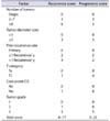
To individualize the prediction of recurrence based on a multivariate analysis of data on 2.596 patients with superficial BC in 7 European Organisation for Research and Treatment of Cancer (EORTC) trials, Sylvester et al. [12] developed a scoring system that was subsequently implemented in European Association of Urology guidelines. The EORTC risk calculator is a powerful tool for clinicians that uses available clinical and pathological data to calculate short-term and long-term risk of recurrence and progression. The variables included are grade, stage, CIS, tumor multiplicity, size, and prior recurrence rate. Each of these variables is assigned a weighted score for determining the end point (recurrence or progression; Table 1). The risk calculator is available at http://www.eortc.be/tools/bladdercalculator.
Although the EORTC risk calculator is one of the most commonly used tools, the reproducibility of pathologic stage and grade is modest, which is a major concern for clinicians.
5. CUETO risk calculator
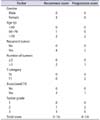
Fernandez-Gomez et al. [22] of the Spanish Urological Club for Oncological Treatment (CUETO) group reported an analysis of 1,062 NMI UC cases from 4 randomized phase 3 trials who received bacillus Calmette-Guérin (BCG). Significant independent predictors for recurrence in the multivariate analysis were multiplicity, prior tumor, female gender, and presence of associated CIS. Of these, multiplicity of tumor was the most important factor for predicting recurrence. In the multivariate Cox regression analysis, age, history of recurrence, high grade, T1 stage, and recurrence at first cystoscopy were identified as independent predictors of progression. Subsequently, a risk stratification model was developed to provide accurate estimates of probability of recurrence and progression after BCG (Table 2). Validation studies showed that the CUETO model is a good predictive model. The Rosevear et al. [23] scoring model is a useful prognostic tool for stratifying recurrence risk in patients with NMI BC who are treated with combined intravesical BCG plus interferon α-2B.
6. Comparison of EORTC and CUETO
The populations studied and the disease characteristics differed between these 2 scoring models. In the EORTC group, only 4.4% of patients presented with CIS, 10.4% had G3 tumors, and only 6% were treated with BCG. An external validation of EORTC tables in 1,062 patients of the CUETO group treated with BCG [24] showed that the former model successfully stratified recurrence and progression in low- and intermediate-risk patients; however, the risk of recurrence and progression after BCG therapy was overestimated in EORTC and its discriminatory ability for progression was decreased. In a large multi-institutional cohort of 4689 patients assessed retrospectively, Xylinas et al. [25] showed that the EORTC risk tables and the CUETO scoring system exhibited poor discrimination for both disease recurrence and progression in NMI BC patients, particularly for high-risk patients. These results underline the need to improve our current predictive tools.
7. Age
The EORTC model does not include age as a prognostic factor. The impact of age has been assessed in many clinical trials. Boorjian et al. [26] and Herr [27] noted an increased risk of recurrence and shorter cancer-free survival with older age at diagnosis in patients with superficial BC treated with BCG.
8. Molecular prediction of recurrence
The role of molecular markers to predict recurrence seems limited. Perhaps the most extensively studied marker is the tumor suppressor gene p53. The p53 protein serves as a "guardian of the genome" by inducing multiple mechanisms of cell cycle arrest after cellular insult. A mutant genotype of p53 was found to be a predictor for recurrence in a study by Shariat et al. [28], whereas the wild-type of p53 was associated with more recurrence in the Moonen et al. [29] study. Oh et al. [30] determined the impact of p53 overexpression on tumor recurrence after BCG intravesical therapy in patients with NMI BC. They noted that strong overexpression of p53 was predictive of recurrence in patients with NMI BC undergoing intravesical BCG treatment. In a series of 80 consecutive patients with pT1N0 urothelial cancer, expression of p53 was altered in one-quarter of patients and p53 was found to be independently associated with BC recurrence (hazard ratio [HR], 3.66; p=0.033) [31].
Analyses of multiple genes or a combination of multiple biomarkers have identified different markers such as Survivin [32], Mcm2 [33], or gene classifiers to discriminate recurrent from nonrecurrent NMI UC [323435]. However, a multicenter validation study of 404 patients from 5 European countries did not show clinical utility of a 26-gene signature for recurrence [36].
9. Clinico-pathological factors for progression
Various clinico-pathological factors have been studied for NMI UC progression [121415161718]. Among these, CIS, high grade, and T1 stage are the most important. The micropapillary variant of urothelial cancer and lympho-vascular invasion are other significant factors related to aggressiveness and can predict progression to muscle invasion [37]. Other important variables for progression are recurrence in bladder at the first follow-up cystoscopy, female gender, and the presence of CIS in the prostatic urethra. For all stages of BC, women have a worse outcome [38]. Saint et al reported that urinary immunological response was more common in men than in women treated with BCG [39]. Thus, it was recommended that women have a more intensive follow-up schedule.
10. Substaging of T1 disease
Substaging of T1 disease is also important in the progression of NMI UC. However, to date, no effective substaging system has been defined [40]. Orsola et al. [41] differentiated T1 disease according to the depth of lamina propria involvement and found significantly different progression rates (34% vs. 8%) for those with deep lamina propria invasion (T1b/T1c) compared with superficial.
To identify the subset of patients with a greater risk of progression, Chang et al. [42] analyzed 406 T1 high-grade cases and stratified T1 stage as 0.5, 1.0, and 1.5-mm depth into the lamina propria. More extensive involvement was associated with unfavorable prognosis.
Van Rhijn et al. [43] introduced a new system to predict the aggressive behaviors of high-grade NMI UC and divided T1 into T1 micro invasive (T1m) and T1 extensive invasion (T1e) disease, with the latter conferring progression of disease. Substage T1m/T1e could potentially be incorporated in future tumor-node-metastasis classifications.
11. Molecular markers for progression
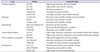
To assess the prognosis of NMI UC, a wide variety of molecular markers including oncogenes, tumor suppressor genes, cell cycle regulators, proliferation antigens, and signaling proteins are studied [174445464748] (Table 3). The value of CK 20, Ki 67, and p53 has been extensively investigated for NMI UC. Bertz et al. [49] investigated these markers in 309 specimens of high-risk BC treated with BCG. In a multivariate analysis, CK 20 expression and Ki 67 were significantly correlated with disease-specific survival.
Shariat et al. [34] determined immunohistochemical staining for p53, p21, p27, and pRB from 74 patients who underwent TURBT for NMI UC and found p53, pRB, and p21 to be independently associated with tumor progression. In addition, this combination of biomarkers also stratified patients into statistically significantly different risk groups for disease recurrence and progression.
12. p53
Tp53 acts as a tumor suppressor protein and induces cell cycle arrest or apoptosis upon DNA damage or other cellular insult. p53 is the marker most frequently investigated to predict progression of BC [505152]. The proportion of altered p53 was shown to increase progressively in specimens from normal urothelium, NMI BC, CIS, muscle-invasive BC, and metastatic lymph nodes in specimens from over 400 patients with BC using tissue microarray [50]. p53 can also help to stratify the heterogeneous population of pT1 patients into risk groups to guide clinical decision-making regarding observation vs. adjuvant therapy [31]; however, its use alone is not yet established in clinical practice. A meta-analysis incorporating 117 studies with over 10,000 patients showed that p53 independently predicted recurrence, progression, and mortality in only 26%, 50%, and 29% of studies, respectively [53].
13. Retinoblastoma
pRB is a tumor suppressor gene involved in cell cycle control. Altered (increased or decreased) RB expression can serve as a predictive marker of outcome in patients with high-risk superficial BC treated with BCG [54].
14. p21
p21 inhibits the activity of cyclin-dependant kinase and thus functions as a regulator of cell cycle progression. Altered p21 expression is independently associated with BC recurrence and progression [55].
15. Caspase 3
Burton et al. [56] evaluated the expression of caspase-3 in patients with CIS and reported that its overexpression in patients with CIS was associated with progression to muscle-invasive BC.
16. Angiogenesis markers
Angiogenesis is a critical event for progression of solid tumors including BC. Microvessel density (MVD) and various other markers are used to quantify angiogenesis. Bochner et al. [57] showed that patients with high MVD (>100 microvessels/HPF) and no p53 abnormalities showed the highest risk of disease recurrence and cancer-specific mortality compared with patients with low MVD.
17. Vascular endothelial growth factor
VEGF is a potent stimulator of endothelial cell proliferation, and increased expression is associated with increasing tumor stage and progression [58].
18. Fibroblast growth factor receptor 3
FGFR3 belongs to the receptor tyrosine kinase family. Approximately two-thirds of NMI UC cases are FGFR3 mutants [59]. Several studies have shown that FGFR3 mutation is a genetic event that leads to favorable pathways in BC with protection against disease progression [606162].
Van Kessel et al. [59] used voided urine samples for analysis of FGFR3 and found this to be a cost-effective strategy for surveillance of patients with NMI UC. They proposed that analysis of FGFR3 mutation could decrease the frequency of cystoscopic surveillance.
20. Molecular grade
Molecular grade (mG1-3) was introduced on the basis of FGFR3 mutation status and expression of the proliferation marker Ki 67. It has been found to be a highly reproducible and prognostic tool in BC progression [46].
Another study comparing the reproducibility of pathologic grading and mG showed reproducibility of the former to be almost perfect (k; 0.76), whereas reproducibility for pathological grade was only fair to substantial (k; 0.17-0.58). The authors concluded that mG is a more reproducible and reliable tool than pathological grade assessment to predict disease progression in NMI UC. Another molecular grading model containing 3 mGs based on combination of Ki 67 LI (labeling index) and VEGF scoring was developed to predict tumor recurrence and progression in NMI UC [7]. Univariate and multivariate analyses were performed that showed this grading model to be effective and accurate for predicting outcome.
21. Her-2 neu
Human epidermal growth factor receptor 2 is a tyrosine kinase in the EGFR family. Detection of amplification of Her-2 by fluorescence in situ hybridization (FISH) is associated with markedly aggressive behavior in NMI UC with high risk of progression [64].
22. Glutathione-S-transferase 1
The enzyme glutathione-S-transferase 1 (GSTT1) causes detoxification of carcinogenic and toxic electrophiles via conjugation with glutathione. The GSTT1 genotype is a strong indicator for predicting recurrence and progression in patients with primary NMI UC. Ha et al. [4] compared this isoenzyme in blood samples of patients with NMI UC with other tissue-based markers and found it to be a better prognostic indicator.
23. Nitric oxide synthase and peroxisome proliferation-activated receptor
Sandes et al. [65] in their study showed that peroxisome proliferation-activated receptor (PPAR)-gamma controls inducible nitric oxide synthase (NOS) expression at early tumor stages. Decreased levels of PPAR (detected by Western blot) and increased inducible NOS (detected by immunohistochemistry) are associated with BC progression.
24. Heme oxygenase 1
Yim et al. [66] analyzed NMI UC tissue specimens with polymerase chain reaction (PCR) and found heme oxygenase 1-isoform (HMOX1) mRNA levels in NMI UC to be significantly high in patients with disease recurrence and progression. These findings suggested that increased HMOX1 expression not only promotes cell proliferation but also contributes to a more aggressive NMI UC phenotype.
25. ABO blood type
Recently, Klatte et al. [67] studied the impact of ABO blood group type on outcome of patients with NMI UC and found in univariate and multivariate analyses that blood group type O exhibits the highest recurrence and progression rates. He concluded that the inclusion of ABO blood types in other models could increase the accuracy of standard prognostic factors.
26. Combination of molecular markers
Bladder carcinogenesis is a multistep process and most intermediary proteins (markers) are connected to each other via various pathways. Considering the complexity of the molecular abnormalities associated with BC, it is unlikely that a single marker will accurately segregate tumors of similar phenotypes into different prognostic categories. Various combinations of molecular markers can be used to predict progression. Examples include the mG described above. Similarly, using the tissue micro assay technique to determine the expression of multiple immunohistochemical markers from one tissue sample using p53, pRB, p21, and p27, Shariat et al. [50] found that the number of altered markers is independently associated with increased risk of progression. Karam et al. [68] found that the number of simultaneously altered apoptosis markers (such as p53, Bcl-2, caspase-3, and survivin) is an important prognostic indicator for recurrence and cancer-specific mortality. It is recommended that multiple molecular markers be used to improve the predictability of future risk stratification models, which can help to guide patient counseling and management decisions [8].
Recently, Ding et al. [69] showed that combining the clinico-pathological factors of EORTC risk scores and expression of Ki 67 by using immunohistochemical studies and scoring for intensity and area of staining could improve the risk stratification of NMI UC. The combination of high-risk EORTC and Ki 67 expression improves the accuracy of progression prediction.
27. Role of urinary markers in NMI UC
Urinary markers have limited value in prognostication of BC and are used (mainly as an adjunct to cytology) for detection and surveillance of urothelial cell cancer recurrence [70]. For the primary detection of BC, the value of using a urinary marker other than cytology is limited in patients who present with hematuria or other symptoms suggestive of BC. For follow-up, a reliable marker has the potential to decrease the frequency of surveillance cystoscopy, thus decreasing the bother for the patients and the overall cost of follow-up.
The sensitivity of urinary markers in surveillance is higher but the specificity is generally lower than for urine cytology [71]. However, none of these markers have been routinely implemented into clinical decision-making, and urinary markers have little added value owing to insufficient evidence for clinical benefit [7072].
28. Bladder tumor antigen
Bladder tumor antigen (BTA) is available in 2 formats, i.e., BTA stat and BTA trak, for detecting the human complement factor H-related protein and complement factor H in urine, respectively [72]. BTA stat can be performed as a point-of-care test because it is based on qualitative assay and can be performed in a few minutes. BTA TRAK is a quantitative standard enzyme-linked immunosorbent assay (ELISA) [73].
Systematic reviews and meta-analyses have revealed a sensitivity of 70% and specificity of 75% for BTA Stat. For BTA TRAK, the sensitivity and specificity are 66% and 65%, respectively [74], in reported literature. False-positive results can arise from benign inflammatory conditions such as hematuria and pyuria, urolithiasis, and recent instrumentation [75].
29. Nuclear mitotic apparatus protein 22
Nuclear mitotic apparatus protein 22 (NMP22) is a nuclear matrix protein responsible for chromatid regulation and cell separation during replication and is available as a quantitative ELISA or as a point-of-care Bladder check test [76].
NMP22 is much more prevalent in malignant urothelial cells than in normal cells. The sensitivity of the original NMP22 immunoassay ranges from 47% to 100% and its specificity from 60% to 90% [77] in reported literature. Because NMP22 protein is released from dead and dying urothelial cells, other benign conditions of the urinary tract (i.e., urolithiasis, infection, inflammation, hematuria, and even concentrated urine) can give rise to false-positive results [78].
30. Ucyt+/ImmunoCyt
Ucyt+ was formally called ImmunoCyt. It is an example of immune-cytology, which combines cytology with immunofluorescence assay [75]. This test detects cellular markers for BC in exfoliated urothelial cells (i.e., carcinoembryonic antigen and two bladder tumor cell-associated mucins) [72]. The reported overall sensitivity of ImmunoCyt is 50% to 100% [7779] and it has a specificity of 69% to 79%. False-positive rates are seen in patients with benign prostatic hyperplasia or cystitis [80].
31. Telomerase
Telomeres are repetition sequences at the end of chromosomes that protect genetic stability during DNA replication. Each cell division is accompanied by a loss of telomeres by the enzyme telomerase causing chromosomal instability and cellular senescence. In a systemic review, telomerase was found to have a sensitivity of 75%; however, it had low specificity compared to cytology [78].
32. Microsatellites
These are tiny, highly polymorphic DNA fragments frequently found throughout the genome. The PCR-based urine test is used to analyze and detect replication errors [81]. It is very sensitive for low- and high-grade lesions with sensitivities of 67%, 86%, and 93% for recurrent G1, G2, and G3 lesions, respectively, and has a specificity of 88% [74].
33. UroVysion
The UroVysion test uses the FISH technique to detect aneuploidy in chromosomes 3, 7, 17, and loss of the 9p21 locus of the p16 tumor suppressor gene. The urinary marker can predict early recurrence in patients with a negative cystoscopy result [80]. This test has been deemed an "anticipatory positive" test, because it can predict early recurrence in patients with negative cystoscopy or cytology results up to 6 to 20 months before the development of a visible malignancy [8283]. The sensitivity has been found to be up to 80% with high specificity of more than 80% in systematic reviews and meta-analyses [84].
34. Aurora kinase A
Aurora kinase A (AURKA) is a gene encoding a key regulator of mitosis and is currently regarded as a novel urinary marker. It is amplified or overexpressed in cancer cells and the level of AURKA amplification is associated with level of aneuploidy. The FISH test for the AURKA gene yields a specificity of 96.6% and sensitivity of 87% [85].
A pooled analysis of studies have revealed a sensitivity of 44% for cytology for all types of BC but higher sensitivity for immune-cytology (ImmunoCyt: 84%; 95% confidence interval [CI], 77%-91%), FISH (UroVysion: 76%; 95% CI, 65%-84%), and NMP22 (68%; 95% CI, 62%-74%) [71].
Horstmann et al. [86] analyzed the combined use of cytology, FISH (UroVysion), immunocytology (ImmunoCyt), and NMP22 (ELISA) for surveillance of NMI BC in a cohort of 106 patients. A considerable increase in sensitivity (>90%) with higher negative predictive value was found for the test combination.
The UroVysion, ImmunoCyt, BTA stat, BTA TRAK, NMP22, and NMP22 bladder CHECK tests are currently the only FDA-approved urine-based bladder tumor markers. All of these tests are approved for use in surveillance of BC, with the exception of ImmunoCyt, which is also approved for the initial diagnosis of BC.
35. Follow-up strategy
Because of broad variation in the incidence of recurrence and progression, NMI UC requires strict surveillance, and various follow-up policies are published using periodic cystoscopy and urine cytology. However, the frequency of the follow-up regimen depends upon risk factors associated with the disease. The conventional strategies are invasive, cause substantial patient discomfort, and are not cost-effective. Addition of potential markers from patients' serum, urinary specimens, and cancer tissue into clinical practice can lead to a more rational surveillance regimen providing cost-effective and noninvasive monitoring.
 XML Download