

 PDF
PDF ePub
ePub Citation
 Print
Print


INTRODUCTION
With recent improvements in laparoscopic techniques, many urologic surgeries can now be performed by using laparoscopy. Moreover, the introduction of robotic systems has simplified minimally invasive surgery. Therefore, many urologic procedures, such as radical prostatectomy, partial nephrectomy, radical cystectomy, and pyeloplasty, have been performed with robotic systems.
Laparoscopic retroperitoneal lymph node dissection (L-RPLND) has been shown to have acceptable morbidity, rapid recovery, and good cosmetic outcomes [1]. Since Davol et al. [2] reported the first case of robot-assisted laparoscopic retroperitoneal lymph node dissection (R-RPLND) in 2006, there have been many reports of R-RPLND for stage I nonseminomatous germ cell tumors (NSGCTs) [34]. However, there has been only one report of R-RPLND applied to post-chemotherapy patients [5].
Here, we report a case of stage IIIb NSGCT treated with R-RPLND after chemotherapy. This is the first description of R-RPLND in Korea.
CASE REPORT
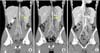
A 17-year-old boy visited Kyung Hee University Medical Center because of a painless, enlarging left scrotal mass. Scrotal ultrasonography revealed an approximately 6.6-cm-sized left testis with a partially cystic portion. Abdominopelvic and chest computed tomography (CT) showed enlarged, conglomerated left paraaortic lymph nodes and right retrocrural lymph nodes (Fig. 1A). The patient underwent left radical orchiectomy. The pathologic report demonstrated a mixed germ cell tumor (immature teratoma 65%, embryonal carcinoma 15%, yolk sac tumor 10%, seminoma 5%, and choriocarcinoma 5%). Tumor markers after orchiectomy were significantly elevated: alpha-fetoprotein, 169.5 ng/mL; beta-human chorionic gonadotrophin, 23,245.16 mIU/mL; and lactate dehydrogenase, 644 U/L. After thorough counseling about treatment for multiple enlarged lymph nodes, the patient decided to receive chemotherapy with bleomycin, etoposide, and cisplatin. Three cycles of chemotherapy were administered, and the levels of all tumor markers decreased; however, the follow-up abdominopelvic CT scan showed increased lymph node size (Fig. 1B). Therefore, we decided to perform R-RPLND for resection of the remnant mass after chemotherapy.
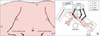
The patient was positioned in a right lateral decubitus position with the left side up and the bed flexed. After pneumoperitoneum was established by use of a Veress needle, a 12-mm camera port was placed at the umbilicus and five more trocars were used in the abdomen: three for robotic arms and two for the assistant (Fig. 2A). The robot was docked at the patient's left side, and the assistant was positioned on the patient's right side (Fig. 2B).
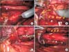
A left-modified template dissection was planned. The procedure was started with an incision at the line of Toldt. After the colon dropped medially, ligation of the gonadal vein was performed, and the spermatic vessel was dissected. The left ureter was easily identified and was isolated with a nelaton catheter. To prevent ureteral injury during dissection of lymph nodes, the ureter was retracted by the third arm of the robot. Paraaortic lymph nodes were dissected from the level of the common iliac artery to the root of the left renal artery along the lateral margin of the aorta (Fig. 3A, B). Dissection of lymph nodes was a difficult and time-consuming process because of severe adhesion between the lymph nodes and great vessels as a result of chemotherapy. Postganglionic sympathetic fibers were detected and preserved to maintain ejaculatory function. Multiple aorto-caval lymph nodes were also observed (Fig. 3C) and were all dissected carefully by using Hem-o-lok clips and small metal clips. The skeletonized aorta and inferior vena cava were observed (Fig. 3D). All of the resected lymph nodes were placed in an EndoCatch bag. A drain tube and a Foley catheter were inserted, and the surgery was completed.
The total operative time, including console time, was 420 minutes. There were no postoperative complications. The Foley catheter was removed on the first postoperative day, and the patient was discharged on the fourth postoperative day. Pathology revealed a count of 20 lymph nodes, 2 of which showed a metastatic immature teratoma. The patient received two cycles of chemotherapy with etoposide and cisplatin. At the time of this writing, his laboratory tests were within normal limits, and there was no evidence of metastasis on radiologic examination (Fig. 1C). Furthermore, the patient had not complained of a decreased amount of semen or a dry ejaculation.
DISCUSSION
Chemotherapy with a cisplatin-based regimen is the initial approach for the treatment of clinical stage IIC or III NSGCTs. However, RPLND is indicated when teratomatous components are found in the testicular mass, incomplete remission is confirmed, or retroperitoneal masses remain without increased tumor markers after primary chemotherapy [6]. Postchemotherapy RPLND provides both staging and therapeutic benefits. However, the procedure of conventional open RPLND is difficult, and a previous study has reported considerable morbidity that results in a long recovery time and a large incision [7].
With the development of minimally invasive techniques, many efforts have been made to perform urologic surgeries with laparoscopy, including RPLND. Since 1992, when the first case of L-RPLND for the treatment of a clinical stage I germ cell tumor of the testis was reported, multiple series of L-RPLND have been reported. Such studies concluded that, although the procedure is difficult and the learning curve must be overcome, L-RPLND is feasible even for postchemotherapy NSGCT patients with residual masses [17]. Nonetheless, L-RPLND seems to have the critical limitation of being an extremely difficult procedure. High conversion rates have been reported in other studies, and minor perioperative complications such as lymphoceles, chylous ascites, vascular lesions, and bowel lesions have also been described [89].
The first report of R-RPLND for clinical stage I NSGCT was published in 2006 [2]. Since then, there have been a few studies of R-RPLND for the treatment of clinical stage I testicular tumors. Using a robotic system, physicians can more easily and safely perform RPLND compared with L-RPLND. Indeed, the da Vinci surgical system (Intuitive Surgical, Sunnyvale, CA, USA) facilitates three-dimensional and expanded visualization so that much more delicate procedures can be performed, especially during the detachment of lymph nodes near the great vessels or for nerve preservation [10]. For these reasons, all previous reports on R-RPLND have concluded that R-RPLND is not only safe and beneficial with regard to cosmetics, but also results in a similar oncologic outcome compared with open or L-RPLND [410]. However, many studies were limited to cases of clinical stage I NSGCTs, and there has been only one report of R-RPLND in postchemotherapy NSGCT patients or in those with clinical stage II or higher disease [5]. In that previous study, although patients treated with postchemotherapy R-RPLND showed a higher rate of complications and open conversion with longer operative times than did patients without preoperative chemotherapy, the lymph node yield, estimated blood loss, and postoperative hospital stay were similar. The investigators reported similar lymph node positivity compared with many open RPLND series. Additionally, the authors described less estimated blood loss and a shorter length of hospital stay compared with a large cohort of open RPLND cases.
In this report, we present a case of R-RPLND performed for postchemotherapy treatment of an NSGCT patient with initial clinical stage IIIb disease. To our knowledge, there have been no reports of R-RPLND in Korea. We found that R-RPLND allowed for meticulous and clear dissection with excellent vision. All masses, even though they adhered to the great vessels as a result of previous chemotherapy, were eliminated successfully. However, dissection of lymph nodes from adjacent organs including great vessels was a time-consuming procedure and we had to concentrate so as not to injure great vessels. When the mass was severely adhering to adjacent organs, we used small clips and then cut with scissors. We always kept in mind not to dissect the lymph nodes by force. In addition, during the operation, we prepared to open in case of emergency such as injury to the aorta. Another advantage of R-RPLND was that owing to the improved field of vision and delicate motion of the robot arms, the postganglionic sympathetic chains were easily preserved. We believe that R-RPLND has many advantages, such as treatment of difficult cases that would be impossible with an open or laparoscopic technique. However, our findings cannot be generalized to all urologists because this is a preliminary report, and all of the procedures were performed by a single surgeon with extensive experience in oncologic radical surgeries and robotic surgeries. Additional long-term studies with numerous cases are needed to establish R-RPLND as a reasonable treatment option.
In conclusion, we have described the first case of postchemotherapy NSGCT with initial stage IIIb disease that was successfully treated by R-RPLND. The robot system facilitated delicate and secure dissection of lymph nodes and remnant masses. Therefore, R-RPLND offers an excellent oncologic and functional outcome with great cosmetic benefit.

 XML Download