

 PDF
PDF ePub
ePub Citation
 Print
Print


INTRODUCTION
Renal stone disease is a common health problem that may lead to significant morbidity and mortality. Since its introduction, percutaneous nephrolithotomy (PCNL) has been accepted as the gold standard treatment modality for large and multiple renal calculi [1]. PCNL can achieve a high stone clearance rate but is also associated with certain complications [23].
Bleeding is a serious complication that occurs during tract dilation and is more dangerous in children than in adult patients. The idea of preventing hemorrhage through the use of less traumatic and finer instruments has led to the introduction of novel devices. Flexible ureterorenoscopy (URS) can be performed through the natural body cavity (the urethra, ureter, and renal pelvis), because the flexible tip of the ureterorenoscope allows it to enter any calyces easily, resulting in minimal damage and fewer complications. However, the ureteral diameter in children is small and sheath placement during flexible URS is difficult. Use of flexible URS to access only the upper urinary system is also difficult, and repeated pulling in and out of the flexible ureterorenoscope will cause ureteral injury [4].
At present, most PCNL instruments used in children were designed for adults [5]. However, children and adults are anatomically different; therefore, equipment designed for adults is not always suitable for most pediatric patients. Gunes et al. [6] reported a higher incidence of complications related to PCNL with the use of adult-type instruments in children younger than 7 years.
Most urologists tend to use smaller-sized instruments to treat pediatric renal stones by PCNL. Previous reports [789] have demonstrated that mini-PCNL combined with rigid or semirigid ureteroscopic lithotripsy in the treatment of pediatric kidney stones is a safer and more effective method than standard PCNL. However, pediatric kidney tissue is fragile, and in cases of multiple renal calculi, especially in those with calculi in more than 2 renal calyces, torquing during ureteroscopy can damage the renal parenchyma and cause bleeding, resulting in blurred vision and leading to residual stones. Ultramini PCNL (Fr10) combined with flexible ureterorenoscopic holmium laser lithotripsy can solve this problem. At present, there is a lack of literature about the use of ultramini PCNL combined with flexible ureterorenoscopic holmium laser lithotripsy in the treatment of pediatric patients. The purpose of this study was to evaluate the effectiveness and safety of this technique in the treatment of multiple renal calculi in pediatric patients.
MATERIALS AND METHODS
1. Patients
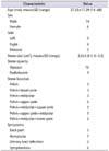
This was a retrospective study. Ethical approval was given by the Medical Ethics Committee of the Affiliated Hospital of Hebei University. Between September 2009 and August 2011, we performed 20 procedures involving ultramini PCNL combined with flexible ureterorenoscopic holmium laser lithotripsy. All cases had multiple renal calculi, and the average patient age was 37.35 months (range, 14-68 months). There were 3 cases of bilateral renal calculi and 17 cases of unilateral stones (9 left-sided and 8 right-sided stones). There were 14 male and six female patients. Twelve stone sites were located in two renal calyces, eight calculi sites were scattered in more than two renal calyces, and three stone sites were limited to one renal calyx. The average stone diameter was 2.0 cm (range, 1-3.0 cm). In this group, five patients presented with lumbar back pain, seven patients had hematuria, two patients had urinary tract infection, and six patients had no obvious symptoms but stones were found on radiological investigation. The preoperative patient evaluation included history taking, clinical examination, and routine laboratory investigations such as blood analysis, urine analysis, renal function tests, biochemical tests, urinary tract ultrasound, and computed tomography (CT) imaging. In patients who did not undergo CT scanning, intravenous urography was performed to evaluate the collecting system. Patients with a positive urinary culture received specific antibiotic therapy; the operation was performed only after urine examinations were found to be normal (Table 1).
2. Surgical technique
The operation was performed under general anesthesia. As the first step, an F4 ureteral catheter was retrogradely inserted in the lithotomic position using a 10-Fr semirigid ureteroscope. The catheter was connected to an irrigation system to establish hydronephrosis. The patient was then placed in a prone position. Before the collecting system was punctured under ultrasound guidance with a puncture needle, a puncture site corresponding to the position of the stone and its largest bulk was identified. The puncture of a posterior middle calyx was preferred in most cases because this region is considered an avascular area, and also because performing flexible URS in this region is easy. Once access was established, a security guidewire was inserted into the collecting system. After insertion of the security guidewire, the tract was dilated to 10 Fr by using nephrostomy dilators (ultramini tract). The dilator was equipped with an Fr10 access sheath. The dilator was then removed, leaving the access sheath in the collecting system. Then, the flexible ureterorenoscope with holmium laser lithotripsy system was introduced into the renal pelvis through the access sheath. Procedures were performed with flexible URS. In all cases, the holmium laser lithotripsy system was used for stone fragmentation.
Generally, the first step was to treat the middle calyx stones in order to obtain good visibility, then the upper calyx calculi, and finally the inferior calyceal stones. In 5 patients, a basket (1.5F NCircle, Cook Medical, Bloomington, IN, USA) was used to pull out large stone fragments.
At the end of the procedure, if no bleeding was observed at the removal of the access sheath, a nephrostomy tube did not need to be placed. In cases of bleeding, an Fr8 nephrostomy tube was placed. In most cases, the ureter was drained with a double-J stent. All patients were catheterized with a Foley bladder catheter for 1 day after the operation.
Radiological examination was undertaken on the first postoperative day to evaluate the position of the nephrostomy tube and double-J stent. If the stones were translucent on radiography, the postoperative examination was performed by CT scanning or ultrasound. If the patient had no large residual stones and did not require another operation, the nephrostomy tube was removed before the patient left the hospital. The double-J stent was removed one week after surgery (hospitalization under general anaesthesia, with Fr10 semirigid ureteroscopy). Treatment success was defined as complete clearance of the stones or the presence of residual fragments <4 mm [10].
3. Statistical analysis
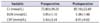
Quantitative data were analyzed and means and standard deviations calculated. Data were analyzed by using SPSS ver. 16.0 (IBM Co., Armonk, NY, USA). The levels of creatinine (Cr), blood urea nitrogen (BUN), C-reactive protein (CRP), and hemoglobin drop before and after the operation were compared by using paired t-tests. A p-value less than 0.05 was considered significant.
RESULTS
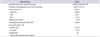
All procedures were performed through a single puncture site, including the three cases of bilateral renal calculi. Puncture was performed through the middle calyx in 19 stone sites and through the lower calyx in four stone sites. Because of bleeding during tract establishment or lithotripsy, the procedure was halted in two patients while retaining the nephrostomy tube, and lithotripsy was resumed after 24 hours [9]. In one case, pus was extracted from the collecting system and reserved nephrostomy tube, and bacterial cultures and drug sensitivity tests were performed. Sensitive antibiotics were selected to control the infection, and after the infection was controlled, the patient underwent an elective operation. The operative time ranged from 60 to 120 minutes, averaging 83.85 minutes. Radiological examination was performed on the first postoperative day. The complete stone-free rate with residual fragments <4 mm was 87% (20/23 sites). Postoperatively, two patients developed a moderate fever (<39℃) that gradually decreased within two days with no bacteriologic evidence of infection. Hemoglobin decreased to a level of 1.0 g/dL (1.0±0.4 g/dL), and no patients required blood transfusion. Levels of BUN, Cr, and CRP were not significantly different before and after the operation (Table 2). There were no intraoperative complications. The duration of hospital stay was 4.85 days (range, 3-7 days) in the whole series. Three sites (13%) received further treatment with extracorporeal shock wave lithotripsy (ESWL). The success rate at 3 months increased to 100% (Table 3).
Two patients were lost to follow-up, and the remaining 18 patients were followed for an average of 9 months (range, 6-12 months). No long-term complications were reported.
DISCUSSION
The incidence rate of renal stones in pediatric patients is lower than in adults [11]. Stone disease in this age group might be associated with anatomical and metabolic abnormalities or infectious diseases, and the risk of recurrence is high [9]. Pediatric renal stones can be successfully managed by PCNL and ESWL [11], but ESWL has many limitations in very young children. Renal function can be affected by repeated ESWL because of a large stone burden or multiple stones. In addition, preschool-aged children exposed to x-rays may suffer from long-term effects such as reproductive system damage, hypertension, and more [12]. Since the use of PCNL in the treatment of children with stone disease was reported, many centers have implemented this method [13]. The main complication of PCNL is bleeding [14], and because the blood volume of children is significantly lower than that of adults, the impact of bleeding is very serious in children. Rodrigues Netto et al. [15] confirmed that 11% to 14% of children who underwent PCNL required blood transfusions. Unsal et al. [16] reported that the size of dilatation and the number of tracts are probably the most important factors that affect blood loss during surgery, leading to nephrectomy. Bleeding complications generally occur during the dilation of the tract and lithotripsy, especially when rigid or semirigid instruments are used. The ideal method to prevent bleeding is the use of small-caliber instruments [17]. Mishra et al. [18] confirmed that mini PCNL significantly reduces bleeding compared with standard PCNL and shortens the time of hospitalization. Therefore, a smaller caliber tract and equipment is very suitable for pediatric patients.
Mini PCNL refers to a tract dilation of Fr14-16. One study [9] demonstrated that when mini PCNL was combined with semirigid ureteroscopy for the treatment of renal calculi, the stone-free rate reached 85%, renal injury was less severe, the operation time was shorter (86.5 minutes), and hemoglobin was reduced to approximately 1.4±1 g/dL [19]. There was only one case of recurrence, and no apparent complications. We used the ultramini PCNL combined with flexible URS in the treatment of children with multiple renal calculi. Hemoglobin decreased insignificantly (1±0.4 g/dL) in our study, and no blood transfusions were needed.
The mini PCNL combined with rigid or semirigid ureteroscopic lithotripsy also has some limitations, especially when the stones are not in the same calyx. Aron et al. [5] stated that torquing a rigid nephroscope is the most important cause of bleeding during PCNL; therefore, they used multiple tracts instead of a single tract in their series. However, multiple tracts increase the risk of complications such as renal infundibulum laceration and hemorrhage [2021]. Flexible URS can lessen the risk of such complications. Flexible URS has become a more efficient and safer means of treating stones throughout all renal calyces though a single tract [22], while reducing risk factors. Nouralizadeh et al. [23] used single tract access in their pediatric PCNL series, which consisted of 24 renal units, and one patient required a blood transfusion. One study has also confirmed that flexible URS also has a better outcome with large renal stones [22]. We used the ultramini tract and flexible URS in the treatment of pediatric multiple renal calculi and found that it had many advantages: the tract was tenuous and damage during dilation was decreased; multiple renal stones could be processed through a single tract, which prevented kidney laceration and hemorrhage; and the perfusion pressure of flexible URS is less than that of the rigid or semirigid URS and can maintain a clear operation field and therefore ensure greater safety. Our study showed that a stone-free rate of 87% could be achieved in treating multiple renal calculi, including three bilateral cases, with no postoperative complications.
The ultrasound-guided procedure is safe and effective. We chose ultrasound real-time monitoring, and the puncture process proceeded smoothly.
Bozkurt et al. [24] reported that 32 patients with pregnancy complicated by upper urinary calculi underwent holmium laser lithotripsy and were followed until the babies were born, and all the newborns were healthy. We found no complications in children after the use of holmium laser lithotripsy, and confirmed that the holmium laser is safe to use in pediatric patients and even for fetal ages.
Most of the cases in this study were successfully treated over the same period, but for bleeding cases, a staged operation is necessary. The operation can be performed through the original tract with less bleeding, clear operational vision, less fluid absorption, a short operation time, and a high rate of stone clearance.
The sequence followed in the renal calices was as follows: first, fragmentation of the stones in the middle calyx to ensure good visibility, then moving into the upper pole calyx, and finally treatment of stones in the lower calyx in which smaller stones are deposited. Khalil [25] reported that if the stone is located in the inferior calyx, more ESWL time may be needed to obtain the desired effect, because the lower calyces possess the special anatomical structure named the infundibulum [26]. Lower calyceal stones are not easily eliminated from the body, and therefore during the operation, the stones should be powdered completely.
CONCLUSIONS
Ultrasound-guided ultramini PCNL combined with flexible URS holmium laser lithotripsy is associated with fewer complications and shorter hospitalization and is a safe and effective method for pediatric patients with multiple renal stones. The main limitation of our study was that this was a retrospective non-case-control study and the sample size was small. Precise data on the postoperative visual analogue scales pain score were also lacking. We need more samples and advanced observation.
 XML Download