Abstract
A patient with a complete right ureteral triplication presented with recurrent pyelonephritis and flank pain that was refractory to medical management. Evaluation showed that the atrophic upper-most renal moiety had been chronically obstructed and was associated with a dilated ureter. Intraureteral and intravenous indocyanine green (ICG) were used as real-time contrast agents intraoperatively to facilitate right robotic partial nephroureterectomy of the diseased system. Intraureteral ICG was used to accurately distinguish the pathologic ureter and associated renal pelvis from its normal counterparts. Intravenous ICG was used to assess perfusion in the right kidney and delineate the margins of diseased renal parenchyma.
Complete ureteral triplication [1], the presence of three individual ureters originating from the kidney and draining into three separate orifices, is a rare congenital anomaly. Patients with such anatomy may be predisposed to recurrent infections, renal colic, and incontinence secondary to associated obstruction and reflux [1,2]. When patients with complete ureteral triplication require surgical management, the surgeon must evaluate the degree of renal compromise attributable to the pathologic portions of the urinary tract, and correctly distinguish the diseased portions of the urinary tract from its healthy counterparts. It is for this reason that the role of perioperative imaging to guide operative treatment cannot be overemphasized.
Indocyanine green (ICG), a fluorescent dye that can be visualized under near-infrared fluorescence (NIRF), has been utilized intraoperatively during robotic urologic procedures to highlight relevant anatomy and assess perfusion [3]. ICG may be visualized using the FIREFLY system, which is integrated into the da Vinci Si robot (Intuitive Surgical, Sunnyvale, CA, USA). Herein, we describe a case in which both intraureteral and intravenous ICG was used to facilitate a complex robotic partial nephroureterectomy in a patient with complete ureteral triplication.
A 31-year-old female with a congenital right ureteral triplication presented with recurrent right-sided pyelonephritis and persistent right-sided flank pain refractory to medical management. Delayed phase computed tomography (CT) scan with intravenous contrast clearly showed that each of the three right-sided ureters was associated with its own collecting system, and emptied separately into the bladder. The uppermost moiety showed sequelae of chronic obstruction: significant parenchymal thinning, delayed contrast excretion, and dilated ureter. The two inferior collecting systems and their associated ureters appeared to be emptying normally (Fig. 1). Mercaptuacetyltriglycine (MAG3) study showed symmetrical renal function with no evidence of obstructive uropathy bilaterally. The uppermost moiety was not well delineated on the MAG3 study.
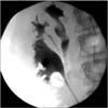
The patient elected for robotic right uppermost pole moiety nephroureterectomy. The patient consented to off-labeled use of ICG after a complete discussion of its risks and benefits. We used cystoscopy and retrograde pyelography to show that the three separate right ureteral orifices were consistent with Weigert-Meyer law, and identify the ureter of interest (Fig. 2). After preparing the IC-Green (Akorn Inc., Lake Forest, IL, USA) by mixing 25-mg sterile ICG in 10-mL distilled water, we used a 6-Fr ureteral catheter to inject a 5-mL solution of ICG retrograde into the lumen of the diseased ureter. The ureteral catheter was then clamped to maximize ICG retention in the ureter. A 16-Fr Foley catheter was then inserted and secured to the ureteral catheter.
The patient was placed in right-side-up flank position with the table in full flexion. We used our standard robotic port placement strategy for robotic nephroureterectomy that was previously described at our center [4]. After mobilizing the right colon, we turned on the FIREFLY modality to locate the diseased ureter. Under NIRF, the diseased ureter fluoresced green while the other two healthy ureters did not fluoresce (Fig. 3). The diseased, fluorescent ureter was circumferentially dissected distally. Two Weck Hem-o-lok clips (Teleflex, Morrisville, NC, USA) were secured on the ureter at its junction with the bladder prior to transecting the ureter. The diseased ureter was then dissected proximally to its associated renal pelvis.
During our dissection of the uppermost pole renal pelvis, we encountered a significant amount of fibrotic tissue, likely attributable to the patient's history of recurrent pyelonephritis. By toggling the NIRF on and off, we were able to clearly distinguish between the green fluorescing renal pelvis and ureter, from the nonfluorescing fibrotic tissue. After identifying the arterial and venous branches of the main renal artery and vein supplying the uppermost pole, we clipped the proximal and distal ends using two Weck Hem-o-lok clips and transected the vessels. We then injected 2-mg ICG intravenously to visualize perfusion to the kidney. Under NIRF, there was a perfusion defect in the uppermost pole moiety of the kidney where we had removed its blood supply; the remainder of the kidney was well-perfused and fluoresced green (Fig. 4). Utilizing NIRF to delineate our margins, we excised the diseased collecting system and its blood supply en bloc. Operative time was 157 minutes.
Ureteral triplication is a rare congenital anomaly; only approximately 100 cases have been described in the literature [5]. Smith classified ureteral triplication into four major variants [1,2]: Type 1, complete ureteral triplication (35%)-three ureters originating from the kidney and emptying into three orifices; type 2, incomplete triplication (21%)-three ureters originating from the kidney, with two of these ureters joining to empty into two orifices; type 3, trifid ureter (31%)-three separate ureters originating from the kidney, with all three ureters joining to empty into a single orifice; type 4, double ureter, one bifurcated (9%)-two separate ureters originating from the kidney, with one ureter birfuricating into an "inverse Y" to empty into three orifices. Our case was a type 1, complete ureteral triplication with all three ureters associated with its own renal collecting system and emptying into three separate bladder orifices.
Surgical management of patients with ureteral triplication, which may involve simultaneous treatment of the upper and lower urinary tracts, is most often indicated when there is a risk of renal compromise as in cases of veiscoureteral reflux, obstruction, ureteral ectopy, and recurrent infections. Given the variations in anatomy and increased predisposition for pathology, prudent use of perioperative imaging to guide surgical management of ureteral triplications is essential. Patel et al. [6] described the role of various preoperative imaging modalities on assessing anatomy and function in a patient with a type 2 ureteral triplication with ectopia. A dimercaptosuccinic acid renal scan showed decreased perfusion and uptake by the right upper pole, suggesting a duplicated anomaly and ectopic ureter. Cystoscopy and retrograde pyelography demonstrated that there were two ureters originating from the right kidney that joined intramurally to form a single bladder orifice. Intravenous urography and CT scan revealed the ectopic third ureter, which was dilated and associated with the upper pole.
Flechsig et al. [7] described using dynamic magnetic resonance nephrography to concomitantly assess anatomic and functional characteristics in a 5-year-old girl with a type 1 left ureteral triplication. Initial evaluation with a MAG3 renal scan suggested poor renal excretion in the left lower pole, but the authors noted that precise localization of the impaired region was difficult due to limited resolution. However, a dynamic magnetic resonance nephrography allowed for a detailed region-of-interest evaluation that showed 45%, 46%, and 9% function in the upper, middle and lower pole, respectively. Furthermore, the authors noted that the correlation between preoperative imaging and intraoperative findings assisted in performing laparoscopic partial nephroureterectomy.
An intraoperative imaging modality that assists in correctly identifying the pathologic ureter and associated renal pelvis, and in precisely determining the margin between compromised versus healthy renal parenchyma would facilitate surgical management of patients with ureteral triplication. Such an imaging modality would be particularly helpful during robotic surgery, as the surgeon relies solely on visual cues to guide dissection due to the lack of tactile feedback. Despite this, a means to intraoperatively image relevant anatomy and evaluate function in real-time during robotic surgical management of ureteral triplications has yet to be described. ICG has recently been increasingly utilized in urologic procedures as an intraoperative contrast agent to highlight relevant anatomy. It is particularly helpful for intraoperative use as it has a low toxicity, high signal-to-noise ratio, and penetrates tissue [3]. Tobis et al. [8] first detailed the use of intravenous ICG during robotic partial nephrectomy to differentiate normal renal parenchyma (bright green fluorescence) from tumors, cysts, and areas of fat necrosis (decreased/no fluorescence). Bjurlin et al. [9] noted that intravenous ICG may also be used to assess tissue perfusion during surgical management of the upper urinary tract. Poorly perfused fibrotic scar tissue appeared dark while well perfused renal pelvis and ureter appeared green. We have found the use of intraureteral ICG to assist in robotic ureteral reconstructions, which was first described at our center, to be particularly helpful in localizing the ureter despite severe periureteral inflammation and in determining margins of ureteral strictures [3].
In our case, we utilized both intraureteral and intravenous ICG intraoperatively to highlight relevant anatomy during robotic right uppermost pole nephroureterectomy in a patient with type 1 ureteral triplication. Intraureteral ICG aided in accurately identifying the pathologic ureter and associated renal pelvis in real-time; the system of interest fluoresced green while the other two systems did not fluoresce. This aided our ability to focus our dissection to the ureter of interest without risking devascularization to the healthy ureters. Intravenous ICG aided in assessing perfusion to the right kidney and delineating the margins of defective renal parenchyma in real-time; the well-perfused middle and lower collecting systems fluoresced bright green, while the upper-most collecting system did not fluoresce as brightly. The use of intraureteral and intravenous ICG to highlight relevant anatomy intraoperatively may be of particular benefit in patients with complex urinary tract anatomy, especially in cases necessitating concomitant operative management of upper and lower tracts.
Figures and Tables
Fig. 1
(A) Coronal view suggesting three ureters, each associated with own renal pelvis. (B) Coronal view showing cortical thinning and right uppermost pole.

References
1. Smith I. Triplicate ureter. Br J Surg. 1946; 34:182–185.
2. Sivrikaya A, Cay A, Imamoglu M, Sarihan H. A case of ureteral triplication associated with ureteropelvic junction obstruction. Int Urol Nephrol. 2007; 39:755–757.
3. Lee Z, Moore B, Giusto L, Eun DD. Use of indocyanine green during robot-assisted ureteral reconstructions. Eur Urol. 2015; 67:291–298.
4. Lee Z, Cadillo-Chavez R, Lee DI, Llukani E, Eun D. The technique of single stage pure robotic nephroureterectomy. J Endourol. 2013; 27:189–195.
5. Hsu TH, Goldfarb DA. Blind-ending ureteral triplication. J Urol. 1998; 159:1295.
6. Patel PM, Stock JA, Hanna MK, Lutzker L. Ureteral triplication with ectopic upper pole moiety. Urology. 2001; 58:279–280.
7. Flechsig H, Fuchs J, Warmann SW, Schaefer JF. Magnetic resonance nephrography for planning of laparoscopic partial nephrectomy in a pediatric case of ureteral triplication. J Pediatr Surg. 2010; 45:2053–2057.
8. Tobis S, Knopf J, Silvers C, Yao J, Rashid H, Wu G, et al. Near infrared fluorescence imaging with robotic assisted laparoscopic partial nephrectomy: initial clinical experience for renal cortical tumors. J Urol. 2011; 186:47–52.
9. Bjurlin MA, Gan M, McClintock TR, Volpe A, Borofsky MS, Mottrie A, et al. Near-infrared fluorescence imaging: emerging applications in robotic upper urinary tract surgery. Eur Urol. 2014; 65:793–801.


 PDF
PDF ePub
ePub Citation
 Print
Print


 XML Download