

 PDF
PDF ePub
ePub Citation
 Print
Print


INTRODUCTION
Ureteral stenting was first described by Zimskind et al. [1] in 1967 and is widely used in contemporary urologic practice. Indications for ureteral stents include the relief of ureteral obstruction with or without stone, stricture, identification of the ureter during a surgical procedure, and protection of ureteral anastomosis in urinary diversion. However, the use of an indwelling ureteral stent can cause many complications, ranging from mild (hematuria, irritative urinary symptom, vesico-ureteral reflex, and recurrent urinary tract infection) to severe (stent migration, encrustation, ureteral perforation, erosion, and fistula) [2]. One extremely rare complication is knot formation of an indwelling stent in the ureter. To our knowledge, only 20 cases of knotted ureteral stent have been described in the literature. We present a case in which a Terumo guidewire was successfully used to remove a knotted stent percutaneously without anesthesia. We also review the current literature on predisposing factors and management strategies for knotted stents.
CASE REPORT
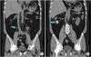
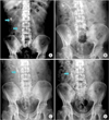
A 53-year-old man with gout and chronic renal disease was admitted to the hospital because of right flank pain, nausea, vomiting, and fever. A 2.0-cm renal stone in the lower calyx and a 0.4-cm stone in the upper ureter without hydronephrosis were identified on computed tomography images (Fig. 1). The patient was treated with antibiotics and underwent ureteroscopic lithotripsy and stent placement to decompress the infected kidney and extracorporeal shock wave lithotripsy (ESWL) of the renal stone (Fig. 2A, B). After the signs of infection subsided and the uropathy improved (changes in high-sensitivity C-reactive protein from 22.2 to 2.75 mg/dL and in creatinine from 3.6 to 1.7 mg/dL), we performed several sessions of ESWL on the right renal stone. Two months later, we found Steinstrasse around the stent (Fig. 2C). We attempted to change the stent but failed to remove it because of resistance. We therefore checked for an encrusted stent by x-ray and computed tomography, which revealed a knotted stent and Steinstrasse (Fig. 2D). The patient refused general or spinal anesthesia, so the stent was removed percutaneously under local anesthesia.
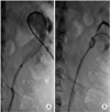
We inserted a 7-French angio sheath using ultrasonographic and fluoroscopic guidance and then inserted a KMP (Beacon Tip Torcon NB advantage angiographic catheter, Cook Medical, Bloomington, IN, USA) catheter. Finally, a folded 0.018 Terumo guidewire was inserted into the area of knot formation through the KMP catheter. The knotted stent was caught in the folded guidewire and removed percutaneously. Subsequently, an 8.5-French pigtail catheter was inserted in the kidney (Fig. 3). After several sessions of ESWL performed within 2 weeks of the surgical procedure, the remnant stone and Steinstrasse were completely eliminated and the pigtail catheter was removed.
DISCUSSION
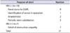
Formation of a knotted ureteral stent was first described by Groeneveld in 1989 [3]. To date, 20 such cases have been reported and several treatment methods have been described. Our analysis of previous reports of knotted stent indicated that the male to female ratio is 3:1 and the mean age is 50 years (range, 4-86 years). The knotted stents were located in the upper ureter in 18 cases, the middle ureter in 2 cases, and the distal ureter in 1 case (Supplementary Table 1) [4,5,6,7,8]. The most common reasons for stent placement were ESWL of a renal stone (9 cases), treatment of upper ureter stone or upper ureter obstruction (7 cases), to aid with anastomosis of the ureter (2 cases), periodic stent substitution (2 cases), and interoperative ureter determination (1 case) (Table 1). In the current case of a 53-year-old man, an indwelling stent was inserted to aid in the removal of a renal stone and to relieve an upper ureteral obstruction.
The results of our analysis suggest that the excessive length of the stent and/or its coil formation facilitated the knot formation in the ureteral catheter. Ureteral catheter knots form during implantation or removal of the catheter. Analysis of cases with and without hydronephrosis before insertion of an upper ureteral stent paradoxically showed that knot formation occurred in 13 cases with no hydronephrosis (Table 1), which suggests that knot formation occurs more often in cases with no hydronephrosis. Therefore, physicians should pay greater attention to insertion or removal of a ureteral stent in patients without than in those with hydronephrosis. If possible, performing the procedure with the aid of fluoroscopic imaging may help prevent knot formation.
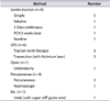
Various techniques have been used to assist with the removal of knotted ureteral catheters, such as gentle traction (9 cases), percutaneous extraction (4 cases), extraction via ureteroscopy (6 cases), extraction using a guidewire (1 case), and open ureterotomy (1 case) (Table 2). Of nine patients treated using a gentle traction method, the stent was gently extracted in five patients and by abdominal pressure under anesthesia in one patient. In one reported case, the knotted stent was extracted by anchoring the end of the stent at the leg. In another case, the stent was extracted after waiting for status of the percutaneous nephrostomy (PCN). In one case, stent removal was achieved by injecting petroleum jelly in the ureterocutaneostomy [5]. However, ureteral avulsion is a potential risk of these techniques.
Cases of ureteroscopic removal of knotted ureteral stents have also been reported. Traction using forceps or a basket was reported in three patients, and cutting with a holmium laser was reported in another three patients (Table 2) [6,7]. When a knotted stent is removed by ureteroscopy, the patient needs to be under general or spinal anesthesia; this procedure has a risk of failure related to ureteral stricture. Hydronephrosis that occurs because of a knotted stent should be resolved in cases of failure by inserting a ureteroscope.
Three cases of percutaneous removal of a knotted ureteral stent were reported: in 1992, 1994, and 2012. In two cases, the knotted stent was removed via PCN [4]. In the remaining case, a nephroscope was used to remove the knotted stent [8] (Table 2). The exact PCN technique used was not specified, but nephroscopy is an invasive technique, requires general anesthesia, and may result in renal injury. Therefore, removal of knotted stents with a simple procedure under local anesthesia is an attractive alternative option.
Reported techniques for removal of a knotted ureteral stent include retrograde gentle traction, untying the knot, cutting the knot, and percutaneous removal. Gentle traction, which is the most common method, can result in ureteral injury. Use of ureteroscopy to untie or cut a knot requires general or spinal anesthesia and possibly a holmiun laser. Percutaneous removal of a knotted stent is a simple and effective method that does not require general or spinal anesthesia and can be performed when the knot is located in the upper portion of the ureteral stent or when hydronephrosis is present. This technique may be a useful treatment option in patients who have difficulties with general or spinal anesthesia and/or in those who do not want to undergo cystoscopy for later removal of a ureteral stent.

 XML Download