

 PDF
PDF ePub
ePub Citation
 Print
Print


INTRODUCTION
In men and women, profound structural and functional alterations occur in the lower urinary tract in association with aging, which may be responsible for lower urinary tract symptoms (LUTS) in the elderly population [1]. Most studies of LUTS in community- or hospital-based populations indicate that an increase of LUTS with age is not gender-specific [2].
In the Korean EPIC study, the overall prevalence of LUTS was 61.4% (53.7% of men, 68.9% of women) and the prevalence increased with age. Among the symptoms, urinary incontinence was reported by 28.4% of women and the most prevalent type was stress urinary incontinence (SUI) [3].
Clinical experience and the literature suggest that women may have an increase in micturition frequency and a decrease in bladder capacity, bladder sensation, detrusor contractility, and urethral sphincter function with increasing age [1,4,5,6,7]. However, the results of these studies were based on questionnaire analysis. Knowledge of ageassociated changes in urodynamic parameters in women is lacking. Thus, we analyzed age-associated changes in urodynamic parameters in women, especially in patients with SUI.
MATERIALS AND METHODS
We analyzed the urodynamic study (UDS) results of patients with urodynamically proven SUI between March 2008 and July 2014. We obtained approval for the study from the Institutional Review Board at our hospital. Exclusion criteria included cerebrovascular accident, dementia or Alzheimer disease, multiple sclerosis or Parkinson disease, spinal cord injury or malformation resulting in gross neuropathy, detrusor-sphincter dyssynergia, and current urinary tract infection. The patients' medical history including diabetes mellitus, hypertension, and parity was checked thoroughly.
Methods and units of UDS conformed to the standards recommended by the International Continence Society [8,9]. Written informed consent for UDS was given to all subjects before the detailed clinical evaluation. UDS was performed preoperatively by a single expert in an exclusive urodynamic room intended for quiet and protected from unnecessary interruptions. UDS consisted of uroflowmetry followed by filling and voiding cystometry and was conducted interactively with the patient. Noninstrumented uroflowmetry was conducted when the patients felt a normal desire to void, and catheterized postvoid residual urine volume (PVR) was measured. Filling and voiding cystometry were conducted with the patient in a sitting position. A 5-Fr rectal balloon catheter was inserted at the anus, and electromyographic electrodes were attached at both sides of the anus. A 6-Fr triple-lumen transurethral catheter was inserted into the urethra and connected to the pressure transducer. Prior to bladder filling, signal quality control was done. We checked that resting values for abdominal, intravesical, and detrusor pressures were in a typical range. Cough was used to ensure that the abdominal and intravesical pressure signals responded equally. Then, the bladder was filled at a rate of 50 mL/min. When artifacts occurred during study, they were immediately corrected. All measured and derived signals displayed according to ICS standards with abdominal pressure, vesical pressure, detrusor pressure, and flow [8]. Filling volume, electromyography, and voided volume were displayed in additional curves. UDS findings and the interpretation of the results were documented immediately after the study was finished. In the uroflowmetry, maximal flow rate (Qmax), time to Qmax, voided volume, PVR, and filling cystometry data including first, strong desire to void and Valsalva leak point pressure (VLPP) were measured. Also, Qmax and detrusor pressure at Qmax (Pdet@Qmax) of voiding cystometry data were analyzed. The bladder contractility index (BCI= pdet@Qmax+5Qmax) was also calculated [8].
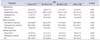
The urodynamic parameters were analyzed and compared between the age groups. The patients were categorized into the following age groups: less than 50, 50 to 59, 60 to 69, and greater than 69 years old.
The Shapiro Wilks test was used to test for normality. We used the Kruskal-Wallis test for comparisons of continuous variables between age groups. A p-value of less than 0.05 was considered statistically significant. For multiple comparisons, we used the Wilcoxon rank sum test, and p-value of less than 0.0083 according to Bonferroni's method was considered statistically significant. All statistical analyses were performed by using STATA 11.0 (StataCorp LP, College Station, TX, USA).
RESULTS
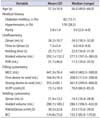
The subjects included 776 patients. Among the patients, 151 were withdrawn owing to incomplete UDS data or because they met the exclusion criteria. A total of 625 patients enrolled in the present study. The patients' clinical characteristics are shown in Table 1. Diabetes mellitus and hypertension occurred in 13.1% and 28.5% of the patients, respectively. The mean parity number was 3.0±1.4. The mean Qmax was 26.2±10.7 mL/s, time to Qmax was 7.5±5.4 s, mean voiding time was 25.7±15.7 s, and the mean voided volume was 292.1±132.2 mL. The mean catheterized PVR was 31.7±46.6 mL (Table 1). Qmax in uroflowmetry and PVR were significantly different between the age groups (p=0.001 and p=0.042, respectively) (Table 2). In filling cystometry, the mean first desire and strong desire to void were 166.9±76.3 mL and 355.2±85.1 mL, respectively. The mean VLPP was 73.1±18.9 cmH2O, and the mean maximal cystometric capacity was 441.3±76.4 mL (Table 1). The first desire to void was significantly different between the age groups (p=0.042) (Table 2). In voiding cystometry, mean Qmax, voided volume, and Pdet@Qmax were 21.0±13.5 mL/s, 280.7±180.15 mL, and 30.2±22.8 cmH2O, respectively. The mean BCI was 134.8±72.6 (Table 1). Pdet@Qmax and BCI were significantly different between the age groups (p=0.016 and p=0.046, respectively). Qmax and Pdet@Qmax were decreased and PVR was increased significantly with age after 60 years (Table 2).
DISCUSSION
The major finding of our study is that Qmax and voided volume showed a significant decrease and PVR and desire to void showed a significant increase with aging. Also, Pdet@Qmax and BCI were significantly decreased with aging. Very few studies have been done of age-associated changes in urodynamic parameters in a large group of women. Valentini et al. [10] reported that a brisk change in the LUTS of women older than 75 years underlined deterioration in bladder function with a high incidence of detrusor hyperactivity with or without impaired contractility, whereas urethral function changed progressively. The effect of aging appears to be predominant compared with the effect of menopause. Detrusor function significantly deteriorated in the oldest group. A progressive decrease of maximum urethral closure pressure occurred with aging. However, the subjects with no detrusor overactivity showed a decrease in detrusor pressure at opening and maximum flow with aging, whereas PVR increased [10]. Zimmern et al. [11] reported that women aged 65 years and older with SUI had significant decreases in Qmax, Pdet@Qmax, and VLPP; increases in the time to Qmax, voiding time, and desire to void; and no difference in PVR. Detrusor hypocontractility increases with age. To the best of our knowledge, that result is the outcome of the most recent study for age-associated change in urodynamic parameters in a large group of women. The results of our study are similar to the existing findings in worsening of urodynamic parameters. The BCI indicated that bladder contractility also decreased with age after 60 years.
A study presented by Basu et al. [12] rebuts the main points of change with aging in urodynamic parameters. Those authors used correlation to analyze the association between age and voiding parameters in 896 datasets. Multivariate analysis was used to further investigate the data among different age groups. The outcomes of the analysis showed a significant effect on voiding volume but did not show significant effects on any other variables studied. According to the data, age was not related to flow rate percentile, maximum flow rate, or Pdet@Qmax. The study concluded that the data suggest that there is no significant change in voiding function related to age. Further studies on a large group of subjects are needed to meticulously analyze age-associated change in urodynamic parameters.
Older and younger women have similar SUI outcomes after primary SUI surgery. However, older women have more persistent urgency symptoms and a worse impression of improvement in their urinary tract condition than do younger women [3,13]. In our opinion, these findings could be the result of decreased voiding function at baseline in the older women.
The limitations of our study include those inherent to a retrospective study design in the primary acquisition of data. In addition, our study did not require the subjects to obtain a voided volume of more than 120 to 150 mL. Although it is known that a valid Qmax requires a voided volume of more than 150 mL [8], some of our subjects had difficulty reaching the minimum range because their health was not in the best condition and they were older females with SUI. However, in order to measure Qmax with functional bladder capacity, noninvasive uroflowmetry was conducted when the patients felt a normal desire to void. Therefore, we intended to study a large group of subjects to practically analyze the general voiding patterns of the subjects. Despite these limitations, the study was strengthened by clear and robust validated outcome measures by UDS, and it provides more information than a questionnaire analysis. To our knowledge, this is the first report of age-associated changes in urodynamic parameters in Asian women with SUI. This information will be helpful for providing more professional counseling to Asian women who suffer from SUI with aging.
 XML Download