Abstract
Purpose
To evaluate the predictive role of the neutrophil to lymphocyte ratio (NLR), platelet to lymphocyte ratio (PLR), mean platelet volume (MPV), and platelet count (PLT) in the diagnosis of testicular torsion (TT) and testicular viability following TT.
Materials and Methods
We analyzed two study groups in this retrospective study: 75 patients with a diagnosis of TT (group 1) and 56 age-matched healthy subjects (group 2). We performed a complete blood count as a part of the diagnostic procedure, and NLR, PLR, MPV, and PLT values were recorded. We compared the patient and control groups in terms of these parameters. Then, TT patients were divided into two subgroups according to the time elapsed since the onset of symptoms. Subsequently, we evaluated the relationship between the duration of symptoms and these parameters.
Results
There were significant differences between groups 1 and 2 in NLR, PLR, and PLT (p<0.001 for all). There was no predictive role of MPV in the diagnosis of TT (p=0.328). We determined significantly high sensitivity and specificity levels for NLR in the prediction of TT diagnosis (84% and 92%, respectively). Furthermore, NLR was significantly related to the duration of symptoms in TT patients (p=0.01).
The annual incidence of testicular torsion (TT) is 1 in 4,000 males younger than 25 years, and its clinical diagnosis remains challenging for urologists and pediatric surgeons [1]. TT is an important diagnosis to consider owing to the risk of infarction and infertility [2]. Testicular recovery is more likely if the intervention begins in the first 6 hours after onset of symptoms. Viability decreases dramatically after 12 hours [3]. Many clinical and experimental studies have been performed to reduce the negative exploration rate and testicular loss [3,4,5]. These studies have been based on clinical variables and color Doppler ultrasonography (CDU) findings. Moreover, spectrometric tests and dual-wavelength systems have been investigated in recent experimental studies [6,7]. However, there is no consensus about the proper method for the exact diagnosis of TT.
The neutrophil to lymphocyte ratio (NLR), platelet to lymphocyte ratio (PLR), and mean platelet volume (MPV) have become popular research topics as inflammatory hematological parameters. The possible predictive role of these markers in cancer (including prostate cancer, renal cell cancer, and urinary tract urothelial carcinomas) diagnosis and prognosis has been reported [8,9,10]. To our knowledge, these parameters have not been used previously to predict TT diagnosis. In the current study, our aim was to evaluate the predictive role of NLR, PLR, MPV, and platelet count (PLT) in the diagnosis of TT and testicular viability following TT.
A total of 75 patients (group 1) who were under 25 years of age and diagnosed with TT by scrotal exploration between July 2007 and May 2014 and 56 age-matched healthy subjects (group 2) were included in this study. A comprehensive medical history was obtained from the patients' medical records. All participants had been examined physically by a urologist resident and attending physician. The database also comprised the side of the affected testis, presence of erythema and swelling, scrotal tenderness, localization of the testis, fever (>38.5℃), pyuria, nausea/vomiting, abdominal pain, and the presence of a normal cremasteric reflex. Furthermore, we performed CDU for each patient as part of the diagnostic process.
A complete blood count was performed by the method of flow cytometry (Beckman Coulter LH 780 Analyzer, Beckman Coulter Inc., Miami, FL, USA) for each individual. Venous blood samples were drawn into tubes containing ethylenediaminetetraacetic acid for the measurement of hematological parameters before surgical intervention from patients and healthy control subjects. All hematological parameters were denominated in 103/µL. NLR and PLR were assessed by using these parameters. The MPV value was denominated in femtoliter. Peripheral blood of patients was obtained at the time of the first application. Patients with perinatal (extravaginal) TT, acute epididymo-orchitis, and torsion of the appendix testis were excluded from the study.
For the statistical analysis, duration of symptoms was categorized as either longer or shorter than 12 hours in group 1. Sensitivity, specificity, positive predictive value (PPV), negative predictive value (NPV), and accuracy of the hematological parameters were compared among these two groups in relation to duration of symptoms. The protocol was approved by the local Ethics Committee at Suleyman Demirel University.
All statistical analyses were performed by using the IBM SPSS Statistics ver. 20.0 (IBM Co., Armonk, NY, USA). The Kolmogorov-Smirnov test was used to determine whether the values were normally distributed. Continuous variables were expressed as means (with standard deviation [SD]) or medians according to the distribution state. Categorical variables were expressed as numbers and percentages. The chi-square test was used to compare proportions in different groups. Student t-test or Mann-Whitney U-test was used to compare the two independent groups according to distribution. The parameters affecting hematologic parameters were investigated by using Spearman/Pearson correlation and Student t-test where appropriate. Linear regression analysis was performed to analyze the correlation of clinical findings and hematological parameters. Receiver operator characteristic curves were used to determine the cutoff values of hematologic parameters.
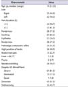
The diagnosis of TT was based on the results of the surgical exploration. Presenting symptoms and clinical findings of group 1 are shown in Table 1. The median ages of groups 1 and 2 were 14 years (range, 3-25 years) and 15 years (range, 5-25 years), respectively.
There were significant differences between groups 1 and 2 in terms of NLR, PLR, and PLT. However, there was no statistically significant difference in MPV between the two groups (Table 2).
We determined cutoff values for these hematological parameters in predicting TT diagnosis. Additionally, the sensitivity, specificity, PPV, and NPV of these parameters for TT diagnosis are shown in Table 3. ROC curve analysis of these parameters is shown in Fig. 1.
We determined a significant relationship between scrotal tenderness and NLR, PLR, and PLT (p=0.001 for all); however, there was no significant difference in terms of other symptoms. Furthermore, we indicated a significant difference between the early-presenting (<12 hours) and latepresenting groups (≥12 hours) in terms of NLR (p=0.01; beta coefficient, 0.380). Surprisingly, the differences in PLR, MPV, and PLT were not significant in these groups.
Most acute scrotal pathologies do not require surgical intervention, except for scrotal trauma. Therefore, efforts are made to distinguish TT from other scrotal pathologies. The similarity of clinical findings (scrotal swelling and erythema, testicular sensitivity) and CDU errors may result in unnecessary scrotal explorations. In recent years, many studies have been reported to introduce distinctive clinical and radiological features of TT [3,4,5,6,7]. However, challenges in the diagnosis of TT remain.
Neutrophils play a crucial role in the inflammatory processes [11]. Hematologic parameters, especially the NLR, have been shown to play a predictive role in the prognosis of acute and chronic inflammatory processes [12,13,14,15,16,17]. TT is an acute inflammatory process and testis viability is strictly related to the presenting time to the hospital. To the best of our knowledge, NLR has not been investigated previously in TT patients. In the current study, we evaluated the predictive role of NLR, PLR, MPV, and PLT in the diagnosis of TT and testicular viability following TT.
Hypertension has deleterious effects on cardiovascular and cerebrovascular systems and increased platelet activation may be one of the important underlying mechanisms of this affect [18,19,20]. Vascular complications in relation to thrombosis may be based on this increased activity [21]. MPV is known as a basic indicator of platelet activity [22]. Platelets with increased volume are supposed to have more capacity to generate inflammatory agents and are more likely to aggregate [23]. As pointed out in the current literature, platelet activity and MPV are related to thrombosis and endovascular processes. Thus, the lack of a significant relationship between MPV and TT may not be surprising.
Leucocyte count and related parameters are commonly used markers for inflammatory processes [24,25]. The potential role of leukocyte subtypes and the ratio of these markers in relation to inflammatory processes have been discussed in the current literature [26,27]. NLR was introduced as a simple and practical inflammatory marker that may have a predictive role in the diagnosis of systemic inflammatory processes [26,27,28,29,30]. Thus, the potential significance of NLR as an indicator of inflammation has been increasing. TT may initiate inflammatory systemic processes and thus the significance of NLR as a novel inflammatory marker was examined in our present report. We found a significant relationship between NLR, PLR, and PLT and the diagnosis of TT. However, only NLR was found to be related to the duration of symptoms in patients with TT. Thus, in relation to the results of the linear regression analysis, only NLR seems to be a helpful parameter in TT prognosis. These results suggest NLR as an accessible, inexpensive, predictive parameter for testicular viability in relation to TT.
One of the limitations of the present study was its retrospective nature and relatively small sample size. Systemic inflammatory markers such as NLR, PLR, and MPV are more valuable for the differential diagnosis of TT and acute inflammatory diseases such as epididymo-orchitis. Another limitation is that we did not include patients with acute inflammatory scrotal diseases in our study. However, we discussed the potential relationship between TT and systemic inflammatory markers. Also, the lack of some acute-phase reactants such as the C-reactive protein level and erythrocyte sedimentation rate is a limitation of the study. Additionally, CDU could be used to check testicular viability after treatment of TT, but the retrospective nature of the current study did not allow this procedure.
Despite these limitations, our clinical observations showed that the NLR may be associated with diagnosis and prognosis of TT. The NLR may be a practical, helpful, and inexpensive test. It can be easily incorporated into routine use as a predictive factor. Despite these results, large-scale, prospective, randomized studies evaluating the role of NLR in the differential diagnosis of TT and the viability of the testis are needed.
Figures and Tables
Fig. 1
Receiver operator characteristic curve for neutrophil to lymphocyte ratio (A), platelet to lymphocyte ratio (B), mean platelet volume (C), and platelet count (D). Diagonal segments are produced by ties.

Table 1
Presenting symptoms and clinical findings (n=75)

Table 2
Hematologic parameters of the study groups

References
1. Barada JH, Weingarten JL, Cromie WJ. Testicular salvage and age-related delay in the presentation of testicular torsion. J Urol. 1989; 142:746–748.
2. Weiss AP, Van Heukelom J. Torsion of an undescended testis located in the inguinal canal. J Emerg Med. 2012; 42:538–539.
3. Boettcher M, Bergholz R, Krebs TF, Wenke K, Aronson DC. Clinical predictors of testicular torsion in children. Urology. 2012; 79:670–674.
4. Boettcher M, Krebs T, Bergholz R, Wenke K, Aronson D, Reinshagen K. Clinical and sonographic features predict testicular torsion in children: a prospective study. BJU Int. 2013; 112:1201–1206.
5. Kalfa N, Veyrac C, Baud C, Couture A, Averous M, Galifer RB. Ultrasonography of the spermatic cord in children with testicular torsion: impact on the surgical strategy. J Urol. 2004; 172(4 Pt 2):1692–1695.
6. Capraro GA, Mader TJ, Coughlin BF, Lovewell C, St Louis MR, Tirabassi M, et al. Feasibility of using near-infrared spectroscopy to diagnose testicular torsion: an experimental study in sheep. Ann Emerg Med. 2007; 49:520–525.
7. Canpolat M, Yucel S, Sircan-Kucuksayan A, Kol A, Kazanci HO, Denkceken T. Diagnosis of testicular torsion by measuring attenuation of dual wavelengths in transmission geometry across the testis: an experimental study in a rat model. Urology. 2012; 79:966.e9–966.e12.
8. Wei Y, Jiang YZ, Qian WH. Prognostic role of NLR in urinary cancers: a meta-analysis. PLoS One. 2014; 9:e92079.
9. Gunay E, Sarınc Ulasli S, Akar O, Ahsen A, Gunay S, Koyuncu T, et al. Neutrophil-to-lymphocyte ratio in chronic obstructive pulmonary disease: a retrospective study. Inflammation. 2014; 37:374–380.
10. Karabacak M, Dogan A, Turkdogan AK, Kapci M, Duman A, Akpinar O. Mean platelet volume is increased in patients with hypertensive crises. Platelets. 2014; 25:423–426.
11. Barbu C, Iordache M, Man MG. Inflammation in COPD: pathogenesis, local and systemic effects. Rom J Morphol Embryol. 2011; 52:21–27.
12. Kahramanca S, Ozgehan G, Seker D, Gokce EI, Seker G, Tunc G, et al. Neutrophil-to-lymphocyte ratio as a predictor of acute appendicitis. Ulus Travma Acil Cerrahi Derg. 2014; 20:19–22.
13. Canpolat U, Aytemir K, Yorgun H, Sahiner L, Kaya EB, Kabakci G, et al. Role of preablation neutrophil/lymphocyte ratio on outcomes of cryoballoon-based atrial fibrillation ablation. Am J Cardiol. 2013; 112:513–519.
14. Bucak A, Ulu S, Oruc S, Yucedag F, Tekin MS, Karakaya F, et al. Neutrophil-to-lymphocyte ratio as a novel-potential marker for predicting prognosis of Bell palsy. Laryngoscope. 2014; 124:1678–1681.
15. Park JJ, Jang HJ, Oh IY, Yoon CH, Suh JW, Cho YS, et al. Prognostic value of neutrophil to lymphocyte ratio in patients presenting with ST-elevation myocardial infarction undergoing primary percutaneous coronary intervention. Am J Cardiol. 2013; 111:636–642.
16. Kahramanca S, Kaya O, Ozgehan G, Irem B, Dural I, Kucukpinar T, et al. Are neutrophil-lymphocyte ratio and plateletlymphocyte ratio as effective as Fournier's gangrene severity index for predicting the number of debridements in Fourner's gangrene? Ulus Travma Acil Cerrahi Derg. 2014; 20:107–112.
17. Tokgoz S, Kayrak M, Akpinar Z, Seyithanoglu A, Guney F, Yuruten B. Neutrophil lymphocyte ratio as a predictor of stroke. J Stroke Cerebrovasc Dis. 2013; 22:1169–1174.
18. Nadar S, Blann AD, Lip GY. Platelet morphology and plasma indices of platelet activation in essential hypertension: effects of amlodipine-based antihypertensive therapy. Ann Med. 2004; 36:552–557.
19. Tsiara S, Elisaf M, Jagroop IA, Mikhailidis DP. Platelets as predictors of vascular risk: is there a practical index of platelet activity? Clin Appl Thromb Hemost. 2003; 9:177–190.
20. Bath P, Algert C, Chapman N, Neal B. PROGRESS Collaborative Group. Association of mean platelet volume with risk of stroke among 3134 individuals with history of cerebrovascular disease. Stroke. 2004; 35:622–626.
21. Boos CJ, Beevers GD, Lip GY. Assessment of platelet activation indices using the ADVIATM 120 amongst 'high-risk' patients with hypertension. Ann Med. 2007; 39:72–78.
22. Park Y, Schoene N, Harris W. Mean platelet volume as an indicator of platelet activation: methodological issues. Platelets. 2002; 13:301–306.
23. Bath PM, Butterworth RJ. Platelet size: measurement, physiology and vascular disease. Blood Coagul Fibrinolysis. 1996; 7:157–161.
24. Thomsen M, Ingebrigtsen TS, Marott JL, Dahl M, Lange P, Vestbo J, et al. Inflammatory biomarkers and exacerbations in chronic obstructive pulmonary disease. JAMA. 2013; 309:2353–2361.
25. Hotchkiss RS, Karl IE. The pathophysiology and treatment of sepsis. N Engl J Med. 2003; 348:138–150.
26. Azab B, Chainani V, Shah N, McGinn JT. Neutrophil-lymphocyte ratio as a predictor of major adverse cardiac events among diabetic population: a 4-year follow-up study. Angiology. 2013; 64:456–465.
27. Dirican A, Ekinci N, Avci A, Akyol M, Alacacioglu A, Kucukzeybek Y, et al. The effects of hematological parameters and tumor-infiltrating lymphocytes on prognosis in patients with gastric cancer. Cancer Biomark. 2013; 13:11–20.
28. Ertas G, Sonmez O, Turfan M, Kul S, Erdogan E, Tasal A, et al. Neutrophil/lymphocyte ratio is associated with thromboembolic stroke in patients with non-valvular atrial fibrillation. J Neurol Sci. 2013; 324:49–52.
29. Celikbilek M, Dogan S, Ozbakir O, Zararsiz G, Kucuk H, Gursoy S, et al. Neutrophil-lymphocyte ratio as a predictor of disease severity in ulcerative colitis. J Clin Lab Anal. 2013; 27:72–76.
30. Dirican N, Anar C, Kaya S, Bircan HA, Colar HH, Cakir M. The clinical significance of hematologic parameters in patients with sarcoidosis. Clin Respir J. 2014; 07. 03. [Epub]. http://dx.doi.org/10.1111/crj.12178.


 PDF
PDF ePub
ePub Citation
 Print
Print


 XML Download