

 PDF
PDF ePub
ePub Citation
 Print
Print


INTRODUCTION
Urinary bladder carcinoma (UBC) is common worldwide with around 330,000 cases diagnosed every year. It has two major types, transitional cell carcinoma (TCC) and squamous cell carcinoma (SCC). TCC is the most common type in Western countries [1], whereas SCC predominates in some countries in Asia and Africa owing to endemic schistosomiasis [2]. SCC differs from TCC in its clinicopathological and molecular characteristics. Most SCC cases present with stage T3 and T4, with a lower incidence of nodal metastasis. Molecular pathogenesis and genetic alteration of TCC have been extensively studied [3]; however, this knowledge in SCC is limited. Available data suggest that SCC arises from transformation of squamous metaplastic lesions, which occurs secondary to inflammation [4]. UBC has a tendency to early relapse in about 70% of patients, independent of clinical prognostic variables. This high rate of recurrence necessitates accurate identification of prognostic factors that identify patients at high risk for disease progression [5]. One of these factors is the expression of survivin.
Survivin is a member of the inhibitors of apoptosis family of antiapoptotic proteins. The gene is expressed during embryonic life but not in terminally differentiated adult tissues [6]. Its expression was reported in UBC but not in normal urothelium. This differential expression has both diagnostic and therapeutic implications. In diagnosis, detection of the protein in urine has been recommended recently as a noninvasive tool for early diagnosis of primary UBC and in follow-up of patients after removal of UBC [7]. In therapy, several research studies are underway to assess the usefulness of survivin as a cancer vaccine that induces the immune system to mount a cancer-specific immune response against tumor cells [8]. The protein molecule has several cellular actions: it inhibits apoptosis, regulates cell division, and promotes angiogenesis [9]. Several studies reported that high expression of survivin is a poor prognostic marker for TCC of the urinary bladder (UB) [6,10]. To our knowledge, assessment of survivin expression in SCC of the UB and comparison with TCC have not been conducted.
The aims of the present study were to compare the immunohistochemical expression of survivin protein in different types of UBC (TCC, TCC with squamous differentiation [TCC-SD], and SCC) and to assess the association between survivin expression and tumor proliferation (assessed by Ki67 expression as used previously [11]) in relation to clinicopathological criteria and disease outcome in the mentioned tumor types.
MATERIALS AND METHODS
1. Patients and specimens
One hundred four randomly selected consecutive primary UBC specimens (52 TCC, 32 SCC, 20 TCC-SD) with complete 12-month follow-up data and 5 specimens of nonneoplastic urothelial tissue were obtained from the archives of the Department of Pathology, Assiut University Hospital, Assiut, Egypt. Patients with non-muscle-invasive disease were treated with maximal transurethral resection (TUR) and immediate postoperative instillations chemotherapy with or without BCG therapy. Patients with muscle-invasive disease were treated with radical cystectomy and pelvic lymph node dissection without neoadjuvant chemotherapy. Disease recurrence was defined as any evidence of tumor in a retained bladder after TUR or detection of new growth by imaging modalities. Disease progression was defined as any increase in tumor stage or occurrence of distant metastasis (DM). Any recurrence or DM was scored as an event. Ethical approval for the study was obtained from the Faculty of Medicine Research Ethics Committee.
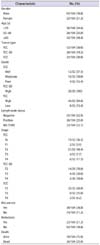
All specimens were fixed in 10% formalin, embedded in paraffin, and stained with hematoxylin and eosin. The age of the patients ranged from 35 to 75 years. Specimens were classified and graded according to the World Health Organization classif ication [12]. Tumor staging was identified according to the American Joint Committee on Cancer/Union for International Cancer Control TNM system [13]. Schistosomal infection was present in 18 of 32 SCC specimens (56%) and in 17 of 52 TCC specimens (32%). Additional clinicopathological characteristics of the examined specimens are shown in Table 1.
2. Immunohistochemical staining
Expression of survivin and Ki67 proteins was assessed by using immunohistochemical (IHC) staining by applying the avidin biotin immunoperoxidase complex technique (Ultravision Plus Detection System antipolyvalent HRP/DAB, ready to use; Thermo Scientific Co., Fremont, CA, USA). IHC was performed according to the manufacturing protocol. Tissue sections were deparaffinized, rehydrated, and immersed in citrate buffer and then microwaved. Sections were incubated overnight at room temperature with diluted primary survivin antibodies in phosphate buffered saline buffer (dilution of 1:200; rabbit monoclonal antibody, Thermo Scientific Co.) and with Ki67 (dilution of 1:200; Clone SP6, rabbit monoclonal antibody, Thermo Scientific Co.). After the application of secondary antibody, diaminobenzidine was applied and the sections were then counterstained with Mayer's hematoxylin, dehydrated, and mounted.
3. Evaluation of expression of survivin and Ki67 expression
The percentage of cells showing nuclear survivin expression was calculated. Specimens were classified according to survivin status into two groups: group 1 specimens with normal expression (no reactivity or few focally positive cells) and group 2 specimens showing nuclear expression in more than 10% of cells. The value of 10% was used as the cutoff level because it was found that when 10% or more of tumor cells express survivin, those tumors gain different biological characteristics resulting in poorer outcome [5,14]. The nuclear Ki67-LI level was expressed as the percentage of positively stained cells in 10 high-power fields that showed highest positivity. Twenty percent was used as a cutoff to categorize specimens into (1) specimens with low Ki67-LI (those with Ki67 positivity in <20% of tumor cells) and (2) specimens with high Ki67-LI (those with Ki67 positivity in >20% of tumor cells) as used previously [15].
4. Statistical analysis
Two-by-two tables and the chi-square test were conducted to examine differences in survivin expression between TCC and SCC and to find associations between survivin expression (normal and altered) and clinicopathological criteria. The value of the Fisher exact test was used when the number in cells was less than 5. Disease-free survival was calculated from the date of diagnosis to the date of recurrence or DM. The Kaplan-Meier method was used to calculate survival function, and the differences were assessed with the log-rank statistic. Multivariate survival analyses were conducted by using the Cox proportional-hazards regression model. Statistical significance was set at p<0.05. All reported p-values were two-sided. Statistical analyses were performed with the SPSS ver. 16.0 (SPSS Inc., Chicago, IL, USA).
RESULTS
1. Staining patterns for survivin and Ki67
Positive nuclear staining for survivin was detected in 92 of 104 UBC samples (88%), in 28 of 32 SCC samples (87.5%), in 18 of 20 TCC-SD samples (90%), and in 46 of 52 TCC samples (88.46%) (Fig. 1). No staining was detected in any of the normal urothelium. Altered survivin expression was detected in 60 of 104 specimens (58%) as a whole. Alteration in survivin status was significantly more frequent in the TCC (78%) than in the SCC (38%) type (p<0.0001) (Table 2).
Percentage of positive Ki67 staining in tumor cells ranged from 10% to 100% with a mean of 47% (standard error, ±2.6) and a median of 45% (standard deviation, ±2.5). Seventy-three specimens (70%) were characterized with high Ki67-LI (35 TCC [48%], 14 TCC-SD [19%], and 24 SCC [33%]; p=0.756) (Fig. 2).
2. Association between survivin status and clinicopathologic criteria
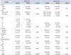
In TCC, altered survivin expression was significantly associated with higher tumor grade (p<0.001), high Ki67-LI (p=0.0001), and recurrence (p=0.001). In SCC, altered survivin status was only significantly associated with tumor stage as shown in Table 3. In TCC-SD, altered survivin expression was detected in 8 of 20 specimens. All eight specimens were high grade and characterized with high Ki67-LI (p=0.017); all eight patients developed metastasis (p<0.0001) and died from the disease (p<0.0001). However, no association was seen in TCC-SD between altered survivin status and patient gender (p=0.648), tumor stage (p=0.057), or presence of lymph node metastasis (p=0.264).
3. Relationship between survivin status and Ki67-LI and patients' outcome
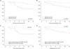
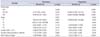
The association between survivin status in TCC and SCC and patient outcome was mentioned above (Table 3). It was difficult to conduct survival analysis by using the log rank test on each type alone because the number of events (recurrence or death) became small when the specimens were subdivided. In all UBC specimens (104 specimens), Kaplan-Meier survival curves showed that specimens with altered survivin expression had a significantly shorter time for development of recurrence and DM compared with patients with normal survivin expression (p=0.002 and p<0.001, respectively) (Fig. 3A, B). Multivariate analysis was conducted after adjusting survivin status for other prognostic factors (histopathological type, stage, lymph node status, and Ki67-LI). The analysis showed that altered survivin status was an independent poor prognostic factor for recurrence and for disease progression in UBC patients. Ki67-LI, however, lost significance in the multivariate analysis for development of recurrence but showed a significant association with the development of DM (Table 4).
To determine if combined high Ki67-LI and altered survivin expression would be a more powerful prognostic tool than each factor separately, specimens were recategorized into two groups: group 1, with low Ki67-LI and/or normal survivin expression, and group 2, with both altered survivin expression and high Ki67-LI. It was found that group 2 has a worse prognosis than did group 1 and that this difference was greater than with the use of altered survivin or high Ki67-LI as a prognostic factor alone. By the end of 12 months, the recurrence-free interval was 56% in group 2, in which 24 of 54 tumors developed recurrence, compared with 86% in group 1, in which only 7 of 50 developed recurrence. The same was found in association with DM. The 12-month metastasis-free interval was 96% in group 1, in which 2 of 50 tumors developed DM, compared with 63% in group 2, in which 40 of 54 tumors developed DM (Fig. 3C, D). In the multivariate analysis, combined altered survivin expression and high Ki67-LI was an independent poor prognostic factor for both recurrence (hazard ratio [HR], 7.55; 95% confidence interval [CI], 2.274-25.096; p<0.001) and DM (HR, 71.584; 95% CI, 9.159-559.462; p<0.001).
DISCUSSION
The prognostic importance of survivin in TCC has been reported in several studies [16,17]. However, the pattern of expression of survivin and its association with tumor characteristics and outcome in SCC have not yet been analyzed. A recent meta-analysis study concluded that "Survivin expression could be used in identifying a subgroup of patients with potential to benefit from a targeted therapy against survivin" [17]. The question we were interested in answering was, Is altered survivin expression in SCC also associated with clinicopathological features and patient outcome similar to TCC?
The current study detected positive staining for survivin in both TCC and SCC tumors but not in normal urothelial tissues, which is similar to results reported previously [18]. Absence of expression in normal UB epithelium makes survivin a diagnostic tumor marker and also a suitable targeted therapeutic agent [19]. When tumor cells express survivin at abnormal levels, a condition known as "altered survivin expression," the cells gain some biological characteristics that make them long survivors and more proliferative [9].
In the TCC specimens studied here, high significant associations were f ound between altered survivin expression with high grade and stage of tumors and Ki67-LI. In SCC, there was an association with increase in tumor stage only. In TCC-SD, significant associations were shown with stage and Ki67-LI. Thus, TCC-SD is similar in its expression pattern to TCC.
Degree of tumor differentiation, which is the basis of histological grading, is dependent upon degree of cell proliferation. When tumor cells proliferate at higher rates, they lose the ability to differentiate similar to their tissue of origin and become higher grade tumors [12]. The same finding was reported in previous studies [18,20]. The difference in survivin expression is significantly high enough between low and high tumor grades that some authors have recommended the use of survivin as an adjuvant diagnostic tool to mark high-grade tumors [10].
Survivin is a unique molecule able to induce cell proliferation on one hand and to inhibit apoptosis on the other [19]. No wonder then to find such a high association between altered survivin expression and tumors with high grade and high proliferative index in TCC. What is not understood is why no such association was seen with SCC, even though its histological grading followed the same rule of cell differentiation and proliferation. Our knowledge of the molecular events in carcinogenesis and progression of SCC is still limited. Studies have found significant differences in the genetic changes between TCC and SCC [21]. Other molecular factors in SCC may interact with survivin and interfere with its action as an inducer for proliferation. Many of our patients (58%) with SCC had associated schistosomal infection. Chronic schistosomal cystitis is known to create an inflammatory environment in the UB that alters some of the molecular characteristics of the SCC compared with TCC [4,22].
A significant association between altered survivin expression and increased probability of recurrence and development of DM in the whole group of UBC was detected in this study. The same findings were reported in studies that evaluated the level of survivin at the protein level [18] and also at the transcription level [23]. It was difficult to do survival analysis by using Kaplan-Meier survival curves in SCC and in TCC subtypes separately. This would have decreased the number of events in each subgroup and would not give reliable statistical results. By use of the chi-square test, altered survivin status was significantly associated with the development of recurrence in TCC, similar to previous reports [24], but not in SCC. Such an association may be related to the effect of survivin in stimulation of cell proliferation on one hand synergized by inhibition of apoptosis on the other [25].
The mechanism behind the high recurrence rate of UBC is complex. Several molecular factors such as tumor proliferation, p53 expression, and matrix metalloproteinase content and angiogenesis play a role [26]. Recently, epigenetic changes such as DNA methylation and histone modification were found to also play an important role in the regulation of tumor genes and hence the overall biological behavior of UBC [27]. When global examination of molecular changes in UBC was done, it was interesting to find that UBC can be subdivided into major subgroups that differ in their clinical outcome and in the major genes that regulate cell cycle, apoptosis, cell adhesion, and growth receptors. In the same study, the genetic changes in SCC were found to differ from those in other types [28]. No reports were found in the literature about the association between altered survivin expression in SCC of the UBC and patient outcome. In oral SCC, altered survivin expression was associated with tumor progression and poorer survival [29].
Research efforts have succeeded in development of a survivin vaccine that has been used successfully to improve survival of patients with metastatic UBC [30]. Although SCC does express survivin, its high expression is not associated with the major clinicopathological features or recurrences. The findings from this study raise an alarm. Are UBC patients with SCC types suitable candidates for survivin vaccine? This is an important question to be answered before approving the survivin vaccine in the management of UBC.
CONCLUSIONS
Alteration of survivin expression occurs more frequently in TCC than in SCC. In TCC but not in SCC, altered survivin status is associated with higher tumor grades, high tumor proliferation, and recurrence. In UBC as a whole, altered survivin is a poor independent prognostic factor; however, more studies on a larger number of patients are recommended to assess the prognostic role of survivin in the TCC and SCC subtypes separately.


 XML Download