

 PDF
PDF ePub
ePub Citation
 Print
Print


INTRODUCTION
Although various approaches to the treatment of ureteral defects have been introduced, renal autotransplantation or ureteral substitution using intestine may be the only modality of treatment in the case of an extensive ureteral defect. We report here on a case of ureteral substitution using appendix to repair a ureteral defect that was caused by resection of a retroperitoneal rhabdomyosarcoma that involved the right lower ureter in a child.
CASE REPORT
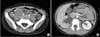
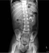
A 7-year-old boy was referred to our outpatient clinic for evaluation of his abdominal mass, which was found at a local clinic by use of abdominal ultrasound. He underwent laparoscopic biopsy of his abdominal mass and the mass was finally diagnosed as an embryonal rhabdomyosarcoma in the retroperitoneum. The size of the mass was 18 cm×16 cm×7 cm, and after 4 cycles of chemotherapy for 5 months, the mass size was reduced to 5 cm×2 cm×3 cm. Surgical resection of the mass was then performed and the patient was followed up for 2 years. Abdominal computed tomography showed no remnant mass of the tumor. We found no evidence of recurrence of the tumor for 1 year after surgery. After 1 year, however, follow-up computed tomography showed a newly developed mass (3.3 cm×3.0 cm in size) surrounding the right lower ureter along the right iliac artery, resulting in hydronephrosis of the right ureter (Fig. 1). We diagnosed the mass as a recurrent retroperitoneal rhabdomyosarcoma and we decided to perform surgical resection. The operation was performed through a midline vertical incision and the anatomy was confirmed by direct inspection. The mass had severe adhesions with the surrounding tissues and it completely encircled the right lower ureter along the right iliac artery. After resection of the mass, direct end-to-end anastomosis of the ureter was not possible owing to the length of the resected segment (Fig. 2A). Therefore, we decided to perform ureteral substitution by using the appendix to repair the ureteral defect. The cecum and the right colon were mobilized. The vermiform appendix was assessed. It was 6 cm in length from the base to the tip and no macroscopic abnormalities were noted. The appendix was divided at its base and the stump was ligated and conventionally inverted. Care was taken to preserve the appendicular artery, which was identified as running in the short triangular mesoappendix. The isolated appendix was mobilized to the retroperitoneum and positioned in an antiperistaltic manner to avoid strangulation. Tension-free end-to-end anastomosis with 5-0 Vicryl was performed between the spatulated end of the ureter and the tip of the appendix (Fig. 2B). A ureteral stent was placed by using a guide wire before completing the anastomosis. The resected mass was 4.5 cm×3.0 cm×1.0 cm in size and it grossly revealed no central necrosis. The mass was finally diagnosed microscopically as retroperitoneal rhabdomyosarcoma. The patient's postoperative recovery was uneventful. The ureteral stent was removed 6 weeks postoperatively and retrograde pyelography revealed a patent appendiceal interposition and no extravasation. Intravenous urography showed no definite stricture point and improvement of the hydronephrosis that was seen before surgery (Fig. 3). This patient died at 6 months postoperatively as the result of multiple metastases of rhabdomyosarcoma.
DISCUSSION
The appendix has an irregular lumen that is approximately 8 French in diameter and the blood supply arises from the appendicular artery, which is a branch of the ileocolic artery, located in the mesoappendix. We believe that there are various advantages to using the appendix as a right ureteral substitute versus ileal replacement or renal autotransplantation. Appendiceal interposition is technically easy and the appendix can be readily mobilized with its blood supply [1]. The length-to-diameter ratio of the appendix, its contractility, and its peristalsis prevent urinary stasis [2]. Another advantage of the appendix over ileal interposition is its small surface area, which results in negligible urine absorption, and this explains the lack of serum electrolyte abnormalities. The appendiceal lumen corresponds in caliber to that of the ureteral lumen and this allows secure anastomosis with the proximal or distal ureter. The location of the appendix facilitates mobilization and replacement of the upper, mid, or lower ureter [1]. The use of the appendix allows complete retroperitonealization of the anastomoses to both the ureter and the bladder. For these reasons, Mitrofanoff [3] in 1980 first described the use of the isolated appendix as an intermittent catheterization route to empty a continent urinary reservoir. Numerous variations on the Mitrofanoff principle have been reported since 1980, which were directed at creating an ideal continence mechanism. Duckett and Snyder [4] used the Penn pouch technique to rotate the isolated appendix 180° before antireflux implantation into the reservoir. Issa et al. [5] avoided the need to isolate and reimplant the appendix by remodeling the in situ appendix via invagination and plication into the cecum. Utilization of the in situ appendix and incorporation of the cecoappendiceal unit in the reconstruction was also reported by Longaker et al. [6]. The appendix was recently adopted for hepatobiliary reconstruction in extrahepatic biliary atresia [7]. However, a few technical problems may limit the use of the appendix. The appendix is unavailable or unsuitable for reconstructive purposes when it is surgically removed beforehand or is too short to bridge the existing anatomic gap or a defect is left-sided. We anticipate that the appendix will be more commonly used in the future as a ureteral substitute as more urologists become comfortable with its use in various reconstructive procedures.

 XML Download