

 PDF
PDF ePub
ePub Citation
 Print
Print


INTRODUCTION
Bladder outlet obstruction is a relatively common urological problem that occurs as the result of congenital anomalies such as posterior urethral valve, benign prostatic hyperplasia, and urethral stricture [1]. Infravesical obstruction results in numerous changes in the upper and lower urinary tract, eventually leading to chronic renal failure [2,3,4]. Increased bladder outlet resistance also impairs the storage function and results in loss of bladder viscoelasticity secondary to fibrous proliferation and mucosa and collagen accumulation [2,3,4]. Although the explanations for bladder dysfunction are not fully understood, fetal urethral obstruction has been reported to result in deposition of an abnormal quantity of collagen in the bladder, which can play a major role in bladder hypocompliance [4]. Furthermore, chronic renal damage due to tubulointerstitial fibrosis is one of the adverse effects of bladder outlet obstruction [5,6,7]. Although the first-line treatment of bladder outlet obstruction is relief of the obstruction by surgery, some patients are not good candidates for operation. Therefore, attempts should be made to find the best medication for prevention of the structural changes of kidney and bladder. The present study aimed to investigate and compare the effects of tamoxifen, captopril, pentoxifylline, and simvastatin on prevention of renal and bladder fibrosis and damage in a rat model of partial urethral obstruction (PUO). Primarily, tamoxifen is an antiestrogen therapy for receptor-positive breast cancer, infertility, gynecomastia, and related diseases [8,9]. However, positive effects of tamoxifen have been observed in the treatment of retroperitoneal fibrosis and also in renal fibrosis [8,9]. Captopril is commonly prescribed as an angiotensin-converting enzyme inhibitor for the treatment of some medical conditions, including hypertension and some types of congestive heart failure [10,11]. The positive effect of captopril in amelioration of renal fibrosis has also been evaluated in previous studies [10,11]. Pentoxifylline mainly improves blood flow through peripheral blood vessels [12,13]. In addition, it has been shown that pentoxifylline can reduce tubulointerstitial fibrosis [12,13]. Simvastatin is primarily used for the treatment of dyslipidemia and the prevention of cardiovascular diseases. However, research has shown that after unilateral ureteral obstruction, renal fibrosis is reduced in simvastatin-treated animals [14,15]. To the best of our knowledge, no studies have compared the effects of these drugs on renal and bladder fibrosis. In addition, the microscopic structure of the kidney and bladder has not been compared quantitatively in PUO animals treated with the above-mentioned drugs. In the present study, this quantitative technique was used to measure the volume or length of the kidney components (its proximal and distal convoluted tubules [PCT and DCT], loop of Henle, collecting duct, and interstitial tissue) and bladder components (epithelium, muscular tissue, interstitial tissue, and number of fibroblasts and fibrocytes).
MATERIALS AND METHODS
1. Animals
This study was conducted on 42 male rats weighting 180±20 g. The animals were treated according to the standard directive recommended and approved by the research authorities of Shiraz University of Medical Sciences, Shiraz, Iran (approval no. 9101015143). The rats were randomly divided into six groups each including seven animals, which is considered to be an acceptable number for stereological studies [16]. The sham-operated animals underwent laparotomy only without undergoing any procedures or receiving any treatments. Other groups, however, underwent a surgery to induce PUO. The PUO groups received normal saline (PUO+NS), tamoxifen (10 mg/kg/d; PUO+TAM) [9], captopril (35 mg/kg/d; PUO+CAP) [10], pentoxifylline (100 mg/kg/d; PUO+PEN) [12], or simvastatin (15 mg/kg/d; PUO+SIM) [14] by gavage for 28 days. The doses selected were based on studies that confirmed the protective effects of these drugs in fibrosis.
2. Surgical procedure
After the rats were anesthetized with ketamine/xylazine (25 mg/kg intraperitoneally and 2.5 mg/kg intraperitoneally, respectively) in the supine position, the abdominal cavity was opened by a midline incision to reach the posterior urethra. Then, a pediatric 22-gauge Angiocath was introduced to the posterior urethra and a silk 4-0 ligature was tied around the Angiocath. Afterwards, the Angiocath was removed and PUO was induced. Finally, the incision was closed. The rats' kidneys and bladders were removed after 28 days.
3. Stereological study
At first, the rats' kidneys and bladders were removed and the renal pelvis was dissected out. Then, the kidney and bladder were weighed. The primary volume of the kidney, V(primary), was measured by using Sherle's immersion technique [17,18]. After that, the organs were fixed in neutral buffered formaldehyde for 1 week. Then, the tissue shrinkage produced by fixation, processing, and staining was estimated. Estimation of the shrinkage and length of the renal tubules as well as the vessels require isotropic uniform random sectioning [19,20]. These sections were obtained through the orientator method [19]. Then, two random circular pieces were punched out from the isotropic uniform random slabs. The estimated shrinkage was also used to estimate the final volume of the kidney or bladder to avoid the consecutive sectioning that is required for the Cavalieri method. Shrinkage degree was estimated by using the following formula [19]:
Shrinkage degree = 1-(AA/AB)1.5
where AA and AB are the area of the circular pieces after and before processing and staining, respectively. The pieces of kidney and bladder were embedded in separate paraffin blocks. After sectioning and staining, the sampled section was analyzed by using a video microscopy system. In doing so, 10 to 12 microscopic fields were sampled in a systematic random manner. Then, the stereological grids were superimposed on the images by means of the stereology software. The volume density of each structure (Vv: the fraction of the unit volume of the kidney or bladder that is occupied by the structural component) was estimated by using the point-counting method [19]. In addition, the volume fraction of the renal structures was estimated at a final magnification of ×1,500 by using the following formula:
Vv(component) = P(components)/P(reference)
where P(components) and P(reference) were the total number of the points hitting the favored components and the reference tissue (kidney or bladder), respectively. The total volume of the structural components was estimated by multiplying the volume density by the kidney or bladder volume [19].
Moreover, the length density (Lv: the length of each tubular structure in the unit volume of the kidney) of the PCTs, DCTs, collecting ducts, Henle's loop, and vessels was estimated by randomly overlaying an unbiased counting frame with an area of 2,700 µm2 on the live monitor images and by using the following formula:
Lv(tubules) = 2ΣQ(tubules)/ΣA
where Q and A were the sectional profiles of the tubules and areas of the counting frames, respectively. The length of the tubular components was estimated by using the following formula:
L(tubules) = V(primary)×Lv(tubules)×(1-shrinkage degree)2/3
The numerical density (Nv: number of cells per unit volume of tissue) of the fibroblasts and fibrocytes of the bladder was estimated by using the optical disector method on 25-µm sections and the following formula [19]:
Nv = [ΣQ/(ΣA×h)]×(T/BA)
where Q, A, h, T, and BA were the counted cell nuclei, area of the counting frames, height of the disector (12 µm), estimated thickness of the microscopic tissue section by use of a microcator (20 µm), and micrometer setting of the microtome for sectioning (25 µm), respectively. Height of the disector was determined after plotting the z-axis distributions of the cell nuclei [21].
N(cells) = V(primary)×Nv(cells)×(1-shrinkage degree)3/3
RESULTS
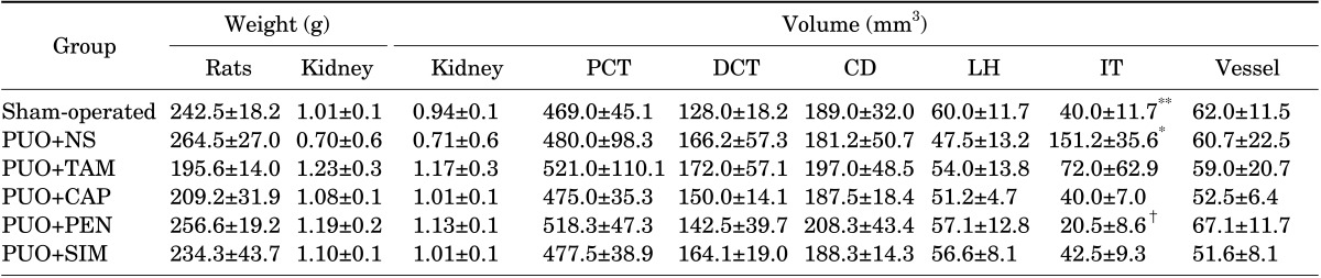
1. Volume of the kidney, tubules, and vessels
No significant differences were observed between the groups regarding the volume of the kidney, PCT, DCT, collecting duct, Henle's loop, and vessels (Table 1).
2. Length of the renal tubules and vessels
The length of the PCT, DCT, collecting duct, Henle's loop, and vessels, respectively, was reduced by 37%, 38%, 40%, 46%, and 23% in the PUO+NS group compared with the sham-operated animals (p<0.01). However, treatment of the PUO animals with pentoxifylline, tamoxifen, captopril, and simvastatin prevented the decrease in tubule and vessel length (Table 2) (p<0.05). Pentoxifylline more significantly preserved the length of the renal tubules and vessels.
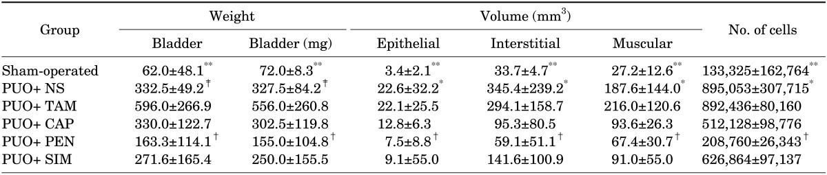
3. Volume of the bladder, epithelial tissue, and muscle tissues
The volume of the bladder, epithelial tissue, and muscle tissue was increased by 5- to 7-fold in the PUO+NS rats compared with the sham-operated animals (p<0.01). Moreover, the volumes were lower in the PUO+PEN and PUO+SIM groups than in the PUO+NS rats (p<0.05) (Table 3). In this respect, pentoxifylline was more effective than the other drugs.
4. Volume of fibrous tissue
The volume of the fibrous tissue was increased by 4- and 10-fold in the kidney and bladder of the PUO+NS rats compared with the sham-operated animals, respectively (p<0.01). In addition, the volume of fibrous tissue in the PUO+PEN, PUO+CAP, and PUO+SIM rats was significantly lower than that in the PUO+NS group (p<0.05) (Tables 1, 3).
5. Number of fibroblasts and fibrocytes
The number of fibroblasts and fibrocytes increased by 7.5-fold in the PUO+NS rats compared with the sham-operated animals (p<0.05). Additionally, the number of cells was lower in the PUO+PEN, PUO+CAP, and PUO+SIM groups than in the PUO+NS rats (p<0.05). Nevertheless, no significant reduction was observed in the number of cells in the PUO+TAM group compared with the PUO+NS animals (Table 3).
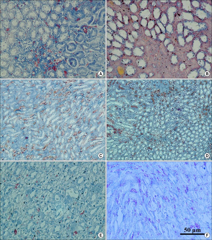
6. Qualitative evaluation of the kidney and bladder
As can be seen from Fig. 1, in the PUO rats who received normal saline treatment, normal tubular cells were lost. Fibrous tissue was formed and replaced the tubular tissue. The tubules seemed dilated. After treatment of the PUO animals, the above-mentioned changes were ameliorated. In the PUO+PEN group, less fibrosis and more normal tubules and vessels could be found. These findings were followed by CAP, SIM, and TAM.
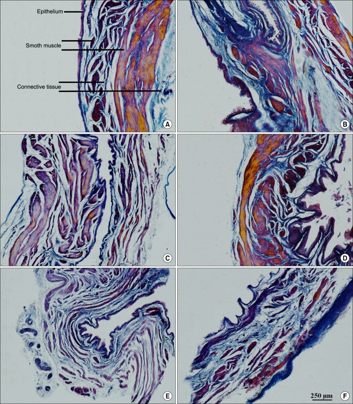
Evaluation of the bladder (Fig. 2) in the PUO group without pharmacological treatment showed that more epithelial tissue can be seen. The muscle layer was hypertrophied. Fibrous tissue was increased in the different layers of the bladder. PUO+PEN ameliorated the changes in bladder structure, followed by CAP, SIM, and TAM.
DISCUSSION
The present study aimed to examine the effects of pentoxifylline, captopril, simvastatin, and tamoxifen on renal and bladder histological changes in rats after PUO. The first step of the study confirmed that PUO led to structural changes in the organs after a period of 28 days. It has been shown that bladder dysfunction secondary to PUO is characterized by bladder wall hypertrophy, decreased capacity and urinary stream, and increased postvoid residual urine [4,5,6,17,22]. If the obstruction is not relieved, the bladder enters the decompensating phase, in which detrusor contractility is decreased, eventually leading to hydronephrosis and renal damage [4,5,6,17,22]. These findings are in line with the findings of the present study. Hypertrophy of the bladder epithelium and muscular layer was seen in PUO rats. In addition, the injury extended to the kidney structure after 28 days. The results of the current study indicated a decrease in the length of the renal tubules and vessels after PUO. Decreased length should be considered a sign of renal tissue loss. Accompanying the loss of renal tissue, the fibrous tissue was increased after PUO, so that the total volume and weight of the kidney did not change.
It appears that two main questions were evaluated here: How much fibrous tissue formation can be prevented? How much intact tissue can be preserved? The current study confirmed that pentoxifylline, captopril, simvastatin, and tamoxifen could prevent fibrous tissue formation and protected the renal tissue in PUO rats.
It has been reported that an increase in angiotensin II levels in obstructive nephropathy up-regulates the expression of several factors, including transforming growth factor (TGF)-β. Local production of TGF-β by macrophages penetrating the kidney is a key mediator of renal fibrosis [4]. Activation of TGF-β stimulates endothelin production, which in turn is a persuasive stimulus for fibrogenesis [4]. Therefore, if these mechanisms could be controlled, fibrous tissue formation would decrease, eventually preserving the normal renal and bladder histology after PUO. It has been shown that after obstruction, captopril not only decreases the angiotensin II and TGF-β levels but also inhibits conversion of procollagen to collagen in the interstitial space of the kidney.
Tamoxifen also has an antifibrotic effect by reducing TGF-β expression by fibroblasts [9]. Pentoxifylline has an inhibitory effect on neutrophil-mediated functions, such as superoxide production, chemotaxis, phagocytosis, and TNF production [18]. TNF-α has a substantial impact on molecular mechanisms, resulting in tubular cell death. Antifibrotic, anti-inflammatory, and anti-ischemic effects of pentoxifylline have also been mentioned in an earlier study [18]. Simvastatin also up-regulates the inhibitors of TGF-β signaling, attenuates epithelial-to-mesenchymal transition, and decreases renal fibrosis [14,15].
Moreover, our previous studies reported that administration of captopril could decrease interstitial fibrosis and preserve renal tubules after PUO [23]. In an animal model, Shirazi et al. [24] showed that captopril preserved the amount of renal tubules and tamoxifen decreased fibrous tissue formation in unilateral obstructed kidney. In another study, Shirazi et al. [12] also demonstrated that pentoxifylline could inhibit interstitial renal fibrosis after PUO.
CONCLUSIONS
In the rat model of PUO, pentoxifylline followed by captopril, simvastatin, and tamoxifen could prevent fibrous tissue formation and protect the renal tissue. Treatment with all the above-mentioned drugs prevented the decrease in tubular length and vessels, with pentoxifylline being the most effective. Moreover, bladder fibrosis was more significantly prevented by pentoxifylline followed by captopril and simvastatin. Also, bladder structure was more significantly protected by pentoxifylline followed by captopril and simvastatin. Therefore, pentoxifylline was most effective in preservation of renal and bladder structure after PUO.
 XML Download