

 PDF
PDF ePub
ePub Citation
 Print
Print


INTRODUCTION
Although endourological management can be a first-line treatment for ureteropelvic junction obstruction (UPJO), its indications are limited and its success rate does not exceed 80% [1]. Open pyeloplasty is also a valuable treatment for UPJO. Open pyeloplasty has a high success rate of 90% in long-term follow-up results [2]. However, it has significant operative morbidities. Laparoscopic pyeloplasty, which was first introduced in 1993 [3], is minimally invasive, like endourological management, and has a high success rate, like open pyeloplasty [4]. However, the long-term follow-up results of laparoscopic pyeloplasty are unclear, as are its indications and limitations. We present our experience with long-term follow-up of laparoscopic pyeloplasty and identify the risk factors for failure of the technique.
MATERIALS AND METHODS
A single surgeon performed 107 laparoscopic pyeloplasties between December 2001 and December 2013. Of these patients, 65 who underwent standard laparoscopic pyeloplasties with transperitoneal approaches and who were followed up for more than 12 months were enrolled in this study. Exclusion criteria were retroperitoneal approaches, concomitant stone surgery, insufficient follow-up study, and short follow-up time. This study was performed under Institutional Review Board approval.
The 65 patients comprised 35 males and 30 females with a mean age of 45.02±19.47 years (range, 11-76 years). The patients' mean body mass index was 23.34±3.14 kg/m2 (range, 17.69-30.59 kg/m2). Chief complaints were flank pain (n=57) and incidental detection (n=8). Twenty-one patients (32.3%) had undergone previous abdominal surgeries, including 10 patients who had undergone open pyeloplasty and endopyelotomy. All patients underwent preoperative imaging and had radiographic evidence of obstruction. Mean stricture length was 1.06±0.82 cm (range, 0.2-3.7 cm) on intravenous pyelography (IVP) or retrograde pyelography. The degree of hydronephrosis on ultrasonography was grade 1/4 (n=5), grade 2/4 (n=10), grade 3/4 (n=36), and grade 4/4 (n=14). All patients had more than 15% of split renal function on a 99mTc mercaptoacetyltriglycine (MAG-3) renal scan, and an obstructive pattern (T1/2 of more than 20 minutes) was present in 53 patients (81.5%).
The type of laparoscopic pyeloplasty was chosen at the surgeon's discretion on the basis of radiologic findings. A 0.038-inch guidewire was inserted preoperatively. A standard transperitoneal laparoscopy was performed with the retrocolic technique. Palpation of the indwelling guidewire helped to identify the course of the ureter to the UPJ. The UPJO area was transected by using scissors and a redundant pelvis could be excised if necessary. The proximal end of the guidewire was pulled from the pelvis, and then a double-J ureteral stent was inserted over the guidewire. A 4-0 or 5-0 Vicryl suture was placed between the spatulated ureter and the renal pelvis from the posterior side to the anterior side. If an anterior lower pole vessel was encountered, it was transposed behind the renal pelvis. All patients had a drain, which was removed postoperatively. Our surgical technique is demonstrated in a supplementary video clip (Supplementary material). The indwelling ureteral stent was removed 8 weeks after surgery.
Operative and follow-up data were collected. Follow-up consisted of both radiologic and symptomatic evaluations. Radiologic success was defined as imaging of a patent UPJ on IVP and resolution of obstruction on a MAG-3 renal scan. Symptomatic success was defined as complete symptomatic relief postoperatively. Overall success was defined by a combination of radiologic improvement and symptomatic relief. Risk factors affecting failure of laparoscopic pyeloplasty were explored. Patient baseline characteristics (age, body mass index, abdominal operation history), anatomical parameters (stricture length, ipsilateral renal function using radioisotope uptake, degree of hydronephrosis, presence of crossing vessel), and operative parameters (operation time, blood loss, drained amount) were evaluated to assess the relationship to failure of laparoscopic pyeloplasty.
Statistical analyses were carried out by using IBM SPSS Statistics ver. 19.0 (IBM Co., Armonk, NY, USA). For the Mann-Whitney test and Fisher exact test, p<0.05 was considered to indicate statistical significance.
RESULTS
Fifty-seven patients were treated by dismembered Anderson-Hynes pyeloplasty and eight patients by Fenger pyeloplasty. During the operation, crossing vessels were found in 27 patients (41.5%). Mean operating time was 159.42±76.98 minutes (range, 57-480 minutes). Mean estimated blood loss was 973.13±335.87 mL (range, 489-2152 mL), and six patients (9.2%) required transfusion. Although there were no cases of open conversion, one patient had an ascending colon injury that was detected postoperatively and repaired. Another patient had a spleen injury, which was treated conservatively. The mean starting time of postoperative ambulation and diet was 1.54±0.64 days (range, 1-3 days) and 1.86±1.41 days (range, 1-3 days), respectively. Mean hospital stay was 8.09±3.29 days (range, 5-20 days). The mean follow-up period was 36.5 months (range, 12-111 months).
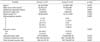
The follow-up IVP and 99mTc-MAG3 renal scans showed improvements in 59 patients; the radiologic success rate was 90.8%. The flank pain disappeared in 54 patients (symptomatic success rate, 94.7%). Eight patients showed failure on radiologic or symptomatic evaluation, and the overall success rate was 87.7%. Among the six patients with radiologic failure, one patient was treated with endopyelotomy at 7 months postoperatively and one patient was treated by use of robotic pyeloplasty at 24 months postoperatively. The remaining four patients were treated with ureteral stenting and conservative management. We studied differences in variables between the success group (group I) and the failure group (group II) to try to identify risk factors for failure. Drained amount was only the risk factor related to overall failure of laparoscopic pyeloplasty (Table 1).
DISCUSSION
UPJO is defined as an anatomic or functional impedance of urine flow from the renal pelvis into the ureter. UPJO is caused by a congenital intrinsic narrowing of the lumen or by external compression. Several reconstructive procedures have been described for the management of UPJO since Trendelenburg's first description. After Anderson and Hynes described a modified dismembered technique in 1949, open pyeloplasty was reported in large series and became a standard treatment option for UPJO because of its high success rate [5,6]. However, the morbidity associated with flank incision was a serious problem and led to the development of minimally invasive surgery.
Endopyelotomy was initially described in the early 1900s, and the concept of Davis intubated ureterotomy was applied [7]. The procedure has evolved dramatically during the past three decades with the advent of minimally invasive treatments for UPJO, compared with standard open pyeloplasty. Cold-knife, electrocautery, and holmium laser incision are used to incise the obstruction, and a ureteral cutting balloon (Acucise, Applied Medical Resources, Laguna Hills, CA, USA) is also used [8,9]. However, antegrade or retrograde outcomes vary from 65% to 94%, which is mainly determined by surgeon's experience and causes of UPJO [10]. Laparoscopic pyeloplasty was introduced for the treatment of UPJO to decrease operative morbidity and maintain the high success rate of open pyeloplasty. It was first performed in 1993 [3,11], and its rate of use has increased dramatically to overtake open pyeloplasty. It is now considered a standard treatment for UPJO and has a success rate of about 90% with less invasiveness [4,12]. However, it remains hampered by technical difficulties, a steep learning curve, and the absence of long-term operative results [13,14].
Although open, endoscopic, and laparoscopic treatment options with acceptable success rates can be applied to most cases of UPJO, long-term follow-up results are needed to confirm the approach as the gold standard of treatment. This is because some of the failures may occur in the late postoperative period. Knudsen et al. [15] reported a success rate of 67% in a 55-month follow-up of antegrade endopyelotomies. DiMarco et al. [16] reported a recurrence-free survival rate of 41% in antegrade endopyelotomies over 10 years. Yanke et al. [14] also reported a 60% success rate in retrograde endopyelotomies at 20 months. Doo et al. [17] recommended that patients who undergo endopyelotomy be observed for at least 36 months, because some failures do not become apparent until that point. Similarly, late failures have occurred in open and laparoscopic pyeloplasties [18]. Some cases of laparoscopic pyeloplasties may not fail in the immediate postoperative period. Madi et al. [19] reported an 81% success rate for the long-term success of laparoscopic pyeloplasty for primary UPJO. In that study, 30% of the failures occurred 2 or more years after pyeloplasty. Varkarakis et al. [20] also emphasized longer follow-up after laparoscopic pyeloplasty. In their failures, 40% occurred after postoperative year 1. They did repeat pyeloplasty, balloon dilatation, and endopyelotomy as a secondary treatment, and these were performed a mean of 18.5 months after the first laparoscopic pyeloplasty. Juliano et al. [21] presented long-term outcomes of laparoscopic pyeloplasty in 132 patients with a follow-up of 18 to 108 months. The average operative time was 127 minutes and the overall success rate was 96%. However, that was a multicenter study with five surgeons and four different techniques.
In the present study, we present the long-term, follow-up results of laparoscopic pyeloplasty in a single surgeon's experience. The mean follow-up period was 36.5 months and radiologic, symptomatic, and overall success rates were 92.3%, 94.7%, and 87.7%, respectively. Among the eight patients with failed laparoscopic pyeloplasty, three patients underwent operative management including open and robotic pyeloplasties, which were performed at a mean of 25.7 months after the primary laparoscopic pyeloplasty. Our long-term, follow-up results confirm the efficacy of laparoscopic pyeloplasty as a standard treatment for UPJO and also confirm the possibility of late failure.
Several factors affect the decision of treatment method for UPJO and the success rate [22]. Although laparoscopic pyeloplasty can be applied to most situations in contrast with endoscopic management, it may have risk factors that affect the operative result. However, knowledge of these risk factors is limited. Madi et al. [19] suggested that the success rate of laparoscopic pyeloplasty was not associated with age, body mass index, American Society of Anesthesiologists (ASA) score, side, gender, horseshoe kidney, coexisting stone, presence of ureteral stent preoperatively, presence of preoperative symptoms, preoperative differential renal function, presence of crossing vessel, type of crossing vessel, attending surgeon, primary operating surgeon, case order, or operative time. Tan et al. [22] revealed that 18 patients with failed laparoscopic pyeloplasty were more likely to have diabetes mellitus, longer length of stay, higher ASA score, a stent placed at the time of pyeloplasty, or ureteral stent malfunction. However, no extremely credible data were presented. We tried to find factors that affected the failure rate of laparoscopic pyeloplasty, such as patient factors, anatomical factors, and operative factors. In our study, drained amount was only risk factor related to failure of laparoscopic pyeloplasty.
Our study had some limitations. It was retrospective and thus may have had some bias. Especially, the definition of symptomatic success was not perfect. The operator's skill could have affected the learning curve. Second, because the data were from a single institution, the number of patients was not large. A multicenter study involving many patients is planned.

 XML Download