

 PDF
PDF ePub
ePub Citation
 Print
Print


INTRODUCTION
Urothelial carcinoma of the bladder is the second most common urological malignancy in both the United States and Korea despite ethical and cultural distinctions between the two distant regions [1,2,3]. Enthusiasm for early detection is highlighted mainly because of its increasing incidence during the recent 10-year period. Although about three-quarters of cases are initially diagnosed as non-muscle-invasive bladder cancer (NMIBC), which can be controlled by transurethral resection of bladder tumor (TURBT) without morbid radical surgery, the formidable 5-year recurrence rate, ranging from 31% to 78%, with a progression rate of 0.8% to 47% remains a clinical dilemma for most physicians [4,5]. Therefore, the role of a regular and integral follow-up protocol in the identification of early tumor recurrence cannot be overemphasized [5].
To achieve this goal, contemporary guidelines recommend the use of diagnostic modalities such as cystoscopy, urine cytology, urinalysis, and computed tomography (CT). Since the initial report nearly five decades ago, urine cytology has been used routinely in the detection of recurrence of NMIBC because it enables direct microscopic investigation of urothelial cells without an invasive procedure [6]. However, besides its low sensitivity, particularly for low-grade tumors [7], further drawbacks of urine cytology include that its interpretation is pathologist-dependent and can be affected by inflammation or cellular yield [8,9]. Although the specificity of urine cytology can be up to 90%, predominantly for high-grade tumors and carcinoma in situ (CIS) of the bladder, clinical decisions based on urine cytology often result in false-negatives. In addition, advances in imaging and radiologic technology have also facilitated detection of recurrence [10,11]. On the basis of these known limitations, recently updated Western guidelines have restricted the usage of urine cytology for the prediction of high-grade tumors before transurethral resection [12]. Urine cytology is no longer recommended for all cases suspicious of NMIBC. However, contemporary regional guidelines still document the routine usage of urine cytology, mainly because of its reported clinical cost-efficacy compared with novel modalities such as bladder tumor antigen (BTA), nuclear matrix protein 22 (NMP22) and fluorescence in situ hybridization (FISH) [13]. The aim of this study was therefore to determine the usefulness of urine cytology in the detection of tumor recurrence, particularly with respect to practicality and cost-effectiveness, compared with other routine components of surveillance modalities including cystoscopy, urinalysis, and CT.
MATERIALS AND METHODS
1. Patients enrolled
From January 2010 to June 2013, a total of 393 consecutive patients underwent TURBT for lesions suspicious of bladder cancer and pathologically proven NMIBC at Yeungnam University Medical Center. All patients had undergone routine cystoscopy, urine cytology, urinalysis, and CT at 3 and 6 months after the initial TURBT for the detection of tumor recurrence. Within 6 months, abnormal bladder lesions were observed on cystoscopy examination in 62 patients with prior NMIBC, and these patients were enrolled in this study. In all enrolled cases, suspicious lesions were surgically resected by TURBT or bladder biopsy. To minimize false-positives of urine cytology, patients with a prior history of radiation, pyuria at urinalysis, and urolithiasis by radiologic evaluation were excluded from this study. Patients with renal insufficiency who underwent magnetic resonance imaging instead of CT were also excluded.
2. Evaluation protocol for recurrence of NMIBC
Cytological evaluation was performed as follows: urine was spun and the resulting cell pellet consisting of the sloughed urothelial cells was resuspended in a fixative agent according to standard operating procedure. This was then mounted on a microscope slide and analyzed under ×400 magnification. Cytological results were considered positive if the presence of tumor cells or atypical cells was observed. Urinalysis was considered positive when microscopic (more than three red blood cells per high-power field) or gross hematuria was detected. Abdominal CT with contrast media was performed by using an identical protocol, which was then interpreted by a single experienced radiologist. Any abnormal enhancing lesions in the bladder were identified as positive findings. Among these three modalities, specific studies that were positive at the time of routine evaluation were inspected and compared with the outcome of pathologic examination. Results of cystoscopy, urinalysis, and CT for detection of tumor recurrence could be confirmed in 1 day. However, confirmation of urine cytology results took more than 1 day.
3. Patient groups
Patients were grouped for assessment of each modality: group I, urine cytology; group II, CT; group III, urinalysis; group IV, urine cytology plus CT; group V, urine cytology plus urinalysis; group VI, CT plus urinalysis; group VII, combination of all three modalities. All three modalities were performed in all patients; however, patients were divided into groups according to the results of the modalities.
4. Costs of each modality for bladder cancer follow-up
The costs were 96,300, 46,300, 3,500, and 217,975 Korean Won (KRW, the currency of South Korea) for cystoscopy, urine cytology (including technical and professional components), urinalysis, and CT, respectively. Particularly in Korea, most of the patients with pathologic proven NMIBC were fully covered by the mandatory medical insurance system, and they paid only 5% of the total medical cost after reimbursement. For follow-up of patients diagnosed with bladder cancer under the Korean insurance system, patient costs were 4,815, 14,665, 175, and 10,899 KRW for cystoscopy, urine cytology, urinalysis, and CT, respectively. Because all patients underwent cystoscopy, costs per modality were recalculated in order to add each modality to cystoscopy. Patient total cost was calculated as [(cost of each modality+cystoscopy cost)×no. of patients enrolled]. Cost per cancer detected was calculated as total cost/number of true positives (recurred tumor with positive result).
5. Ethical concerns and statistical analysis
This retrospective study was conducted in accordance with good clinical practice guidelines. All patients signed an informed consent form before treatment. The principles of the Helsinki Declaration were followed.
By use of the final pathologic outcomes as a reference, the sensitivity, specificity, positive predictive value (PPV), negative predictive value (NPV), and accuracy of each group were assessed and compared, respectively. PASW Statistics ver. 18.0 (SPSS Inc., Chicago, IL, USA) was used for statistical analysis, and p-values less than 0.05 were considered significant.
RESULTS
1. Demographic characteristics of the patients
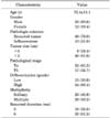
The demographic characteristics of the patients are summarized in Table 1. A total of 49 patients were confirmed to have tumor recurrence and 13 patients were confirmed to have inflammation according to the final pathologic outcome. Among 62 subjects finally enrolled, 44 had received prior intravesical instillation therapy after the initial TURBT. The overall tumor recurrence rate was 12.5% (49/393), and all recurrent cases were revealed as NMIBC. The mean interval from initial TURBT to recurrence was 4.8±1.5 months. The mean size of the tumor mass was 1.7±0.9 cm at the initial TURBT and 1.3±0.9 cm for the recurred NMIBC.
2. Comparison of each modality
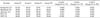
Of all 62 patients, 13, 33, and 32 cases showed positive results of urine cytology, CT, and urinalysis, respectively. The sensitivity of CT and urinalysis was 55.1% and 57.1%, which was higher than the 24.5% sensitivity for urine cytology (p=0.001 and p<0.001). The specificity of CT and urinalysis was 53.9% and 69.2%, which was lower than the 92.3% specificity for urine cytology (p=0.03 and p=0.43). The accuracy of CT and urinalysis was 54.8% and 59.7%, which was higher than the 38.7% accuracy for urine cytology (p=0.02 and p=0.018). However, PPV and NPV did not differ significantly between modalities (Table 2).
In patients with or without prior intravesical instillation therapy after initial TURBT, the sensitivity (29.7% and 23.8%, p=0.09) and specificity (85.7% and 93.1%, p=0.21) of urine cytology did not differ significantly. The sensitivity (56.8% and 58.3%, p=0.68) and specificity (71.4% and 66.7%, p=0.45) of urinalysis were also maintained similarly regardless of prior intravesical instillation therapy.
The sensitivity of group VI (75.5%) was higher than that of group V (59.2%, p=0.021) and group IV (61.2%, p=0.016). Other individual components except PPV differed significantly between groups IV and VI and between groups V and VI. However, in group VII, the sensitivity (77.6%) was statistically similar to that of group VI, a group without urine cytology (75.5%, p=0.87), and no significant difference in any component was observed between groups VI and VII (Table 3).
3. Cost analysis
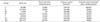
The results of the cost analysis are summarized in Table 4. Total cost and cost per cancer detected did not differ significantly between groups VI and VII (532,488 KRW and 594,017 KRW, p=0.807). After reimbursement of total cost, costs per cancer detected for the seven screening modalities were as follows: group I, 100,647 KRW; group II, 36,084 KRW; group III, 11,049 KRW; group IV, 62,783 KRW; group V, 42,021 KRW; group VI, 26,625; and group VII, 49,851 KRW, respectively. Under the Korean insurance system, the cost per cancer detected by using the combination modalities of urine cytology, urinalysis, and CT was almost doubled that of CT and urinalysis excluding urine cytology (group VI vs. group VII, p=0.041).
DISCUSSION
Because of the high recurrence and progression rates of bladder cancer, early diagnosis through regular clinical follow-up is critical. Early detection and treatment of bladder cancer increases the survival rate of patients. If bladder cancers are detected and treated while the cancer is confined within the bladder's inner lining but has not invaded the muscular bladder wall, the 5-year survival rate is 88%. If the cancer is detected after it has invaded the bladder wall but is still confined to the bladder, the 5-year survival rate drops to 63%. However, only about 50% of patients are diagnosed before the cancer has invaded the muscular bladder wall [14]. As a result, various modalities, such as urine cytology, urinalysis, CT, and cystoscopy, are required for early detection of bladder cancer.
Cystoscopy is considered the gold standard in the initial diagnosis or follow-up of bladder cancer. Although cystoscopy is regarded as the reference standard for the detection of tumor recurrence, a disadvantage of the method is its inability to detect recurrences in the upper tract. In addition, cystoscopy is an invasive procedure, which can be associated with subsequent complaints of anxiety, dysuria, and urinary tract infection [15]. Thus, for follow-up of patients, other modalities such as urine cytology, urinalysis, and CT are performed with cystoscopy.
Among these modalities, urine cytology has been used extensively as a noninvasive investigation in the diagnosis and follow-up of patients with urothelial malignancy [16]. Urine cytology is a valuable tool for detection of high-grade bladder cancer, particularly in a flat lesion that is difficult to detect on cystoscopy [17,18]. It has excellent specificity, but shows low sensitivity (30%-50%) depending on disease stage and differentiation grade [19]. However, the findings on cytological examination of urine are influenced by several factors, including proper specimen collection, inter-laboratory variation in specimen processing, subjective interpretation, and lack of a standard method of reporting [17]. An additional problem in urine cytology is that stones, infections, urinary tract instrumentation, and radiotherapy can all cause changes in urine cytology that mimic malignancy [20,21,22].
With this background, several novel studies were recently reported. A systematic review by Mowatt et al. [23] showed that involving cytology was considered to be the least worthwhile option and suggested that urine cytology is perhaps going to become even less valuable because of having the lowest sensitivity among other modalities such as FISH, Immunocyt, and NMP22 and in terms of cost-effectiveness. In addition, Lotan and Roehrborn [13], who reported a low sensitivity of urine cytology (20%-53%), suggested that other urine-based bladder tumor markers such as BTA, NMP22, and FISH could replace routine cytology during follow-up.
Today, there is a need to redefine the role of urine cytology and review the guidelines for its use in the evaluation of urological malignancies [24]. This is reflected by the change in the 2013 European Association of Urology guidelines for NMIBC, in which cytology is no longer recommended for all patients, but rather the role of voided urinary cytology is restricted to "prediction of high grade tumor before transurethral resection" [12].
In our study, sensitivity was highest when urine cytology was performed with CT and urinalysis (group VII). However, sensitivity was statistically similar when CT and urinalysis were performed in combination excluding urine cytology (group VI). There were also no statistically significant differences in PPV, NPV, or accuracy between group VI and group VII. In addition, cost and cost per cancer detected were reduced almost by half when all modalities excluding urine cytology were performed. Another study reported similar results. A systematic review and economic evaluation by Rodgers et al. [25] suggested that urine cytology has no application in ruling out malignancy or excluding patients from further investigation.
Our study had some limitations, most of which were innate to the retrospective design and limited number of patients with tumor recurrence. Because of this, we could not analyze the data according to the baseline risk stratification, which may have affected the reliability of the results for urine cytology. Furthermore, direct comparison of medical cost between nations is difficult owing to the complexity of the charging system. Under the Korean insurance system, most patients with pathologically proven NMIBC were fully covered by the mandatory medical insurance system, and they paid only 5% of the total medical cost after reimbursement. This difference may cause discordance in terms of cost-effective aspects.
Despite these limitations, at the time of the change in guidelines for urine cytology, this study was a new trial that evaluated practical and cost-effective aspects of urine cytology for the follow-up of bladder cancer. Conduct of more confirmatory studies that are prospective and include a large number of patients are required to determine the usefulness of urine cytology in the detection of tumor recurrence.
CONCLUSIONS
In our study, when performed in combination with urinalysis and CT in the investigation of patients for follow-up of bladder cancer, urine cytology showed low efficacy in terms of practicality and cost-effectiveness under the Korean insurance system. We consider that routine urine cytology may not be useful for follow-up of bladder cancer in terms of practicality and cost. Application to all patients may need to be adjusted.

 XML Download