

 PDF
PDF ePub
ePub Citation
 Print
Print


INTRODUCTION
Posterior urethral valve (PUV) is the most common cause of congenital bladder outlet obstruction in boys and causes renal failure in 25% to 30% of cases before adolescence. PUV is associated with considerable morbidity, including urinary tract infection (UTI), chronic renal failure, urinary incontinence, and even death [1-3]. The diagnosis is made on average in 1 in 1,285 fetal ultrasound screenings [4]. As a result of recent prenatal diagnosis, improvements in respiratory support and resuscitation at birth, and adequate management of end-stage renal disease, the mortality rate in patients with PUV has significantly decreased in the past four decades [5]. Endoscopic ablation of a PUV is the current gold standard of therapy, but approximately 10% to 30% of patients require a second procedure to achieve satisfactory valve ablation [3,6-8]. As animal models have proven, even partial outlet obstruction can lead to structural and functional deterioration in the detrusor muscle and bladder if the obstruction is not released soon enough [9-11], which indicates the need for close follow-up after valve ablation.
Although studies about prognostic factors for outcome of renal function after PUV ablation are available [8,12-15], to date, the effects of preoperative factors on the rate of residual valves have not been precisely addressed. In this study, therefore, we sought to evaluate any possible relationship between preoperative clinical and imaging findings of patients with PUV and remnant leaflets after their valve ablation to identify high-risk patients and achieve purposive follow-up.
MATERIALS AND METHODS
We evaluated 64 patients with clinical evidence of PUVs, confirmed by voiding cystourethrography (VCUG), who were admitted between 2008 and 2012 at the Shiraz University of Medical Sciences. Consent was obtained from patients or the patient's parents or legal guardian. Demographic information and medical history were taken. Preoperative evaluation included clinical examination, history of UTI, serum electrolytes, urinalysis, complete blood count, urine culture, serum creatinine, and radiographic evaluation (VCUG/ultrasonography and dimercaptosuccinic acid [DMSA] scan if necessary). All patients underwent endoscopic ablation of a PUV by the same pediatric urologist. Of these patients, 9 did not participate in follow-up sessions and were excluded from our study. The median patient age at the time of diagnosis was 10.0 months (range, 5 days to 120 months). Patients underwent control cystoscopy and, if needed, valve ablation was performed. Surgeon preference was to use an 11-Fr pediatric resectoscope (Karl Storz GmbH & Co. KG, Tuttlingen, Germany). When the urethra was too small for this instrument, a 3-Fr ureteric catheter with a metal stylet was used. Valves were ablated mainly at the 5, 7, and (in most cases) 12 o'clock positions. The end point of primary ablation was determined by visual assessment of destruction of the valve. A Foley catheter was left in place and was removed 24 to 48 hours after valve ablation. Patients were discharged on oral antibiotics and were observed every 3 to 6 months. We performed cystoscopy in all patients at a follow-up session, at least 3 months after valve ablation (range, 3-12 months). If there was significant outlet obstruction, further ablation was done. Patients were divided into two groups on the basis of observation of obstructive residual leaflets in a second cystoscopy to analyze the possible preoperative factors that were related to obstructive remnant leaflets. Group A had no evidence of obstructive remnant leaflets and in group B a second ablation was done owing to obstructive residual leaflets. We evaluated age at surgery, history of UTI, level of serum creatinine and blood urea nitrogen, specific gravity of urine, hemoglobin, presence of scarring in the DMSA scan, vesicoureteral reflux (VUR) and grade of reflux, renal parenchymal thickness, echogenicity of kidney, bladder wall thickness, renal cortical thickness, anteroposterior diameter of the renal pelvis, diameter of the distal ureter, and hydroureteronephrosis and side between groups.
For statistical analysis, Student t-test and Mann-Whitney test for quantitative numeric data comparison and chi-square test for categorical variables were adopted by using IBM SPSS ver. 19.0 (IBM Co., Armonk, NY, USA). A p-value of less than 0.05 was considered as significant.
RESULTS
Follow-up cystoscopy was performed at least 3 months (range, 3-12 months) after primary valve ablation in 55 boys with a 6.75-F pediatric cystoscope (Richard Wolf GmbH, Knittlingen, Germany). A total of 37 patients (67.3%) had no significant remnant leaflets (group A), whereas 18 boys (32.7%) required a second ablation as a result of valve remnants. The valve remnants were ablated by use of an 11-F pediatric resectoscope (Karl Storz GmbH & Co. KG) or a 3-Fr ureteric catheter with a metal stylet.
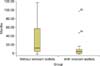
Preoperative findings were reviewed retrospectively for both groups. A pediatric resectoscope was used in 25 boys (67.6%) in group A and in 11 boys (61.1%) in group B, whereas in the other children a 3-Fr ureteric catheter was applied. In both groups of patients, no significant statistical relationship was observed between method of ablation (resectoscope versus urethral catheter method) and remnant leaflets (p=0.764). The median ages at the time of ablation for groups A and B were 15 and 7 months, respectively (Fig. 1). The Mann-Whitney test revealed a significant difference between the groups (p=0.017). Other clinical data that we assessed demonstrated no significant differences in children with and without remnant leaflets after valve ablation and are summarized in Table 1.
Radiographic characteristics of patients as shown by ultrasonography, DMSA scan, and VCUG were collected retrospectively. Sixteen patients had a DMSA scan and scarring was detected in all except one boy, with no significant predictive value for valve remnants (p=1). In VCUG, VUR and side and grade [16] of reflux were compared in the two groups. Significant differences were shown in the presence of VUR and grade 4 or 5 reflux (p<0.05). Our review of the ultrasound studies showed that the echogenicity of the renal parenchyma separated into normal echogenicity and hyperechogenicity [8], and echogenicity was higher in 94.4% of the preoperative ultrasounds of patients with residual leaflets (p=0.000).
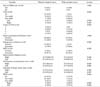
Data on renal parenchymal thickness, bladder wall thickness, renal cortical thickness (in the sagittal plane over a medullary pyramid, perpendicular to the capsule), hydroureteronephrosis and side of hydroureteronephrosis, anteroposterior diameter of the renal pelvis, and distal ureteral diameter demonstrated no influence on PUV ablation outcome in our patients (Table 2).
DISCUSSION
Most previous studies have focused on prognostic clinical and imaging factors in relation to the long-term outcome of renal function [6,8,13,17]. To our knowledge, this is the first study to compare preoperative factors regarding the presence of postoperative obstructive leaflets.
Although endoscopic primary valve ablation is the preferred initial surgical treatment in most patients with PUVs, the absence of obstructive residual valve remnants should be confirmed by careful clinical, radiological, and endoscopic evaluation after surgery [7,18]. Some investigators suggest VCUG to confirm the adequacy of valve ablation [3,19], whereas others recommend cystoscopy follow-up in all patients [1]. Both methods have advantages and disadvantages such as transient dysuria, enuresis, hematuria, and toileting anxiety after VCUG [20] and the need for general anesthesia in cystoscopy and the more invasive nature of cystoscopy than repeated VCUGs [3]. Whatever the method of follow-up, the necessity of post-ablation evaluation cannot be undervalued owing to the high incidence rate of remnant leaflets of 10%-30% in most studies [3,6-8], even as high as 51.6% in one case series [1].
To decrease the effect of technical components of the initial resection in the presence of residual valves, all surgeries in our study were performed by the same pediatric surgeon. Smeulders et al. [1] reported that micturating cystourethrography alone is inexact for excluding residual valve tissue (positive and negative predictive value of 56% and 50%, respectively), so we routinely perform cystoscopy in all of our patients. Given that there are no quantitative guidelines for the adequacy of valve ablation [3,7], our criterion was no visible obstructive residual valves in follow-up cystoscopy. The slightly higher rate of residual valves in our patients (32.7%) than in most cited studies (10% to 30%) [3,6-8] may have been because we applied follow-up cystoscopy in all patients after primary valve ablation, whereas others used follow-up cystoscopy only in cases with abnormal clinical or radiological findings [3,6,8].
Even though controversy exists about the role of age (at diagnosis and time of PUV surgery), it has been mentioned as one of the predictive factors for renal outcome after valve ablation [8,13,21]. We found no published studies that assessed age as a prognostic factor for remnant leaflets, but in our patients, age (at the time of surgery) showed a significant effect on the presence of residual leaflets in follow-up cystoscopy. Specifically, younger patients showed a higher probability of remnant leaflets (p=0.017). The cause of this difference may be the narrower urethra in younger children, which limits the surgeon's visibility and complicates the endoscopic maneuver for PUV ablation owing to fear of causing stricture. In any event, careful and closer follow-up in younger children seems necessary to achieve better long-term renal outcomes.
Besides age at diagnosis, of the other factors that we analyzed, echogenicity of kidney, presence of reflux, and grade of reflux differed significantly between the two groups (p<0.05). Other studies have shown the hyperechogenicity of renal parenchyma as a factor that may help to predict long-time prognosis of renal insufficiency [8,13] but did not comment on it as a factor affecting the consequences of valve ablation. In our patients, 17 boys (94.4%) with residual leaflets in follow-up cystoscopy showed increased renal echogenicity in comparison with 6 patients (16.2%) in the other group, with a p-value less than 0.05. In our patients, 33 children showed evidence of VUR (16 boys in group A and 17 in group B) and the incidence of grade 4 and 5 reflux was significantly higher in patients with obstructive remnant leaflets (Table 2).
To exclude the tendency in infants to have more echogenic kidneys separate from renal function [22], we analyzed this factor in three age groups (under 12, 12 to 48, and over 48 months), which revealed a significant difference in the first two groups (p<0.05). We suppose that this statistical relationship may be due to longer and thicker valves preoperatively that caused more obstruction initially, which was followed by high grades of VUR as well as hyperechoic kidney that persisted after the first ablation. The presence of VUR and hyperechogenicity of the kidney can affect the long-term prognosis of renal function and should be considered in the management of patients with PUV.
Method of ablation, renal parenchymal thickness, bladder wall thickness, renal cortical thickness, hydroureteronephrosis, anteroposterior diameter of the renal pelvis and ureter, serum creatinine, and history of UTI demonstrated no relationship with residual leaflets in our patients. Considering the lack of studies in this field, it seems that more research is inevitable to facilitate proper screening and the detection of patients with residual leaflets.
The need for confirmation of complete resection of PUV is undeniable because valve remnants may result in persistent outflow obstruction or renal failure through time and should be detected quickly to minimize the deterioration of kidney or bladder function. The timing of follow-up sessions in most studies is 3 to 6 months after ablation. However, owing to decreases in function and structural deformities in the bladder as a result of partial obstruction, even after as short a period as 2 weeks in animal models [9-11], earlier follow-up in the first month after ablation, especially in patients at high risk of residual valves, seems more advantageous.
There may be other operative or preoperative factors, in addition to those we evaluated in our study, with the potential to affect the end result of initial valve ablation and residual leaflets. These should be evaluated in other studies with a larger number of cases. This may add to and confirm the prognostic factors for remnant valves. The results of such research can help to provide a better algorithm for management of PUVs and achieve purposive follow-up for high-risk patients instead of invasive procedures.
CONCLUSIONS
Younger age at surgery time, hyperechogenicity of the renal parenchyma, presence of VUR, and grade of reflux before surgery in our patients had a significant relationship with residual valves, but more research is needed to confirm these results. More studies may result in enhanced management of patients at high risk of residual valves after PUV ablation, because the sooner the obstruction is entirely resolved, the better the long-term outcome.
 XML Download