

 PDF
PDF ePub
ePub Citation
 Print
Print


INTRODUCTION
Prostatic stromal sarcoma (PSS) is quite rare, accounting for only 0.1% to 0.2% of all prostate cancers [1]. In 1998 Gaudin et al. [2] grouped sarcoma and related proliferative lesions of the specialized prostatic stroma into two clinicopathologic categories: prostatic stromal proliferation of uncertain malignant potential (PSPUMP) and PSS. We recently experienced two cases of PSS that were surgically treated by use of robot-assisted laparoscopic radical prostatectomy (RALP) and open radical cysto-prostatectomy with an ileal conduit.
CASE REPORTS
1. Case 1
A 29-year-old man was referred from another urologic clinic for severe perineal pain with a weak urine stream and dysuria. When he visited Kyungpook National University Medical Center, he reported a history of acute urinary retention and had a Foley catheter in place for about 1 month. A digital rectal examination (DRE) showed a relatively enlarged prostate with tenderness on pressure. An abdominal computed tomography (CT) scan revealed an 8×7×4.5-cm-sized moderately enlarged prostate. The estimated volume of the prostate by transrectal ultrasound (TRUS) and the prostate-specific antigen (PSA) level were 80 mL and 8.0 ng/mL, respectively. Considering the relatively young age of the patient, acute bacterial prostatitis was suspected, but the patient's clinical symptoms did not exactly line up with acute bacterial prostatitis. To relieve the patient's symptoms and drain the urine, we performed a suprapubic cystostomy and started the patient on intravenous antibiotics, but his symptoms were not relieved. Repeated TRUS and DRE suggested a prostate cold abscess, but prostate malignancy could not be ruled out. Thus, we performed prostate magnetic resonance imaging (MRI).
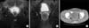
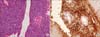
The prostate MRI showed a huge prostatic mass that obstructed the bladder neck (Fig. 1). According to the results of the MRI, we performed transurethral resection of the prostate (TURP) to relieve the bladder outlet obstruction and to confirm the pathologic nature of the mass. The pathology report from TURP indicated high-grade sarcoma with extensive necrosis. We performed chest CT and a whole-body bone scan to evaluate the extensiveness of the disease to determine the feasibility of surgery. No metastatic lesions were found on the chest CT or whole-body bone scan. In the prostate MRI, the whole prostate was shown to be involved by the malignant lesion and rectal wall invasion was suspected. Considering the particularly young age of the patient and the lack of evidence of bladder invasion, we decided to perform RALP and extended pelvic lymph node dissection, with an expectation of sparing the bladder. This decision was made with the informed consent of the patient and the patient's legal guardian. During the operation, rectal wall invasion of sarcoma was noted. Accordingly, the anterior wall of the rectum was resected and repaired. Gross examination of the radical prostatectomy specimen showed a 12×8.0×7.0-cm-sized relatively well-demarcated mass containing hemorrhage. The final histopathologic analysis revealed PSS with invasion to both seminal vesicles. The rectal mucosa and prostatic resection margins were positive for tumor, but none of the lymph nodes had tumor present (Fig. 2). After 1 month of follow-up, the PSS had recurred and metastasized to the lung and liver. The patient's symptoms and the recurred mass in the pelvis became aggravated at 45 days after the operation. The patient was transferred to another hospital for hospice care.
2. Case 2
A 45-year-old man was referred from another urology clinic for voiding difficulty lasting about 2 months. He had a history of gross hematuria and hematospermia lasting for 7 months until the present. A DRE showed a relatively enlarged prostate without tenderness on pressure. An abdominal CT scan revealed a 10×12×6.4-cm-sized huge solid mass with focal cystic change in the pelvic cavity that was displacing the right ureter and with periprostatic fat infiltration and probable invasion into the urinary bladder, seminal vesicle, and rectum (Fig. 1). His estimated volume of the prostate by TRUS and his PSA level were 575 mL and 1.19 ng/mL, respectively.
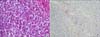
TRUS-guided prostate biopsy was performed to determine the nature of the tumor, which was revealed as a malignant spindle cell tumor with rhabdoid features, most likely PSS. We performed chest CT and a bone scan to determine the stage of the tumor and whether it had metastasized to other organs. The chest CT and bone scan revealed no metastasis. Considering the patient's age and the aggressiveness of the disease, open radical cysto-prostatectomy with an ileal conduit was performed. During the operation, rectal wall invasion of the sarcoma was noted and segmental resection of the rectum was performed with ileostomy. Gross examination of the radical cysto-prostatectomy specimen showed a 10×8.5×6.0-cm-sized prostate containing a bright yellow friable soft mass with central necrosis and a 9.0×6.0×5.0-cm-sized bladder with grossly noted PSS invasion. The final histopathologic analysis revealed PSS with invasion to both seminal vesicles and the subserosal layer of the rectum, but none of the lymph nodes had tumor present and the resection margins were negative (Fig. 3). The patient is now undergoing adjuvant radiotherapy and there have been no other complications or signs of recurrence after 3 months of follow-up.
DISCUSSION
Primary prostate sarcomas are rare tumors that account for less than 0.1% of primary prostate malignancy in adults [3]. Leiomyosarcoma is the most common histopathological type of prostate sarcomas in adults [3]. PSS is an even rarer form of sarcomas and the published cases number less than 30 [4]. Generally, a prostate stromal lesion is classified into two categories: PSPUMP and PSS. This division was made by Gaudin et al. [2] in 1998 and by Herawi and Epstein [5] in 2006 on the basis of the degree of cellularity, mitotic figures, necrosis, and stromal overgrowth. Local recurrence and distant metastasis are quite common in PSS; however, the natural history of PSPUMP is not well known. Hossain et al. [6] reported that PSPUMP is a benign disease entity and that in their series no patients developed evidence of sarcomatous transformation or malignancy. PSS is an aggressive disease with a poor prognosis. The median survival time is less than 15 months, and only 10% of patients survive for more than 3 years [1]. Sexton et al. [3] reported that the overall 5-year actuarial survival rate for adult patients with prostate sarcoma was 38%. In our cases, the patient who was treated with RALP experienced recurrence of the tumor at 30 days after the surgery and was discharged for hospice care. The recurrence was aggressive in a short period with massive tumor formation in the pelvic cavity at 45 days after the operation.
Few prostate malignancies except conventional adenocarcinoma have been treated with RALP owing to the aggressiveness and poor prognosis of such malignancies. Especially in the case of PSS, the low prevalence of the disease, the highly advanced state at the time of diagnosis, and the low survival rate are among the reasons why few surgeons make RALP their first choice. In our first described case, however, after considering several aspects such as the difficulty of anastomosis after wide excision, functional issues like continence and sexual potency, the relatively young age of the patient, the lack of evidence of bladder invasion, and the strong wishes of the guardian of the patient, we decided to perform RALP. We experienced some difficulties resulting from the large prostate size and adhesions; however, en bloc resection of the tumor was feasible. The tumor in the second case was larger than the tumor in the first case and CT revealed invasion to multiple other adjacent organs, especially the bladder. Thus, considering the extent of the tumor, open radical cysto-prostatectomy with an ileal conduit was performed with adjuvant radiotherapy.
Early diagnosis and complete surgical resection are the only means by which to expect a cure [3]. Our recent experiences suggest that, in the case of PSS, surgeons should approach the disease from the viewpoint of extensive surgical resection of the cancer rather than considerations of quality of life for the best chance of cure.
 XML Download